Want to create or adapt books like this? Learn more about how Pressbooks supports open publishing practices.
Chapter 29 – Review
29.1 Chromatography Basics
What is the purpose of chromatography? Check answer[1]
Describe the purpose of the stationary phase and the mobile phase. Check answer[2]
Why do substances travel at different rates? Check answer[3]
29.2 Thin Layer (TLC) and Paper Chromatography (PC)
Try your own paper chromatography using household items. Stationary phase – paper towel or coffee filter, Mobile phase – water or rubbing alcohol, Mixture – water soluble markers, water soluble wet paint, food colouring. Describe the results and how you might adjust the experiment to get different results.
Calculate the Rf values for the following separation: Solvent travelled 5.8 cm, Compound A travelled 2.1 cm, Compound B travelled 2.3 cm, Compound C travelled 4.0 cm, and Compound D travelled 5.6 cm. What conclusions can you make about Compounds A through D? Check answer[4]
Describe some of the factors that will influence a component’s Rf value.
29.3 Chromatographic Columns
Research some uses of ion exchange chromatographic (IEC) columns and size exclusion chromatographic (SEC) columns.
What do all types of chromatographic columns have in common? Check answer[5]
What are some benefits of chromatographic columns over paper chromatography or thin-layer chromatography? Check answer[6]
29.4 Chromatography Technology
Explain the principles of gas chromatography (GC).
Explain the principles of high-performance liquid chromatography (HPLC).
Consider what a scientist may need to consider when choosing a chromatography method. Complete this chart. Research may be needed.
Chart 1: What to consider when choosing a chromatography method. Columns are left blank for you to fill in.
Chromatography Method
PC/TLC
IEC
GC
HPLC
Can the method be used to physically separate sample into different containers?
Can the method be used to check purity of sample?
Can the method be used in combination with another method to determine compound identity?
What type of samples are required? Consider states, amount of sample, polarities, charges.
Is the method inexpensive or expensive to run?
Is the method readily available or does it require specialized lab equipment?
How long does the method take to get a result? (quick or slow)
Using the video IR spectra practice | Spectroscopy | Organic chemistry | Khan Academy – YouTube, predict which molecule has the shown IR spectrum. Spectrum 1 – stop the video at 0:16. Determine which of the three molecules is the correct one. Watch the video for an explanation of the answer. Spectrum 2 – starts at 2:00, stop at 2:05. Determine which of the three molecules is the correct one. Watch the video for an explanation of the answer. Spectrum 3 – starts at 3:34, stop at 3:42. Determine which of the three molecules is the correct one. Watch the video for an explanation of the answer.
What are three things that can be determined using MS? Check answer[15]
Assume that you have two unlabeled samples, one of methylcyclohexane and the other of ethylcyclopentane. How could you use mass spectrometry to tell them apart? The mass spectra of both are shown. Check answer[16] (credit: Organic Chemistry (OpenStax),CC BY-NC-SA 4.0)
The sex hormone testosterone contains only C, H, and O and has a mass of 288.2089 amu, as determined by high-resolution mass spectrometry. What is the likely molecular formula of testosterone? Check answer[17]
How many non-equivalent hydrogens are in the following molecules? How many different signals will you see in a H1 NMR spectrum? Check answer[20]
CH3CH2CH2Br
CH3OCH2C(CH3)3
Ethyl Benzene
2-methyl-1-hexene
Methyl 2,2-dimethylpropanoate (CH3)2CCO2CH3 has two peaks in its 1H NMR spectrum. What are their approximate chemical shifts? Check answer[21]
Each of the following compounds has a single 1H NMR peak. Approximately where would you expect each compound to absorb? Check answer[22]
a)
(credit: Organic Chemistry (OpenStax),CC BY-NC-SA 4.0)
How many peaks would you expect in the 1H NMR spectrum of 1,4-dimethylbenzene (para-xylene, or p-xylene)? What ratio of peak areas would you expect on integration of the spectrum? Check answer[23] (credit: Organic Chemistry (OpenStax),CC BY-NC-SA 4.0)
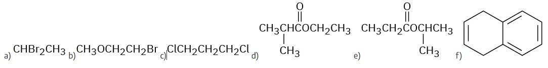
Predict the splitting patterns you would expect for each proton in the following molecules: Check answer[24]
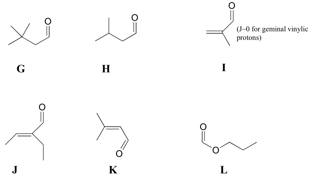
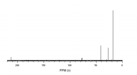
Predict the number of carbon resonance lines you would expect in the 13C NMR spectra of the following compounds: Check answer[27] a) Methylcyclopentane b) 1-Methylcyclohexene c) 1,2-Dimethylbenzene d) 2-Methyl-2-butene e) (credit: Organic Chemistry (OpenStax),CC BY-NC-SA 4.0)
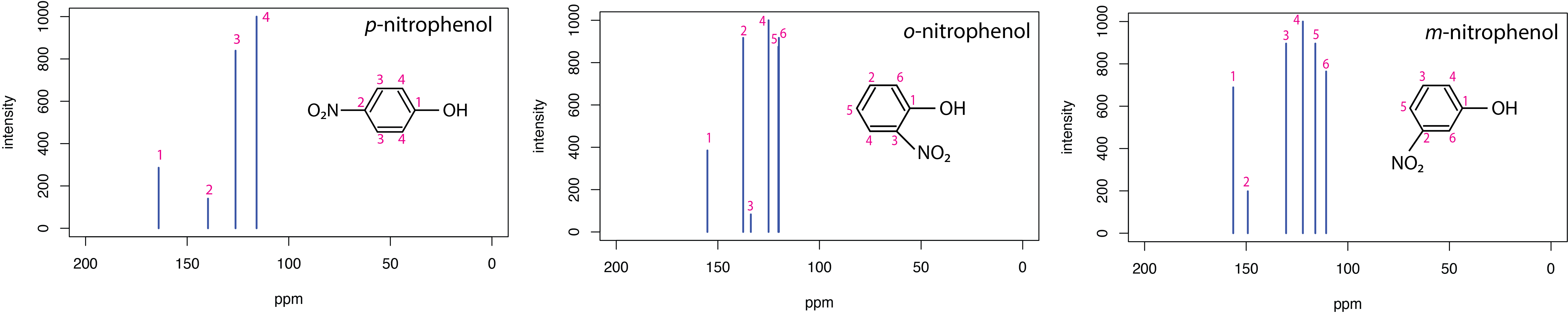
This figure shows 13C NMR spectrum for three related molecules: p-nitrophenol, o-nitrophenol, and m-nitrophenol. Identify some 13C NMR differences between these isomers. Try to explain the differences using the molecule structure.
The Chemical Detectives app on Chemical Detectives – Apps on Google Play [New tab] and Chemical Detectives on the App Store [New tab] allows users to browse various types of spectra (IR, MS, 1H NMR, 13C NMR, and elemental microanalysis) for simple organic compounds and also complete quizzes about various compounds based on their spectra. Customize of types of functional groups is possible (e.g. only hydrocarbons or only alcohols).
A solution of analyte, with molar absorptivity of 676 cm-1 M-1, is placed in a sample cell that has a pathlength of 1.00 cm. At a wavelength of 490 nm, the solution’s absorbance is 0.228. What is the analyte’s concentration? Check answer[30]
Chromatography separates a mixture into it's dissolved components. ↵
The stationary phase (typically a solid) does not move and holds onto the mixture and it's components. The mobile phase moves (typically a gas or liquid) and drags the mixture's components with it at varying rates. ↵
The rate of travel depends on the components affinity for the stationary phase over the mobile phase. More affinity to the stationary phase means the component will travel slower. ↵
Compound A Rf = 0.36; Compound B Rf = 0.40; Compound C Rf = 0.69; Compound D Rf = 0.97. Compound D has high affinity to the mobile phase and low affinity for the stationary phase. Compounds A and B have similar affinities for the stationary phase. ↵
All columns require a stationary phase and a mobile phase of different properties to separate the applied mixture. ↵
Columns allow for collection of the mixture's components whereas TLC/PC are only visual and can't capture the components. PC/TLC are typically much quicker and cheaper to perform. ↵
The pattern in which matter absorbs or emits radiation. ↵
A) carbonyl group in aldehydes, ketones, carboxylic acids, amides, esters B) aromatics, amines, nitro, C) carboxylic acids ↵
A) A OH peak will be present around 3300 cm-1 for methanol and will be absent in the ether. B) 1-pentene will have an alkene peak around 1650 cm-1 for the C=C and there will be another peak around 3100 cm-1 for the sp2 C-H group on the alkene C) Cannot distinguish these two isomers. They both have the same functional groups and therefore would have the same peaks on an IR spectra. ↵
1680 cm-1 for carbonyl group in carboxylic acid, 2820 cm-1 for OH in carboxylic acid, and 2925 cm-1 for CH in aromatics. ↵
Strategy: Identify the functional groups in each molecule, and then check an IR table of values. (a) Absorptions: 3400 to 3650 cm–1 (O–H), 3020 to 3100 cm–1 (=C–H), 1640 to 1680 cm–1 (C═C). This molecule has an alcohol O–H group and an alkene double bond. (b) Absorptions: 3300 cm–1 (≡C–H), 2100 to 2260 cm–1 (C≡C), 1735 cm–1 (C═O). This molecule has a terminal alkyne triple bond and a saturated ester carbonyl group. ↵
Strategy: All IR spectra have many absorptions, but those useful for identifying specific functional groups are usually found in the region from 1500 cm–1 to 3300 cm–1. Pay particular attention to the carbonyl region (1670 to 1780 cm–1), the aromatic region (1660 to 2000 cm–1), the triple-bond region (2000 to 2500 cm–1), and the C–H region (2500 to 3500 cm–1). Solution: The spectrum shows an intense absorption at 1725 cm–1 due to a carbonyl group (perhaps an aldehyde, –CHO), a series of weak absorptions from 1800 to 2000 cm–1 characteristic of aromatic compounds, and a C–H absorption near 3030 cm–1, also characteristic of aromatic compounds. In fact, the compound is phenylacetaldehyde.
↵
Strategy: Look at the possible structures and decide on how they differ. Then think about how any of these differences in structure might give rise to differences in mass spectra. Methyl cyclohexane, for instance, has a –CH3 group, and ethylcyclopentane has a –CH2CH3 group, which should affect the fragmentation patterns. Solution: Both mass spectra show molecular ions at M+ = 98, corresponding to C7H14, but they differ in their fragmentation patterns. Sample A has its base peak at m/z = 69, corresponding to the loss of a CH2CH3 group (29 mass units), but B has a rather small peak at m/z = 69. Sample B shows a base peak at m/z = 83, corresponding to the loss of a CH3 group (15 mass units), but sample A has only a small peak at m/z = 83. We can therefore be reasonably certain that A is ethylcyclopentane and B is methylcyclohexane.
↵
Strategy: Identify the types of hydrogens in the molecule, and note whether each is alkyl, vinylic, or next to an electronegative atom. Then predict where each absorbs. Solution: The –OCH3 protons absorb around 3.5 to 4.0 δ because they are on carbon bonded to oxygen. The (CH3)3C– protons absorb near 1.0 δ because they are typical alkane-like protons. ↵
Strategy: Identify the distinct carbons in the molecule, and note whether each is alkyl, vinylic, aromatic, or in a carbonyl group. Then predict where each absorbs. Solution: Ethyl acrylate has five chemically distinct carbons: two different C=C, one C=O, one O–C, and one alkyl C. The likely absorptions are:
The actual absorptions are at 14.1, 60.5, 128.5, 130.3, and 166.0 δ. ↵
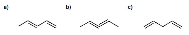
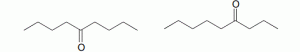
UV because the molecule on the left has conjugation whereas the molecule on the right does not. Their IR spectra would be very similar as their functional groups are similar. ↵
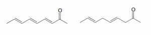
MS because both molecules have the same functional groups and neither have conjugation. The MS spectra will show different fragments due to the location of the carbonyl group. ↵


↵
The actual absorptions are at 14.1, 60.5, 128.5, 130.3, and 166.0 δ. ↵
