11. Biochemistry of the Genome
11.3 Structure and Function of RNA
Learning Objectives
- Describe the biochemical structure of ribonucleotides
- Describe the similarities and differences between RNA and DNA
- Describe the functions of the three main types of RNA used in protein synthesis
- Explain how RNA can serve as hereditary information
Structurally speaking, ribonucleic acid (RNA), is quite similar to DNA. However, whereas DNA molecules are typically long and double stranded, RNA molecules are much shorter and are typically single stranded. RNA molecules perform a variety of roles in the cell but are mainly involved in the process of protein synthesis (translation) and its regulation.
RNA Structure
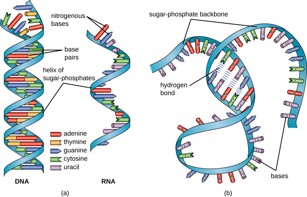
RNA is typically single stranded and is made of ribonucleotides that are linked by phosphodiester bonds. A ribonucleotide in the RNA chain contains ribose (the pentose sugar), one of the four nitrogenous bases (A, U, G, and C), and a phosphate group. The subtle structural difference between the sugars gives DNA added stability, making DNA more suitable for storage of genetic information, whereas the relative instability of RNA makes it more suitable for its more short-term functions. The RNA-specific pyrimidine uracil forms a complementary base pair with adenine and is used instead of the thymine used in DNA. Even though RNA is single stranded, most types of RNA molecules show extensive intramolecular base pairing between complementary sequences within the RNA strand, creating a predictable three-dimensional structure essential for their function (Figure 11.20 and Figure 11.21).
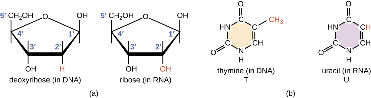
- How does the structure of RNA differ from the structure of DNA?
Functions of RNA in Protein Synthesis
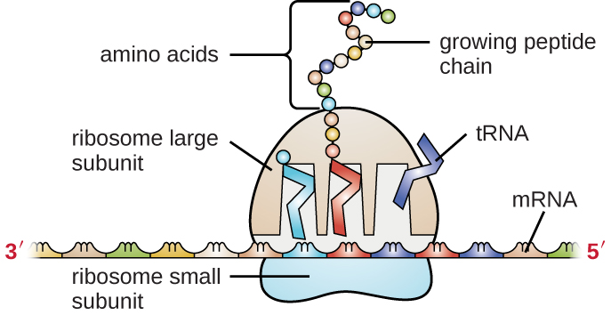
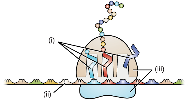
Cells access the information stored in DNA by creating RNA to direct the synthesis of proteins through the process of translation. Proteins within a cell have many functions, including building cellular structures and serving as enzyme catalysts for cellular chemical reactions that give cells their specific characteristics. The three main types of RNA directly involved in protein synthesis are messenger RNA (mRNA), ribosomal RNA (rRNA), and transfer RNA (tRNA).
In 1961, French scientists François Jacob and Jacques Monod hypothesized the existence of an intermediary between DNA and its protein products, which they called messenger RNA.[1] Evidence supporting their hypothesis was gathered soon afterwards showing that information from DNA is transmitted to the ribosome for protein synthesis using mRNA. If DNA serves as the complete library of cellular information, mRNA serves as a photocopy of specific information needed at a particular point in time that serves as the instructions to make a protein.
The mRNA carries the message from the DNA, which controls all of the cellular activities in a cell. If a cell requires a certain protein to be synthesized, the gene for this product is “turned on” and the mRNA is synthesized through the process of transcription (see RNA Transcription). The mRNA then interacts with ribosomes and other cellular machinery (Figure 11.22) to direct the synthesis of the protein it encodes during the process of translation (see Protein Synthesis). mRNA is relatively unstable and short-lived in the cell, especially in prokaryotic cells, ensuring that proteins are only made when needed.
rRNA and tRNA are stable types of RNA. In prokaryotes and eukaryotes, tRNA and rRNA are encoded in the DNA, then copied into long RNA molecules that are cut to release smaller fragments containing the individual mature RNA species. In eukaryotes, synthesis, cutting, and assembly of rRNA into ribosomes takes place in the nucleolus region of the nucleus, but these activities occur in the cytoplasm of prokaryotes. Neither of these types of RNA carries instructions to direct the synthesis of a polypeptide, but they play other important roles in protein synthesis.
Ribosomes are composed of rRNA and protein. As its name suggests, rRNA is a major constituent of ribosomes, composing up to about 60% of the ribosome by mass and providing the location where the mRNA binds. The rRNA ensures the proper alignment of the mRNA, tRNA, and the ribosomes; the rRNA of the ribosome also has an enzymatic activity (peptidyl transferase) and catalyzes the formation of the peptide bonds between two aligned amino acids during protein synthesis. Although rRNA had long been thought to serve primarily a structural role, its catalytic role within the ribosome was proven in 2000.[2] Scientists in the laboratories of Thomas Steitz (1940–) and Peter Moore (1939–) at Yale University were able to crystallize the ribosome structure from Haloarcula marismortui, a halophilic archaeon isolated from the Dead Sea. Because of the importance of this work, Steitz shared the 2009 Nobel Prize in Chemistry with other scientists who made significant contributions to the understanding of ribosome structure.
Transfer RNA is the third main type of RNA and one of the smallest, usually only 70–90 nucleotides long. It carries the correct amino acid to the site of protein synthesis in the ribosome. It is the base pairing between the tRNA and mRNA that allows for the correct amino acid to be inserted in the polypeptide chain being synthesized (Figure 11.23). Any mutations in the tRNA or rRNA can result in global problems for the cell because both are necessary for proper protein synthesis (Table 11.1).
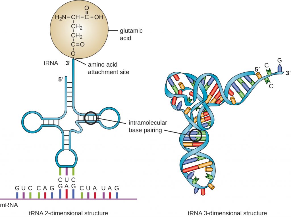
Table 11.1. Structure and Function of RNA
| Structure and Function of RNA | |||
|---|---|---|---|
| mRNA | rRNA | tRNA | |
| Structure | Short, unstable, single-stranded RNA corresponding to a gene encoded within DNA | Longer, stable RNA molecules composing 60% of ribosome’s mass | Short (70-90 nucleotides), stable RNA with extensive intramolecular base pairing; contains an amino acid binding site and an mRNA binding site |
| Function | Serves as intermediary between DNA and protein; used by ribosome to direct synthesis of protein it encodes | Ensures the proper alignment of mRNA, tRNA, and ribosome during protein synthesis; catalyzes peptide bond formation between amino acids | Carries the correct amino acid to the site of protein synthesis in the ribosome |
- What are the functions of the three major types of RNA molecules involved in protein synthesis?
RNA as Hereditary Information
Although RNA does not serve as the hereditary information in most cells, RNA does hold this function for many viruses that do not contain DNA. Thus, RNA clearly does have the additional capacity to serve as genetic information. Although RNA is typically single stranded within cells, there is significant diversity in viruses. Rhinoviruses, which cause the common cold; influenza viruses; and the Ebola virus are single-stranded RNA viruses. Rotaviruses, which cause severe gastroenteritis in children and other immunocompromised individuals, are examples of double-stranded RNA viruses. Because double-stranded RNA is uncommon in eukaryotic cells, its presence serves as an indicator of viral infection. The implications for a virus having an RNA genome instead of a DNA genome are discussed in more detail in the sections on the viruses: Sec. 6.1, 6.2 & 6.3.
Key Takeaways
- Ribonucleic acid (RNA) is typically single stranded and contains ribose as its pentose sugar and the pyrimidine uracil instead of thymine. An RNA strand can undergo significant intramolecular base pairing to take on a three-dimensional structure.
- There are three main types of RNA, all involved in protein synthesis.
- Messenger RNA (mRNA) serves as the intermediary between DNA and the synthesis of protein products during translation.
- Ribosomal RNA (rRNA) is a type of stable RNA that is a major constituent of ribosomes. It ensures the proper alignment of the mRNA and the ribosomes during protein synthesis and catalyzes the formation of the peptide bonds between two aligned amino acids during protein synthesis.
- Transfer RNA (tRNA) is a small type of stable RNA that carries an amino acid to the corresponding site of protein synthesis in the ribosome. It is the base pairing between the tRNA and mRNA that allows for the correct amino acid to be inserted in the polypeptide chain being synthesized.
- Although RNA is not used for long-term genetic information in cells, many viruses do use RNA as their genetic material.
Multiple Choice
Drag and Drop
True/False
Short Answer
- What are the differences between DNA nucleotides and RNA nucleotides?
- How is the information stored within the base sequence of DNA used to determine a cell’s properties?
- How do complementary base pairs contribute to intramolecular base pairing within an RNA molecule?
- If an antisense RNA has the sequence 5ʹAUUCGAAUGC3ʹ, what is the sequence of the mRNA to which it will bind? Be sure to label the 5ʹ and 3ʹ ends of the molecule you draw.
- Why does double-stranded RNA (dsRNA) stimulate RNA interference?
Critical Thinking
- Why does it make sense that tRNA and rRNA molecules are more stable than mRNA molecules?
- Identify the location of mRNA, rRNA, and tRNA in the figure.
Media Attributions
- OSC_Microbio_10_03_RNAStruct
- OSC_Microbio_10_03_RNAStruct2
- OSC_Microbio_10_03_RNAFunct
- OSC_Microbio_10_03_tRNA
- OSC_Microbio_10_03_RNA_img
