5. The Eukaryotes of Microbiology
5.2 Fungi
Learning Objectives
- Explain why the study of fungi such as yeast and moulds is within the discipline of microbiology
- Describe the unique characteristics of fungi
- Describe examples of asexual and sexual reproduction of fungi
- Compare the major groups of fungi in this chapter, and give examples of each
- Identify examples of the primary causes of infections due to yeasts and moulds
- Identify examples of toxin-producing fungi
- Classify fungal organisms according to major groups
The fungi comprise a diverse group of organisms that are heterotrophic and typically saprophytic. In addition to the well-known macroscopic fungi (such as mushrooms and moulds), many unicellular yeasts and spores of macroscopic fungi are microscopic. For this reason, fungi are included within the field of microbiology.
Fungi are important to humans in a variety of ways. Both microscopic and macroscopic fungi have medical relevance, with some pathogenic species that can cause mycoses (illnesses caused by fungi). Some pathogenic fungi are opportunistic, meaning that they mainly cause infections when the host’s immune defences are compromised and do not normally cause illness in healthy individuals. Fungi are important in other ways. They act as decomposers in the environment, and they are critical for the production of certain foods such as cheeses. Fungi are also major sources of antibiotics, such as penicillin from the fungus Penicillium.
Characteristics of Fungi
Fungi have well-defined characteristics that set them apart from other organisms. Most multicellular fungal bodies, commonly called moulds, are made up of filaments called hyphae. Hyphae can form a tangled network called a mycelium and form the thallus (body) of fleshy fungi. Hyphae that have walls between the cells are called septate hyphae; hyphae that lack walls and cell membranes between the cells are called nonseptate or coenocytic hyphae). (Figure 5.18).
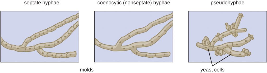
In contrast to moulds, yeasts are unicellular fungi. The budding yeasts reproduce asexually by budding off a smaller daughter cell; the resulting cells may sometimes stick together as a short chain or pseudohypha (Figure 5.18). Candida albicans is a common yeast that forms pseudohyphae; it is associated with various infections in humans, including vaginal yeast infections, oral thrush, and candidiasis of the skin.
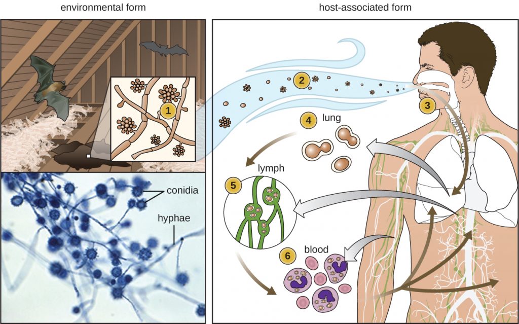
Some fungi are dimorphic, having more than one appearance during their life cycle. These dimorphic fungi may be able to appear as yeasts or moulds, which can be important for infectivity. They are capable of changing their appearance in response to environmental changes such as nutrient availability or fluctuations in temperature, growing as a mould, for example, at 25 °C (77 °F), and as yeast cells at 37 °C (98.6 °F). This ability helps dimorphic fungi to survive in diverse environments. Histoplasma capsulatum, the pathogen that causes histoplasmosis, a lung infection, is an example of a dimorphic fungus (Figure 5.19).
There are notable unique features in fungal cell walls and membranes. Fungal cell walls contain chitin, as opposed to the cellulose found in the cell walls of plants and many protists. Additionally, whereas animals have cholesterol in their cell membranes, fungal cell membranes have different sterols called ergosterols. Ergosterols are often exploited as targets for antifungal drugs.
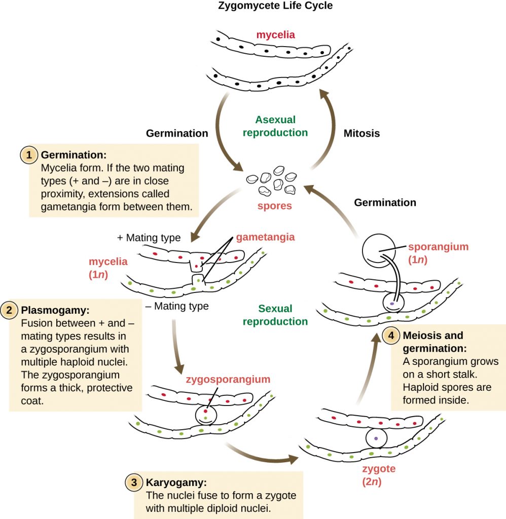
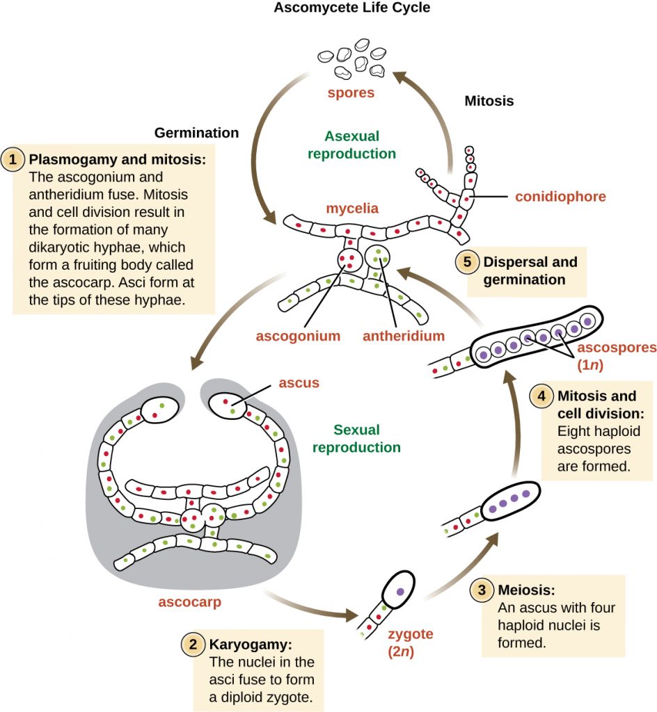
Fungal life cycles are unique and complex. Fungi reproduce sexually either through cross- or self-fertilization. Haploid fungi form hyphae that have gametes at the tips. Two different mating types (represented as “+ type” and “– type”) are involved. The cytoplasms of the + and – type gametes fuse (in an event called plasmogamy), producing a cell with two distinct nuclei (a dikaryotic cell). Later, the nuclei fuse (in an event called karyogamy) to create a diploid zygote. The zygote undergoes meiosis to form spores that germinate to start the haploid stage, which eventually creates more haploid mycelia (Figure 5.20). Depending on the taxonomic group, these sexually produced spores are known as zygospores (in Zygomycota), ascospores (in Ascomycota), or basidiospores (in Basidiomycota) (Figure 5.21).
Fungi may also exhibit asexual reproduction by mitosis, mitosis with budding, fragmentation of hyphae, and formation of asexual spores by mitosis. These spores are specialized cells that, depending on the organism, may have unique characteristics for survival, reproduction, and dispersal. Fungi exhibit several types of asexual spores and these can be important in classification.
- Is a dimorphic fungus a yeast or a mould? Explain.
CLINICAL FOCUS: Part 2
Sarah is relieved the ringworm is not an actual worm, but wants to know what it really is. The physician explains that ringworm is a fungus. He tells her that she will not see mushrooms popping out of her skin, because this fungus is more like the invisible part of a mushroom that hides in the soil. He reassures her that they are going to get the fungus out of her too.
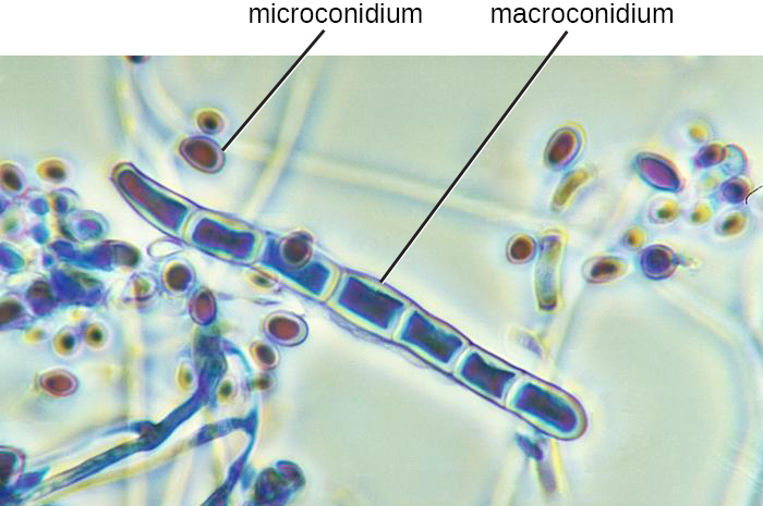
The doctor cleans and then carefully scrapes the lesion to place a specimen on a slide. By looking at it under a microscope, the physician is able to confirm that a fungal infection is responsible for Sarah’s lesion. In Figure 5.26, it is possible to see macro- and microconidia in Trichophyton rubrum. Cell walls are also visible. Even if the pathogen resembled a helminth under the microscope, the presence of cell walls would rule out the possibility because animal cells lack cell walls.
The doctor prescribes an antifungal cream for Sarah’s mother to apply to the ringworm. Sarah’s mother asks, “What should we do if it doesn’t go away?”
- Can all forms of ringworm be treated with the same antifungal medication?
Go back to the previous Clinical Focus box. Go to the next Clinical Focus box.
Fungal Diversity
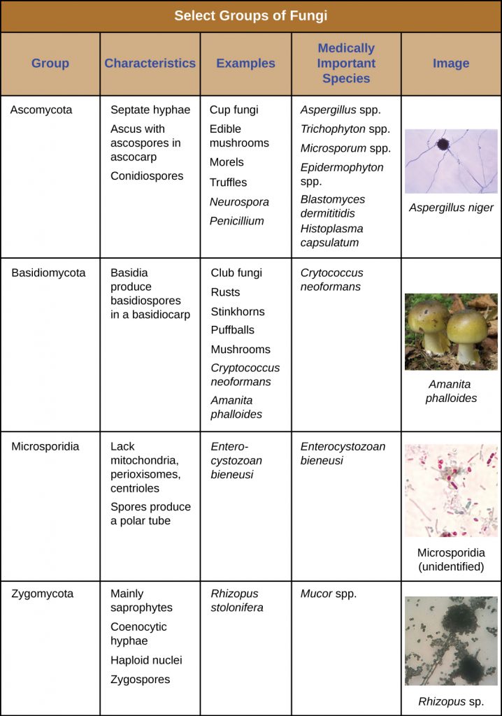
The fungi are very diverse, comprising seven major groups. Not all of the seven groups contain pathogens. Some of these groups are generally associated with plants and include plant pathogens. For example, Urediniomycetes and Ustilagomycetes include the plant rusts and smuts, respectively. These form reddish or dark masses, respectively, on plants as rusts (red) or smuts (dark). Some species have substantial economic impact because of their ability to reduce crop yields. Glomeromycota includes the mycorrhizal fungi, important symbionts with plant roots that can promote plant growth by acting like an extended root system. The Glomeromycota are obligate symbionts, meaning that they can only survive when associated with plant roots; the fungi receive carbohydrates from the plant and the plant benefits from the increased ability to take up nutrients and minerals from the soil. The Chytridiomycetes (chytrids) are small fungi, but are extremely ecologically important. Chytrids are generally aquatic and have flagellated, motile gametes; specific types are implicated in amphibian declines around the world. Because of their medical importance, we will focus on Zygomycota, Ascomycota, Basidiomycota, and Microsporidia. Table 5.2 summarizes the characteristics of these medically important groups of fungi.
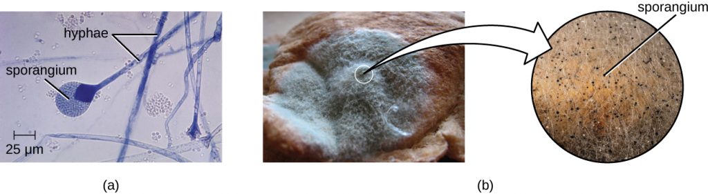
The Zygomycota (zygomycetes) are mainly saprophytes with coenocytic hyphae and haploid nuclei. They use sporangiospores for asexual reproduction. The group name comes from the zygospores that they use for sexual reproduction (Figure 5.20), which have hard walls formed from the fusion of reproductive cells from two individuals. Zygomycetes are important for food science and as crop pathogens. One example is Rhizopus stolonifer (Figure 5.21), an important bread mould that also causes rice seedling blight. Mucor is a genus of fungi that can potentially cause necrotizing infections in humans, although most species are intolerant of temperatures found in mammalian bodies (Figure 5.21).
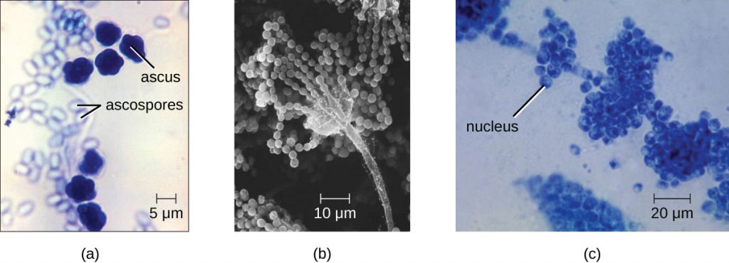
The Ascomycota include fungi that are used as food (edible mushrooms, morels, and truffles), others that are common causes of food spoilage (bread moulds and plant pathogens), and still others that are human pathogens. Ascomycota may have septate hyphae and cup-shaped fruiting bodies called ascocarps. Some genera of Ascomycota use sexually produced ascospores as well as asexual spores called conidia, but sexual phases have not been discovered or described for others. Some produce an ascus containing ascospores within an ascocarp (Figure 5.22).
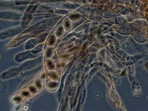
Examples of the Ascomycota include several bread moulds and minor pathogens, as well as species capable of causing more serious mycoses. Species in the genus Aspergillus are important causes of allergy and infection, and are useful in research and in the production of certain fermented alcoholic beverages such as Japanese sake. The fungus Aspergillus flavus, a contaminant of nuts and stored grains, produces an aflatoxin that is both a toxin and the most potent known natural carcinogen. Neurospora crassa is of particular use in genetics research because the spores produced by meiosis are kept inside the ascus in a row that reflects the cell divisions that produced them, giving a direct view of segregation and assortment of genes (Figure 5.23). Penicillium produces the antibiotic penicillin (Figure 5.22).
Many species of ascomycetes are medically important. A large number of species in the genera Trichophyton, Microsporum, and Epidermophyton are dermatophytes, pathogenic fungi capable of causing skin infections such as athlete’s foot, jock itch, and ringworm. Blastomyces dermatitidis is a dimorphic fungus that can cause blastomycosis, a respiratory infection that, if left untreated, can become disseminated to other body sites, sometimes leading to death. Another important respiratory pathogen is the dimorphic fungus Histoplasma capsulatum (Figure 5.19), which is associated with birds and bats in the Ohio and Mississippi river valleys. Coccidioides immitis causes the serious lung disease Valley fever. Candida albicans, the most common cause of vaginal and other yeast infections, is also an ascomycete fungus; it is a part of the normal microbiota of the skin, intestine, genital tract, and ear (Figure 5.22). Ascomycetes also cause plant diseases, including ergot infections, Dutch elm disease, and powdery mildews.
Saccharomyces yeasts, including the baker’s yeast S. cerevisiae, are unicellular ascomycetes with haploid and diploid stages (Figure 5.24) This and other Saccharomyces species are used for brewing beer.
The Basidiomycota (basidiomycetes) are fungi that have basidia (club-shaped structures) that produce basidiospores (spores produced through budding) within fruiting bodies called basidiocarps (Figure 5.25). They are important as decomposers and as food. This group includes rusts, stinkhorns, puffballs, and mushrooms. Several species are of particular importance. Cryptococcus neoformans, a fungus commonly found as a yeast in the environment, can cause serious lung infections when inhaled by individuals with weakened immune systems. The edible meadow mushroom, Agricus campestris, is a basidiomycete, as is the poisonous mushroom Amanita phalloides, known as the death cap. The deadly toxins produced by A. phalloides have been used to study transcription.

Finally, the Microsporidia are unicellular fungi that are obligate intracellular parasites. They lack mitochondria, peroxisomes, and centrioles, but their spores release a unique polar tubule that pierces the host cell membrane to allow the fungus to gain entry into the cell. A number of microsporidia are human pathogens, and infections with microsporidia are called microsporidiosis. One pathogenic species is Enterocystozoan bieneusi, which can cause symptoms such as diarrhoea, cholecystitis (inflammation of the gall bladder), and in rare cases, respiratory illness.
Table 5.2 – Characteristics of select groups of fungi
- Which group of fungi appears to be associated with the greatest number of human diseases?
MICRO CONNECTIONS: Eukaryotic Pathogens in Eukaryotic Hosts
When we think about antimicrobial medications, antibiotics such as penicillin often come to mind. Penicillin and related antibiotics interfere with the synthesis of peptidoglycan cell walls, which effectively targets bacterial cells. These antibiotics are useful because humans (like all eukaryotes) do not have peptidoglycan cell walls.
Developing medications that are effective against eukaryotic cells but not harmful to human cells is more difficult. Despite huge morphological differences, the cells of humans, fungi, and protists are similar in terms of their ribosomes, cytoskeletons, and cell membranes. As a result, it is more challenging to develop medications that target protozoans and fungi in the same way that antibiotics target prokaryotes.
Fungicides have relatively limited modes of action. Because fungi have ergosterols (instead of cholesterol) in their cell membranes, the different enzymes involved in sterol production can be a target of some medications. The azole and morpholine fungicides interfere with the synthesis of membrane sterols. These are used widely in agriculture (fenpropimorph) and clinically (e.g., miconazole). Some antifungal medications target the chitin cell walls of fungi. Despite the success of these compounds in targeting fungi, antifungal medications for systemic infections still tend to have more toxic side effects than antibiotics for bacteria.
CLINICAL FOCUS: Resolution
Sarah’s mother asks the doctor what she should do if the cream prescribed for Sarah’s ringworm does not work. The doctor explains that ringworm is a general term for a condition caused by multiple species. The first step is to take a scraping for examination under the microscope, which the doctor has already done. He explains that he has identified the infection as a fungus, and that the antifungal cream works against the most common fungi associated with ringworm. However, the cream may not work against some species of fungus. If the cream is not working after a couple of weeks, Sarah should come in for another visit, at which time the doctor will take steps to identify the species of the fungus.
Positive identification of dermatophytes requires culturing. For this purpose, Sabouraud’s agar may be used. In the case of Sarah’s infection, which cleared up within 2 weeks of treatment, the culture would have a granular texture and would appear pale pink on top and red underneath. These features suggest that the fungus is Trichophyton rubrum, a common cause of ringworm.
Go back to the previous Clinical Focus box.
Key Takeaways
- The fungi include diverse saprotrophic eukaryotic organisms with chitin cell walls
- Fungi can be unicellular or multicellular; some (like yeast) and fungal spores are microscopic, whereas some are large and conspicuous
- Reproductive types are important in distinguishing fungal groups
- Medically important species exist in the four fungal groups Zygomycota, Ascomycota, Basidiomycota, and Microsporidia
- Members of Zygomycota, Ascomycota, and Basidiomycota produce deadly toxins
- Important differences in fungal cells, such as ergosterols in fungal membranes, can be targets for antifungal medications, but similarities between human and fungal cells make it difficult to find targets for medications and these medications often have toxic adverse effects
Multiple Choice
Fill in the Blank
Short Answer
- Which genera of fungi are common dermatophytes (fungi that cause skin infections)?
- What is a dikaryotic cell?
Critical Thinking
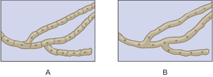
- Which of the drawings shows septate hyphae?
- Explain the benefit of research into the pathways involved in the synthesis of chitin in fungi.
Media Attributions
- OSC_Microbio_05_03_hyphae
- OSC_Microbio_05_03_struct
- OSC_Microbio_05_03_zygo
- OSC_Microbio_05_03_spores
- Web
- OSC_Microbio_05_03_asclife
- OSC_Microbio_05_03_sexspores
- OSC_Microbio_05_03_ascomyLC
- OSC_Microbio_05_03_basidioLC
- OSC_Microbio_05_03_TaxaTable
- OSC_Microbio_05_03_Hyphae_img
