2. How We See the Invisible World

2. Introduction


On the left, brightfield microscopy was used to visualize purple rods-shaped bacteria and larger stained white blood cells. The right image was taken using darkfield microscopy, and shows thin, brightly-lit spiral-shaped bacteria against a dark background.
Figure 2.1. Different types of microscopy are used to visualize different structures. Brightfield microscopy (left) renders a darker image on a lighter background, producing a clear image of these Bacillus anthracis cells in cerebrospinal fluid (the rod-shaped bacterial cells are surrounded by larger white blood cells). Darkfield microscopy (right) increases contrast, rendering a brighter image on a darker background, as demonstrated by this image of the bacterium Borrelia burgdorferi, which causes Lyme disease. [Credit left: modification of work by Centers for Disease Control and Prevention; credit right: modification of work by American Society for Microbiology]

When we look at a rainbow, its colours span the full spectrum of light that the human eye can detect and differentiate. Each hue represents a different frequency of visible light, processed by our eyes and brains and rendered as red, orange, yellow, green, or one of the many other familiar colours that have always been a part of the human experience. But only recently have humans developed an understanding of the properties of light that allow us to see images in colour.

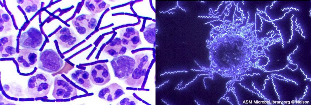
Over the past several centuries, we have learned to manipulate light to peer into previously invisible worlds—those too small or too far away to be seen by the naked eye. Through a microscope, we can examine microbial cells and colonies, using various techniques to manipulate colour, size, and contrast in ways that help us identify species and diagnose disease.

Media Attributions

  • Web

License

Icon for the Creative Commons Attribution 4.0 International License

Microbiology: Canadian Edition Copyright © 2019 by Wendy Keenleyside is licensed under a Creative Commons Attribution 4.0 International License, except where otherwise noted.

Share This Book