Team Think Tank 2 – Designing PCR primers
Team Think Tank (T3) examples:
T3-2: Designing PCR primers.
The purpose of this think tank, is to provide students with the opportunity to collaborate to design their own primers to amplify their gene of interest (folA). In our course we actually do order these primers and have students use them alongside an optimized set. Overall, this exercise serves to give students more ownership and autonomy in the experimental process. Typically, we would complete this exercise during scheduled tutorial/class time one week in advance of when the primers are actually needed (Chapter 1: Part B).
As a team, please complete the following questions. Please tackle the questions in the order they are shown.
Question 1:
- What are Restriction Enzyme compatible cohesive ends? Provide an example.
- What is an Isoschizomer? Provide an example.
- You are using 2 restriction enzymes, NdeI and XhoI, in your cloning project. Please write out their DNA recognition sequence (show the cut site for each enzyme and specify the 3’ and 5’ DNA ends) for each of these enzymes. Please write both the sense and antisense strands for each.
- What are the optimal conditions for the NdeI/XhoI double digestion reaction using Buffer R? (use the Thermo Scientific DoubleDigest program: Click here to access the double digest calculator
- What does “Cleavage efficiency close to the termini of PCR fragments” mean? Why are we concerned about this? Do NdeI/XhoI require special considerations when designing the primers?
Question 2:
- The folA DNA sequence is shown below
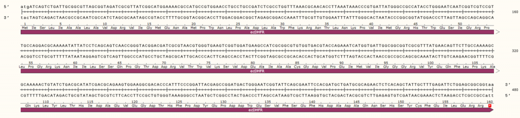
Using this sequence information, please design 2 PCR oligonucleotides (~18-24 bp each, not including any 5’ extensions) containing an NdeI site and an XhoI site engineered at each end of the folA gene as 5’ extensions.
- The NdeI site will be part of the sense primer and the XhoI site will be part of the antisense primer. Please pay close attention to the direction of your promoter and the His-tag, which is already built into your plasmid (which is C-terminal; therefore, remove your stop codon). Use the provided folA and pET26b sequence information to ensure your primers maintain the folA gene in frame with the His-tag.
- Write out the forward and reverse primer sequences. Underline the 5’ extension sequences.
Question 3: Use the GenScript oligo calculator to report the following:
- Melting temperature of each primer (the melting temperatures need to be within 0-4 ºC of one another).
- Annealing temperature for each primer (remember that the annealing temperature is typically calculated as follows: melting temp – 5 ºC)
Question 4:
- Complete the schematic below to show where on the folA gene these primers will anneal (please include the 5’ extensions and sizes, label the 2 strands both with correct termini, 5’ or 3’, and “sense” and “antisense” labels, etc.). Continue this schematic to show the first 2 cycles of PCR. A cycle, by definition, includes annealing of primers, extension of primers and resulting products from the extension reaction. After cycle 1 you should have 4 DNA fragments (including parental strands) and after cycle 2 you should have 8 DNA fragments (including parental strands).
P.S. – do not write the actual DNA sequences: just lines will suffice.

Question 5:
- Using your lab manual (Chapter 1) list the PCR conditions you would use to amplify your folA gene from the pMAC1 plasmid. Be specific about:
- the concentration of reagents
- the temperature and duration of each PCR step
- what is a typical concentration range for: template DNA, forward primer, reverse primers, magnesium, dNTP, Taq DNA polymerase)?
- Using the lab manual background information (Chapter 1) as a reference guide, briefly describe the temperature and duration of each PCR step. Consider what your system-specific temperatures/durations are.
-
-
-
- Initial DNA denaturation
- Denaturation
- Primer annealing
- Extension
- Final extension
-
-
Question 6
- You are given a 1/1000 dilution of your template pMAC1-folA DNA. The initial concentration of undiluted pMAC1-folA is 811 µg/mL. You will need to add a total of 1 µL of the diluted pMAC1-folA stock to your PCR reaction. Please calculate the total amount of pMAC1-folA DNA (in ng) you are adding to your PCR reaction (show your math).
Question 7
- Report the size of your amplified product, folA (in base pairs) as well as the size of your NdeI/XhoI digested folA fragment. Please make sure you explain how you obtained these numbers. Draw the expected band sizes of the hypothetical agarose gel shown below and briefly explain each lane. Given this information, will you be able to tell the difference between the digested and undigested folA DNA fragment?
