Ankle Brachial Pressure Index
Ankle Brachial Pressure Index
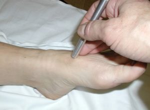
Figure 2: How to Perform an ABPI
, Public domain, via Wikimedia Commons")
The illustration shows the ankle-brachial index test. The test compares blood pressure in the ankle to blood pressure in the arm. As the blood pressure cuff deflates, the blood pressure in the arteries is recorded.
Interpretation of ABPI values
Vascular assessment
Palpation of the dorsalis or posterior tibial arteries with a palpable pulse indicates at least 80mmHg in the foot. A normal ABPI is 0.9 or greater with some compromise of the arterial circulation between 0.7 – 0.9. An ABPI between 0.5- 0.7 indicates mild to moderate disease and a referral to a vascular surgeon for a more comprehensive assessment is recommended. The absence of a palpable pulse is the most sensitive clinical sign of arterial insufficiency (Morley, 2018).
In 2015, Alavi, Sibbald and Mayer (Senior author Swiss Vascular Surgeon) et al. conducted a study to determine the accuracy of audible arterial foot signals with an Audible 8 MHz Handheld Doppler ultrasound for identification of significant peripheral arterial disease as a simple, quick, and readily available bedside screening tool (Alavi et al., 2015). Two hundred consecutive patients referred to an interprofessional wound care clinic underwent audible handheld Doppler (AHHD) ultrasound of both legs. As a control and comparator, a formal bilateral lower leg vascular study including the calculation of Ankle Brachial Pressure Index and toe pressure (TP) was performed at a certified vascular lab. Their study conclusion determined that audible handheld Doppler ultrasound proved to be a reliable, simple, rapid, and inexpensive bedside exclusion test of peripheral arterial disease in diabetic and nondiabetic patients (Alavi et al., 2015).
Table 5. Ankle Brachial Pressure Index Values and Interpretation
| Ankle Brachial Pressure Index | Diagnosis/Interpretation for Arterial Disease |
|---|---|
| >1.40 | Concern for non-compressible arteries (example diabetes mellitus, renal diseases) Further vascular tests should be performed |
| 1 to 1.40 | Normal |
| 0.91 to 0.99 | Borderline |
| ≤ 0.90 | Abnormal range Peripheral arterial disease |
| 0.7 to 0.9 | Mild disease |
| 0.41 to 0.69 | Moderate disease |
| < 0.4 | Severe disease (critical limb ischemia) |
Reproduced from Wounds Canada with permission
Any multiphasic sound (biphasic, triphasic) on the AHHD can be used as an indicator of adequate blood supply to heal. The AHHD can be performed with the client seated or in any position and does require them to lay flat for 20 minutes prior (as with the ABPI). With the AHHD, there is no potential for pain caused by inflation of a blood pressure cuff around the ankle. Also, the results are not influenced by calcified vessels, especially when calculating the ABPI in patients with diabetes. The audible recording can be documented as a MP3 or WAV file on a smartphone. If the signal is monophasic or absent, segmental lower leg duplex Doppler studies need to be performed and vascular referral is advised.
Unfortunately, venous leg ulcers have a long healing trajectory with approximately 30% unhealed at 24 weeks (Evans et al., 2019). The Venous Leg Ulcer Risk Assessment tool is a validated tool to predict the risk of a VLU not healing at week 24.11 www.vlur-risk-tools.org.au/
