67 7.3 Quantifying the Effects of Neurotropin
Learning Objectives
- Understand why the APP/PS1 transgenic mouse is used to model AD
- Discuss the primary methods in which researchers quantified the effects of Neurotropin
- Understand experimental methods typically used in studies involving AD drug testing
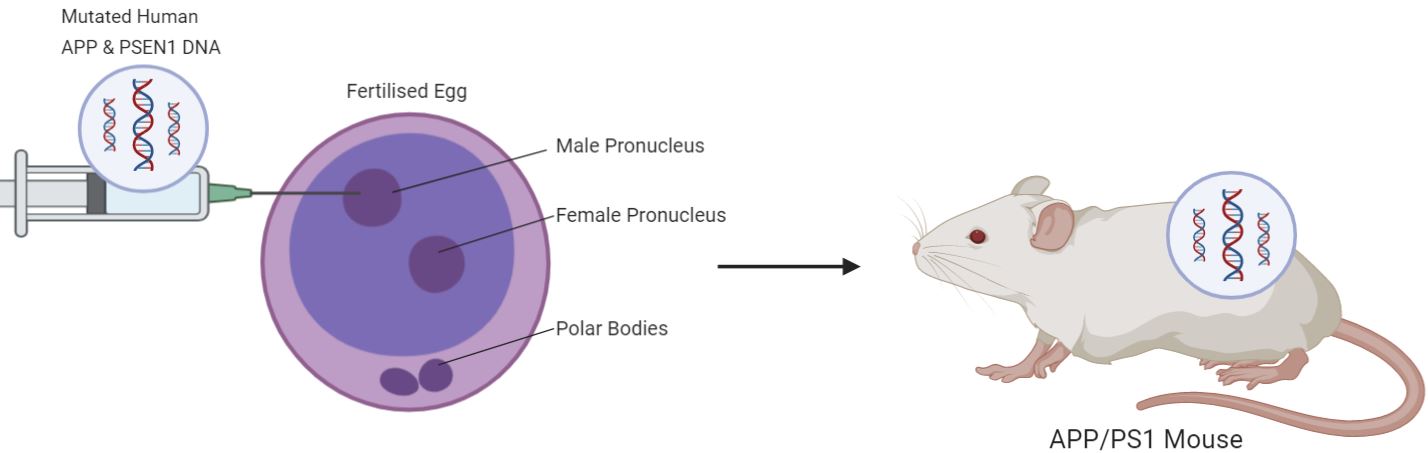
APP/PS1 Mice
The APP/PS1 mouse is a transgenic mouse model commonly used in the study of AD. The mouse co-expresses two human transgenes: the mutated human amyloid precursor protein (APP) and L166P mutated human presenilin 1 (PSEN1) genes. This causes the mouse to over express human APP, leading to the accumulation of Aβ40 and Aβ42 and subsequently plaque deposition as the mouse ages. The physiological and cognitive impairments typically seen in human AD patients are also seen in these mice, such as decreased synaptic plasticity, neuronal loss, and memory loss. The ability to treat these mice with drugs, behavioral tests, and molecular quantification methods making them extremely useful in the study of AD (Gengler et al., 2010).
Morris Water Maze
The Morris water maze (MWM) is a test of spatial learning and memory where animal models are placed in a pool of opaque water. The animal can freely swim around until they discover a hidden platform beneath the surface of the water, allowing them to rest. The animals are then repeatedly placed back into the pool, measuring several variables that indicate their ability to learn from previous attempts and memorize their previous paths (Nunez, 2008). Watch the video below to get a better understanding of how this experiment works:
Immunofluorescent Staining
Immunofluorescent staining is a highly versatile immunochemical technique that allows the visualization of proteins, antigens, and many other components that may be found in various tissues. This is done through the use of fluorophore-antibodies that are specific to the component of interest (Im et al., 2019). For example, in the study of NTP researchers employed immunofluorescent stating used GFAP and Iba1 specific antibodies to identify microglia and astrocytes in brain tissue. Watch the video below to learn more about the process of immunofluorescent staining:
Enzyme-Linked Immunosorbent Assay (ELISA)
ELISA is widely used in research to detect the concentrations of various substances including proteins and peptides. It uses antibodies that bind antigens specific to the substance of interest, allowing researchers to identify the abundance of antigen-antibody complexes, and therefore the concentration of that substance (Aydin, 2015). Samples from the hippocampus and cortex of the WT and TG mice were used in ELISA to observe changes in the concentrations of Aβ40/42, BDNF, and pro-inflammatory cytokines before and after treatment with NTP. Watch the video below to learn more about how ELISA works:
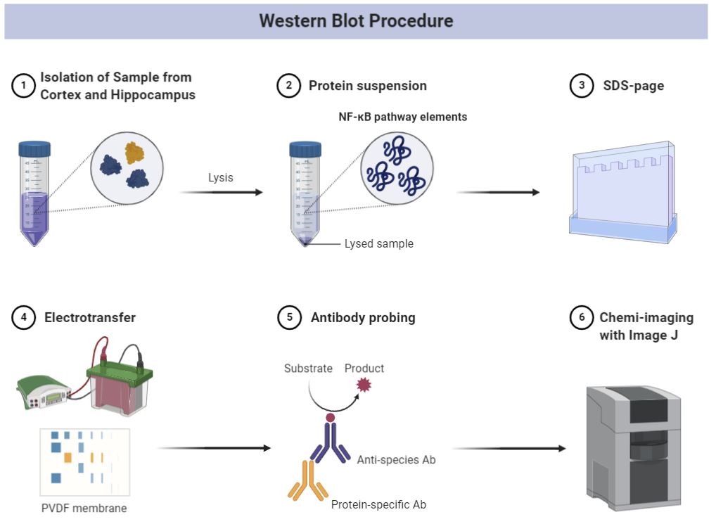
Western Blot
The Western blot is a common lab technique used in the identification and quantification of specific proteins. The process generally involves the separation of proteins based on weight and charge using gel electrophoresis. Antibodies are then used to bind the proteins, forming complexes that are detected with chemiluminescence imaging. These complexes show up as “bands” at varying opacity depending on molecular weight and are compared to a reference protein such as β-actin. (Kim, 2017). The research by Fang et al. used a sample from the hippocampus and cortex of mice to detect different levels of NF-κB pathway elements before and after NTP treatment. The diagram below outlines their procedure:
Key Takeaways
- The APP/PS1 mice is commonly used in AD research due to their molecular, physiological, and behavioral similarity to human AD patients.
- The mice were subjected to a spatial learning test known as the Morris water maze which assessed changes in learning and memory before and after NTP treatment
- Immunofluorescent staining was used to visualize tissue components in brain samples from the APP/PS1 mice, which included various proteins and markers for glial cells
- ELISA was used to quantify changes in concentration of Aβ40/42, BDNF, and pro-inflammatory cytokines
- Western blot analysis was used to specifically measure and visualize the changes in concentration of NF-κB pathway elements
References:
Xiao, Songhua et al. “Graphene quantum dots conjugated neuroprotective peptide improve learning and memory capability.” Biomaterials vol. 106 (2016): 98-110. doi:10.1016/j.biomaterials.2016.08.021