7.3 Work of Breathing and the Need for Mechanical Ventilation
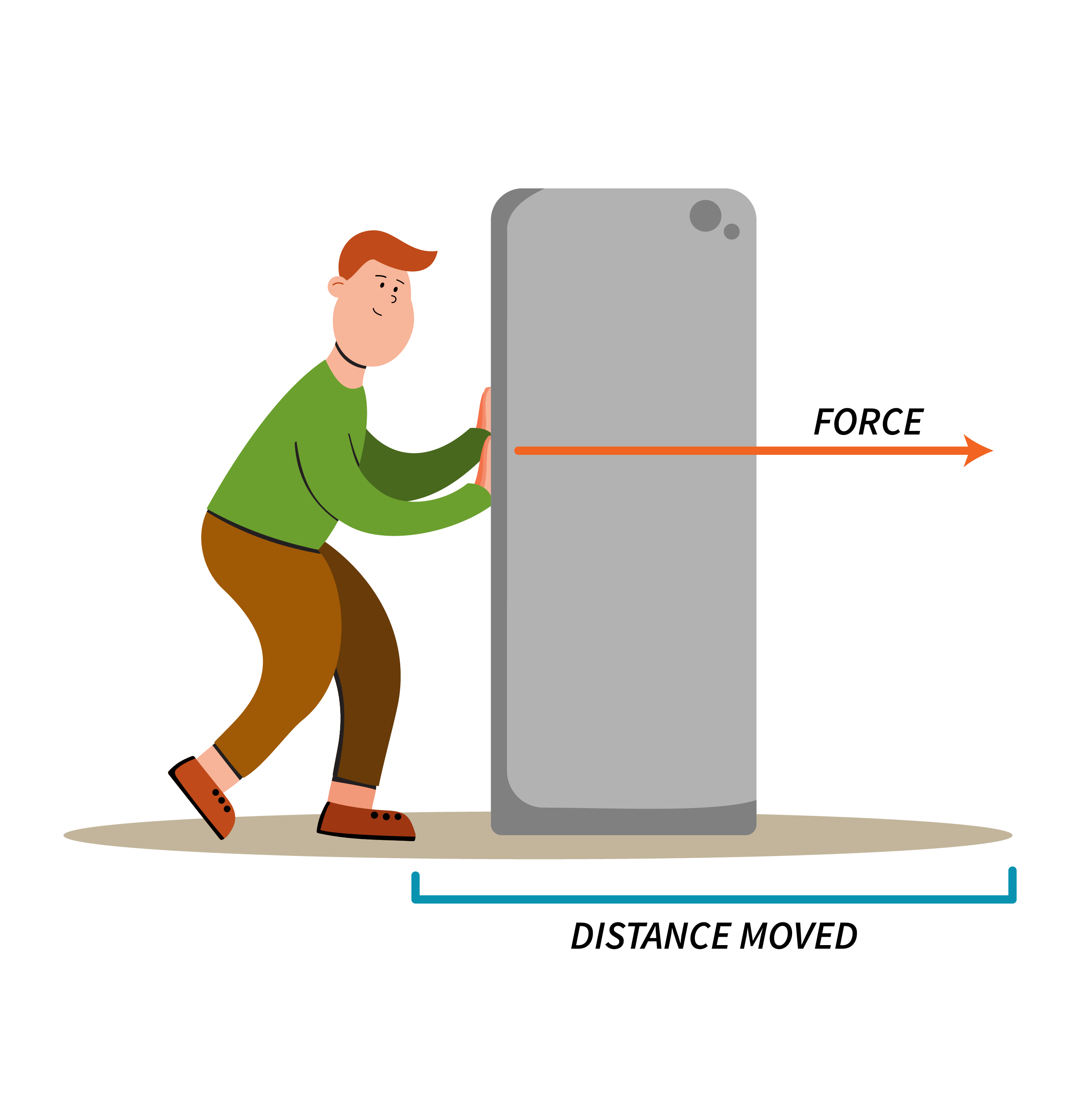
In physics, Work is described as the force required to move an object over a certain distance.
[latex]\text{Work}=\text{Force}\times \text{Distance}[/latex]

In respiratory physiology, work of breathing (WOB) is performed by the respiratory muscles. WOB can be determined by the pressure required to move tidal volume during the breathing process.
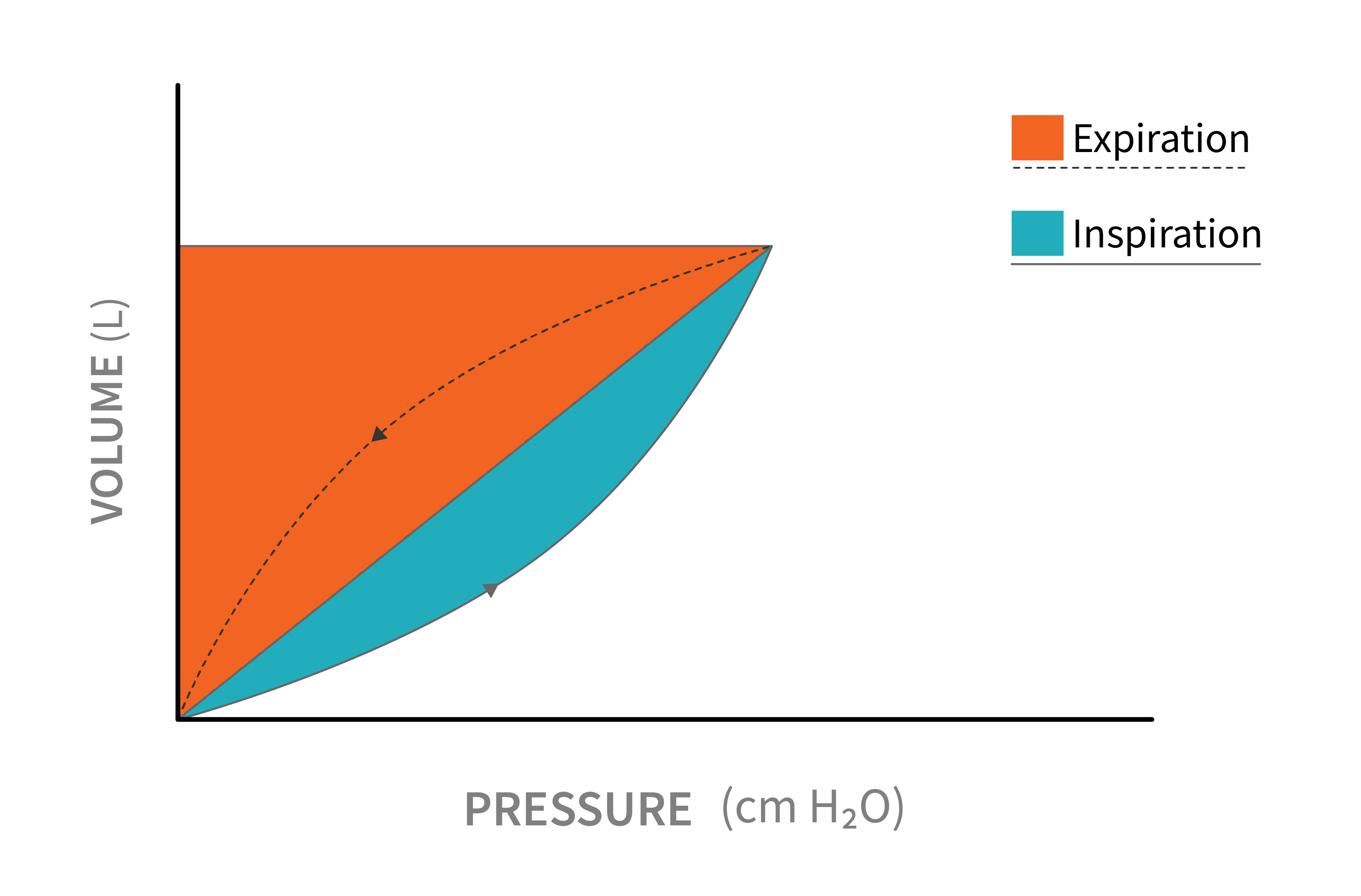
[latex]\begin{eqnarray*}\text{WOB}&=&\text{Pressure}\times\text{Volume}\\\text{WOB}&=&P\times V\end{eqnarray*}[/latex]
The equation above represents the mechanical work performed by the respiratory muscles. The pressure represents the force generated by the muscles, and the volume represents the amount of air moved in each breath.
The respiratory muscles need to overcome elastic and frictional forces that oppose lung inflation (distension), which requires energy. This energy is provided through metabolic work of breathing, which is the amount of energy or oxygen consumption needed by the respiratory muscles to produce sufficient ventilation and meet the metabolic demands of the individual. WOB depends on breathing patterns, both under normal circumstances or disease process.
A graphic representation is helpful in assessing work of breathing. Shaded area represents the work of breathing.

Key Takeaway
Work of breathing is determined by the pressure-volume characteristics (compliance and resistance) of the respiratory system. Work of breathing will increase in diseases that increase resistance or decrease compliance or when respiratory rate increases.
An increase in work of breathing can lead to respiratory failure secondary to respiratory muscle fatigue and weakness. As respiratory therapists, it is extremely important that we recognize signs of increased work of breathing. Several assessment measurements are available to assess work of breathing and respiratory muscle strength and are discussed below.
Object Lesson
There are several factors that can influence WOB.
- Metabolic requirements. As the body requires more energy, WOB must increase in order to ensure sufficient ventilation.
- The effort required by the respiratory muscles to overcome airflow resistance, enabling air movement in and out of the airways and compliance, to maintain appropriate lung volumes.
- The respiratory rate and pattern. The intensity and speed of contraction in the respiratory muscles determine WOB.
- Presence of V/Q. Mismatching and impaired diffusion resulting from various diseases.
How do we determine increased work of breathing and respiratory muscle fatigue?
Respiratory muscle weakness and fatigue are commonly observed in patients with neuromuscular disease, as well as chronic respiratory conditions like COPD or restrictive lung disease. While this is often a sign of disease progression, it is important to recognize it and promptly address any increase in work of breathing caused by muscle weakness to prevent complications.
The following physiological measurements are commonly used to assess respiratory muscle strength, and degree of work of breathing.
Maximum inspiratory pressure (MIP) is the lowest pressure generated during inspiration with the airway occluded.
Remember, the pressure measured during inspiration is considered negative.
This measurement is sometimes called negative inspiratory force (NIF).
This measurement can be performed at the bedside using a manometer.
Normally, MIP ranges between [latex]-50[/latex] to [latex]-100\text{ cmH}_2\text{O}[/latex]. Generally, an MIP of at least [latex]-20[/latex] is required to inhale an adequate tidal volume.
A higher than [latex]-20\text{ cmH}_2\text{O}[/latex] MIP signals inspiratory muscles weakness.
Object Lesson
Measuring MIP is equivalent with gauging the strength of a person's lung muscles through a fitness test. Just as a fitness test measures the maximum force exerted by muscles, measuring maximum inspiratory pressure assesses the maximum force generated by the respiratory muscles during inhalation.

Maximum Expiratory Pressure (MEP), sometimes called peak expiratory pressure, is similar to MIP and is a measurement used to assess the strength of the expiratory muscles. It determines the maximum force generated by these muscles during forced expiration. Normal value for MEP is [latex]100\text{ cmH}_2\text{O}[/latex].
Object Lesson
MEP similar to an athlete's final sprint in a race, demonstrating the athlete's leg muscles power and endurance.

Vital Capacity (VC) is the volume of air that can be exhaled following a maximal inspiration. Similar to MIP and MEP, VC provides information on a person's ability to inhale a large tidal volume in order to produce a strong cough to clear the airway.

Peak Expiratory Flow provides information on airway resistance and a person's ability to maintain airway patency.
Tidal volume, respiratory rate and minute ventilation are also used often as indicators of respiratory muscle strength. A respiratory rate greater than [latex]\text{30 - 35 bpm}[/latex] is usually an indication of increased work of breathing. Without intervention, it will lead to respiratory failure.

