1.1 Anatomy of Breathing
In order to fully comprehend the principles of mechanical ventilation, it is important to have a thorough understanding of the intricate mechanisms that govern human respiration. This chapter serves as a brief yet comprehensive overview of the respiratory anatomy and physiology that is crucial for comprehending the nuances of mechanical ventilation. The respiratory system is responsible for breathing. The lungs are the major organ responsible for this. We have two lungs in the human body. The left lung has two lobes, or sections, while the right lung has three lobes.
Thinking of the lung as a balloon is a great simple analogy but it is important to review the physiology of the respiratory system to fully explain how the lungs behave. The lungs are not simply one big balloon. It is like they are made up of millions of little balloons all stuck together—similar to bubble wrap. These little balloons are referred to as alveoli.
The millions of “small balloons” are still able to take in the same overall volume of air as a single balloon of the same size but by dividing the volume of air into alveoli versus one big balloon, the total combined surface area of the lungs goes up exponentially. Why does this matter? Well, each individual alveolus is surrounded by blood vessels called “capillaries”. Where the alveoli and capillaries meet is where the actual exchange of gases occurs. The large increase in surface area where this exchange can occur allows for more active areas of gas exchange, and this exchange can happen at a more efficient and rapid pace. Oxygen diffuses in from the alveoli to the bloodstream, and carbon dioxide diffuses out of the blood into the alveoli to be exhaled out of the lungs.
Apply Your Learning
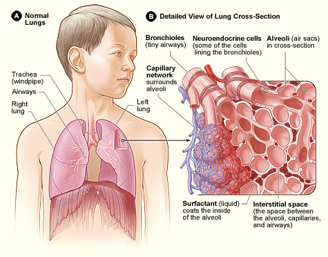
Can you locate the lungs, the alveoli and the capillary network in the diagram below? Don’t worry if the rest of the labels look confusing to you—you’ll learn a lot more about the lungs as you progress through this course!
“The Anatomy of Breathing” from Basic Principles of Mechanical Ventilation by Melody Bishop, © Sault College is licensed under a Creative Commons Attribution-NonCommercial-ShareAlike 4.0 International License, except where otherwise noted.
