19 6.2 – FMD Methods Used in Experimental Research
Learning Objectives
In this methods section, you will learn:
- How FMD is used in different organisms
- How experimental research methods tracks changes from cyclic dietary restrictions
Changes in the diet can increase the longevity and overall healthspan in various organisms. According to a recent study by Brandhorst et al., the fasting mimicking diet (FMD) can be applied to different models: mice and humans. In order to achieve favourable outcomes, each group had their own specific FMD regimen to adhere to. Different analysis techniques were used to track the progression of bodily changes and functions throughout the duration of the study.
Mice
The mouse animal model, specifically C57BL/6, is frequently used in restriction diet experiments. Calorie restriction in C57BL/6 mice can reduce the negative health effects caused by medium-high fat diets (Rusli et al. 11) and changes in the gut microbiome maintains the positive effects of the diet restriction (Wang et al. 8).
Study by Brandhorst et al.
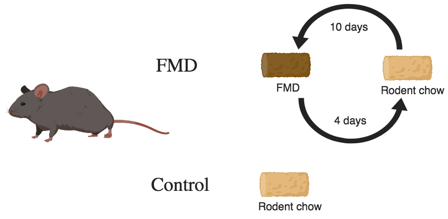
To observe the effects of FMD, a sample size of 110 C57BL/6 mice were used. The FMD for mice consisted of low-calorie and low-protein broth powders (Brandhorst et al. 96). The measurements used to track changes in the mice included standard blood draws, body weight, longevity, and autopsies were performed to analyze the weights of organs and cancer incidence (Brandhorst et al. 96).
Micro-CT Scans
The changes of body composition were observed using micro computed tomography (micro-CT) scans, an X-ray that produces high-resolution three-dimensional images (Clark and Badea 620). Applications for micro-CT scans are tracking the differences of bone density and structure (Clark and Badea 623) and body fat composition including measurements of total fat volume, abdominal fat, visceral fat, and lean body mass (Brandhorst et al. 87).
Other applications that currently being used with micro-CT scans are analyses of tissues and organs such as the lungs and heart/cardiac tissue as well as the tracking of cancers and tumours (Clark and Badea 624). However, there is a limitation to micro-CT scans that may reduce clarity to distinguish different parts of the image. To improve this, a spectral micro-CT can increase the contrast sensitivity to be able to identify the internal and external parts of the body (Clark and Badea 627). Micro-CT scans are quite effective in scanning small animals and can be combined with other techniques such as the Positron emission tomography (PET) scans (Clark and Badea 626).
Humans
Study by Brandhorst et al.
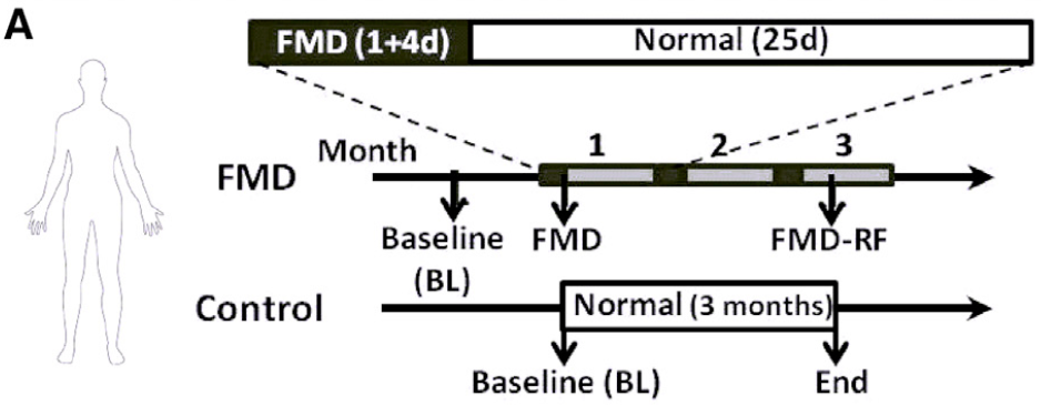
To test the feasibility of FMD in humans, a pilot randomized clinical trial was conducted with a sample size of 39 individuals (Brandhorst et al. 93). The FMD for humans consisted of low-protein and high-carbohydrate, high-fat foods such as soups, energy bars, energy drinks, and chip snacks (Brandhorst et al. 95). Further measurements included blood tests, height, weight, and body composition using Dual-energy X-ray absorptiometry (DEXA/DXA) scans.
DEXA/DXA Scans
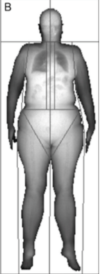
DEXA/DXA scans are X-rays to be primarily used for measuring bone material density and body composition, producing two-dimensional images (Borga et al. 888). In the cases of monitoring changes of body composition, DEXA/DXA scans are more effective than body mass index (BMI) measurements as it does not take into account the amount of muscle mass a person has (Borga et al. 888). With DEXA/DXA scans, one is able to track changes in total body fat volume, trunk fat, and lean body mass (Brandhorst et al. 94).
Widely used and named by the World Health organization as one of the more useful diagnostic tools for osteoporosis, there is a limitation to DEXA/DXA scans (El Maghraoui and Roux 605). The alignment and scanning procedures of these scans must be done correctly, otherwise, it can result in poor diagnoses (El Maghraoui and Roux 608). It is imperative for the physician or operator of the scan to correctly perform it.