9.2 Model Organisms for Cancer Detection
Learning Objectives
What are some commonly used model organisms for laboratory experiments?
Why are Zebrafish good model organisms?
What are some novel imaging techniques to visualize cancer in it’s early stages ?
Model Organisms: Why Zebrafish are good model organisms?
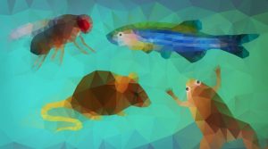
Model organisms are non-human species that are used in the laboratory for experiments that help scientists understand various biological processes. They are chosen on the basis of various factors like maintenance, lifespan, number of offspring, and genetic similarity to humans. (National Institute of General Medical Sciences) Commonly used model organisms for cancer detection include rodents, fruit flies, nematodes, and zebrafish. (Fields, 1) For instance, rodents have been invaluable to our understanding of human cancer since they are genetically similar to us, they are specifically used for defining the different genetic mutations that drive cancer initiation and progression. (National Institute of General Medical Sciences)
The SCID model of immunocompromised mice can engraft a wide range of human cancers (xenotransplantation), and have been used to study human cancer growth in vivo. (Mestas, 5) On the other hand, fruit flies and nematodes can be used to do quick genetic screening, but due to their limited complexity it is tough to directly correlate it to humans. (National Institute of General Medical Sciences)
Zebrafish is a fast-growing tropical fish about 2.5 cm to 4 cm long, making them small and easy to maintain. They produce hundreds of offspring at weekly intervals, with nearly all embryos being transparent allowing researchers to easily examine development of internal structures. (Chakraborty et al., 1) Most importantly, zebrafish share 70% of genes with humans, and 84% of genes associated with human diseases have a zebrafish counterpart. Lastly, the zebrafish genome has been fully sequenced to high quality. (Kari et al., 1, 2) This has enabled scientists to incorporate mutations in zebrafish genes to study their function.
Imaging techniques and practices to visualize cancer initiation
Current imaging techniques to visualize cancer are not successful in showing a live progression of cancer from its initiation to its progression. (Kaufman et al., 1) Identification of cancer initiation events can be very beneficial for targeted treatments and therapeutic interventions, helping treat precancerous lesions or prevent cancer progression. Although, studying the process of cancer initiation has been difficult due to the rarity of these events, the difficulty of visualizing initiating clones in living organisms, and the transient nature of the emergence of an initiating clone before it expands to form an early tumour. (Kaufman et al. 1)
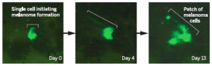
Researchers at Washington University Medical School created a reporter transgene in zebrafish models of melanoma which allowed them to track embryonic neural crest cells as well as melanoma cells in vivo, potentially from its earliest onset. To aid visualization, the researchers amplified and cloned elements of interest upstream of an enhanced green fluorescent protein (EGFP) promoter. Using fluorescence microscopy and time lapse videos, researchers were able to demonstrate the emergence and wide migration of certain clones that went on to become cancer. (Kaufman et al. 1) Thus the use of zebrafish models in melanoma, and fluorescence microscopy have been good practices in advancing the field of cancer initiation visualization.