LAB 9: Bacteriological Analysis of Water
Learning Objectives
- Detect and enumerate total coliforms and E. coli in a water sample.
Introduction
Many illnesses are caused by waterborne bacteria. Testing water for the quantity and types of bacteria is essential to preventing these illnesses. In Canada, a lab performing these tests must be accredited by a governing body, for example, the Standards Council of Canada (SCC). We will be performing the same techniques in our lab, however since we are not accredited, the results are not valid.
If you can safely sample outside water (pond, stream, rain barrel), or if you live in a rural area with well water, please feel free to bring in a water sample. A sample of 500 ml will be more than enough for this lab. If you cannot, we will have water samples in the lab.
In Ontario, drinking water is regulated provincially by the Ministry of the Environment and Climate Change (MoECC), while bottled water is regulated federally by the Food and Drug Act. See page 23 of the Public Health Ontario document posted on FOL to read about drinking water tests done in the province.
Water quality testing uses the principle of indicator organisms. The presence of certain groups of bacteria suggest the water may be contaminated with pathogens and should not be consumed.
Coliforms are Gram negative, non-spore forming, facultatively anaerobic rods that ferment lactose to acid and gas in 48 hours. Coliforms are members of the family Enterobacteriaceaae and include Escherichia, Serratia, Proteus, Enterobacter, Klebsiella and Citrobacter.
The presence of coliforms is common in soil and plants, and do not necessarily indicate unsafe water.
Fecal coliforms (also known as Thermotolerant coliforms) are bacteria associated only with the intestines of mammals. They can grow in the presence of bile salts, a common molecule in the intestine. It is added to selective media to isolate fecal coliforms. These bacteria can produce acid and gas at 44 ⁰C within 48 hours. The presence of these bacteria in water suggests a contamination event, and the water should not be consumed.
E. coli is a fecal coliform. Its presence is detected using selective and differential media to indicate potential fecal contamination.
The allowable limit of total coliforms is 0 CFU/100 ml and for E. coli it is 0 CFU/100 ml. In this lab, we will use two common techniques to detect and enumerate coliforms and fecal coliforms in water samples: most probable number and membrane filtration.
Most Probable Number
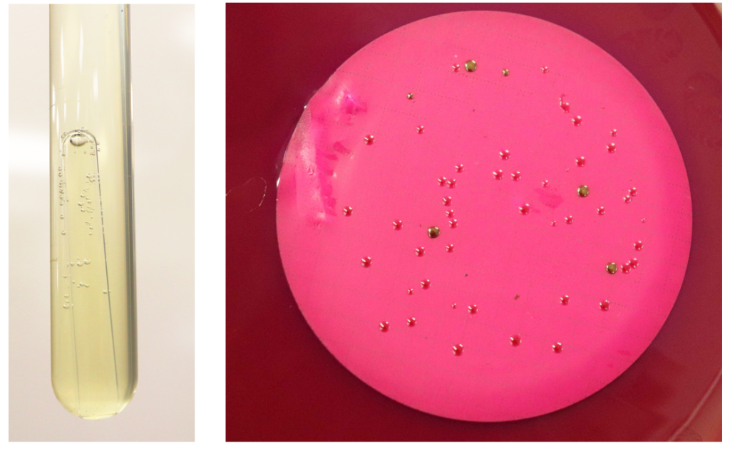
This is a statistical test based on dilution of a water sample until there are no bacteria present in the sub-sample used to inoculate media. The medium is lactose broth (LB, Figure 1). If coliforms are present, they consume the lactose and make gas, which is trapped in the inverted Durham tube. Three ten-fold dilutions are made (not in series) and each dilution is inoculated in three to five replicates.
- Gas production in LB is a presumptive positive result for coliforms
- The number of positive tubes is recorded and this number is used to refer to a table, which gives a probable range of CFU/100 ml. The number of positive tubes is written by descending volume: 10 ml – 1 ml – 100 ul.
Example: There are 3 positive 10 ml tubes, 2 positive 1 ml tubes, and 5 positive 100 ul tubes. The number used to determine the MPN on the reference table is 3-2-5.
To confirm fecal coliform presence, a sample is taken from LB and used to inoculate brilliant green lactose bile (BGLB). This medium contains inhibitors of Gram positive (brilliant green) and bile which selects for coliforms.
Gas production in BGLB is a confirmed positive result for fecal coliforms
To test for fecal coliform presence, we can also detect E. coli. A positive BGLB tube is used as inoculum for an EMB streak plate.
Presence of E. coli on EMB is a completed E. coli test result
There are other methods for detecting E. coli specifically, such as the use of substrates that become fluorescent after enzymatic activity.
Membrane Filtration
This method is rapid and allows precise quantification of total coliforms and E. coli in water samples. A vacuum is fitted with a 0.45 um membrane. The water sample passes through the membrane while bacteria remain trapped on top. The membrane is placed on an agar plate, bacteria-side facing upwards. The medium is selective and differential. It contains bile salts and dyes, which inhibit Gram positive bacteria, and lactose as the main carbon source, which selects for coliforms. When bacteria consume lactose, aldehyde is made, which reacts with dyes to produce a red colour. If the reaction is intense (as for E. coli), the dye crystallizes and creates a metallic sheen. We will use mENDO agar:
- Culture media selects for gram negative bacteria able to grow on bile salts
- coliforms will have a green metallic sheen
- atypical coliforms with less vigorous lactose metabolism may appear red
- Non-coliforms will have colourless, white, red or pink colonies

Water Exercise
Materials
Day 1
- Water sample
- 3 double strength LB (dsLB)
- 6 single-strength LB (ssLB)
- 10 ml pipette and pipette bulb
- P1000 pipette and blue tips
- 2 agar plates
- Inoculating loop
- Vacuum pump
- Filter apparatus
- 2 membrane filters, 0.45 um pore size
- 1 bottle of 90 ml sterile water
- Forceps in 70% ethanol
Day 2
- 1 EMB plate
- 1 BGLB tube
- Inoculating loop
Method
- Most probable number: Label 3 dsLB tubes “10 ml”, 3 ssLB tubes “1 ml” and 3 ssLB tubes “100 ul”.
- Aseptically transfer 10 ml of the water sample into each of 3 dsLB tubes.
- Aseptically transfer 1 ml into 3 ssLB tubes.
- Aseptically transfer 100 ul into 3 ssLB tubes.
- Incubate 24±2 hours at 37 C.
Day 2
-
- Record the number of tubes showing gas and turbidity. Use this MPN table to determine the CFU/100 ml.
- Select one tube with gas use this as inoculum for BGLB. Use a loop to transfer from the LB tube to BGLB. Incubate at 37 C for 24 hours.
Day 3
-
- If the BGLB tube has gas and is turbid, use a loop to streak an EMB plate from the BGLB tube. Incubate the EMB plate at 37 C for 18-24 hours.
- If the tube has no gas but is turbid, streak an EMB plate to confirm that E. coli is not present
- Every group will streak an EMB plate
- If the BGLB tube has gas and is turbid, use a loop to streak an EMB plate from the BGLB tube. Incubate the EMB plate at 37 C for 18-24 hours.
Day 4
-
- Record the EMB result.
- Membrane filtration: on two agar plates and, write “10 ml” and “100 ml” and your group name.
-
- Dilute the water sample: 10 ml water into 90 ml sterile water. Shake well by inverting the bottle at least 25 times. This is bottle 1. Bottle 2 is 100 ml of the original water sample.
- Soak the forceps for two minutes in alcohol. Set up the vacuum pump and filter apparatus, flame the alcohol off the forceps then aseptically place the membrane filter on the apparatus. Secure the funnel on the base.
- While flaming, keep forceps angled downwards to prevent flaming alcohol from dripping on your gloved hands.
- Aseptically transfer the white membrane from the package to the filter apparatus.
- The blue disc is the backing; it is resistant to water. Discard this.
- Add the entire volume of bottle 1 into the funnel and membrane filter. Remove the membrane filter and place on the DC plate labeled with the sample name (10 ml).
- By starting with the most dilute sample, there is no risk of bacterial carry-over between samples, and you don’t need to disinfect the filter apparatus between samples.
- Make sure you place the membrane filter ‘bacteria side up’ so the colonies can form properly.
- Using a graduated cylinder, add 100 ml of your water sample. Filter as above. Place the membrane filter on the mENDO plate labelled with the sample name (100 ml).
- Incubate for 24 hours at 37 C. Count the number of total coliforms (metallic sheen) and atypical coliforms (red colonies). When recording your results, use ‘TFTC’ for membranes with less than 20 colonies and ‘TNTC’ for membranes with over 150 colonies.
-

