LAB 6: Bacterial Media
Learning Objectives
- Test bacterial reactions on selective and differential media.
- Use controls in quality control of culture media.
Introduction
In the labs so far, we have been using general, nonselective media (Luria Bertani broth, LB; trypticase soy broth, TSB; nutrient agar, NA). The growth medium you use depends on the organism you are trying to grow; the organisms we have been growing in the lab grow well on these rich, complex media.
What would happen if you were given a sample of pond water and asked to isolate one genus of bacteria? If you grew this sample on rich, complex media, such as LB, you would grow many species from many genera. One way to narrow the types of bacteria you culture to target one species or genus of interest is to use selective and differential media.
We will be these three types of media in this lab:
Selective media allows the target organism to grow while preventing the growth of other organisms that may be present in the sample.
Differential media allow visual distinctions to be made between growth of different types of organisms.
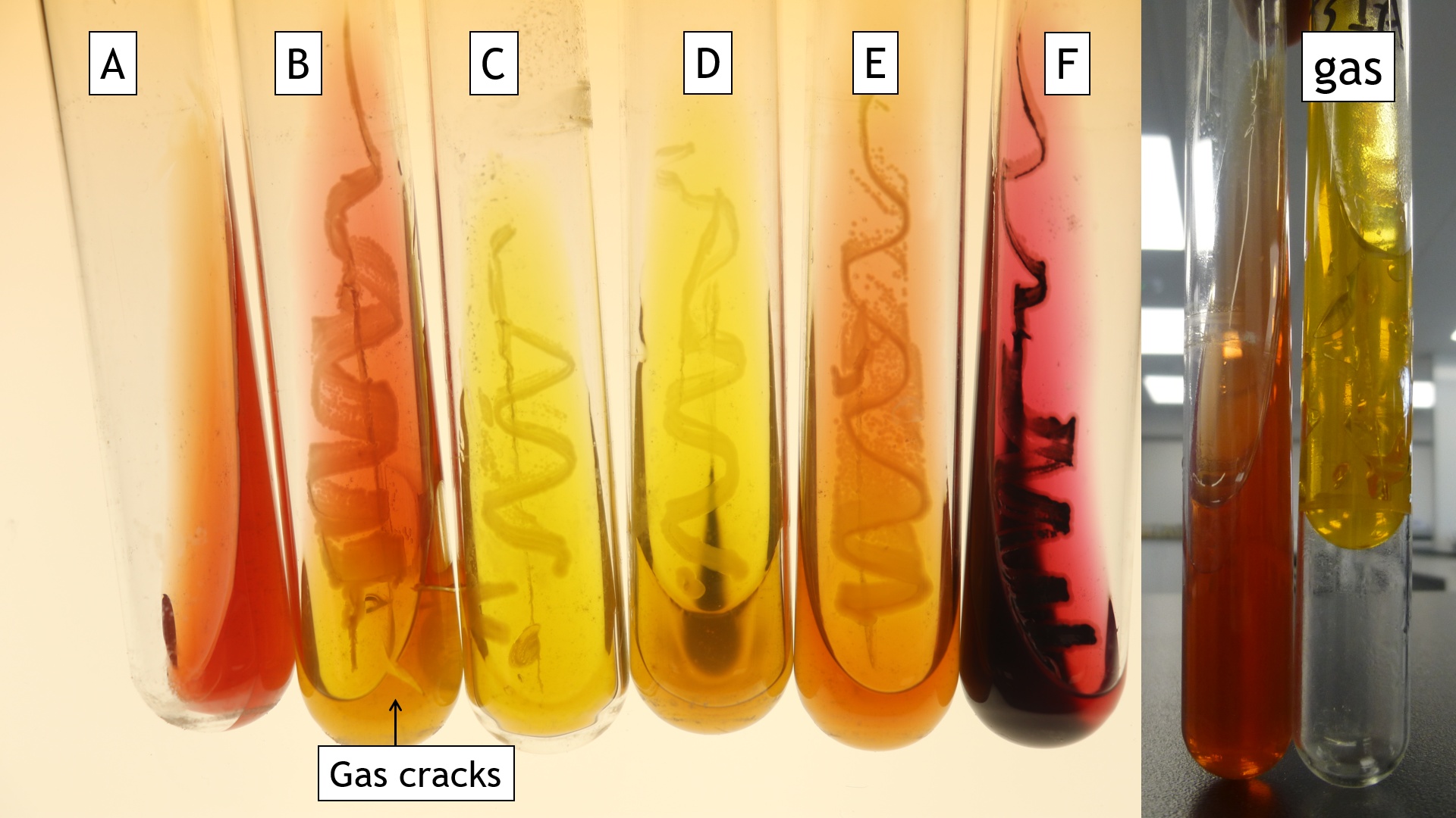
Multitest media possesses multiple tests within one medium (Figure 6.1).
Let’s consider components of media to help you understand differences between media.
Carbon and Nitrogen Sources
All media needs to provide the macronutrients needed for cell growth. Carbon and nitrogen are required in the greatest amounts. In complex media, these are added as complex biomolecules. This approach also ensures hydrogen, phosphorus and sulfur are also added as part of the biomolecules. Common carbon and nitrogen sources are a proteinaceous source that has been partially degraded by enzyme or acid digestion. In a media recipe, these ingredients can be found below.
Carbon and nitrogen sources
Peptones, enzymatic digest of casein (e.g. pancreatic digest), enzymatic digest of soy meal, proteose peptone, tryptone
- These are all found in complex media since their exact chemical composition isn’t known.
Carbon source only
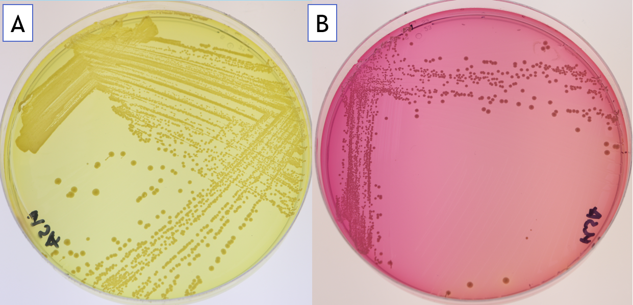
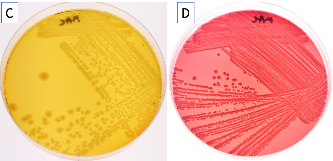
Sugars: lactose, sucrose, glucose, mannitol (Figure 6.2)
Nutrient Extracts
These can be found in some, but not all media. They are a good general nutrient base, including vitamins and minerals with some carbon and nitrogen. Extracts can be made from almost anything, by soaking a substance in water. These can be tailored to the type of organism you are trying to isolate. For example, if you are trying to isolate soil bacteria, you could add a soil extract to your media to mimic the soil environment.
Typical extracts include beef and yeast.
Solidifying Agent
If a solid or semi-solid medium is being made, the liquid is solidified. Typically, agar is used although other gelling agents exist and are used when agar is toxic to the organism of interest. In the petri plates we have been using, 15 g/L of agar is in the medium. By altering the amount of agar in the medium, bacterial motility (the ability of the bacteria to propel themselves) can be tested.
- By reducing the agar to 3 g/L, the medium becomes semi-solid; such plates are termed “swim plates” because they allow bacterial swimming to be examined.
- At 10 g/L agar in the medium, the ability of bacteria to swarm or glide can be tested
Inhibitors in Selective Media
These are found in selective media and they act by inhibiting the growth of certain bacteria. Inhibitors can be bactericidal (kill bacteria) or bacteriostatic (prevent the growth of bacteria).
- If the inhibitor is bacteriostatic, when you pick a colony off the plate, you may have passenger cells of the undesired group, which can resume growth when placed in nonselective media. Thus, it is always good practice to streak for purity after selecting colonies from selective and differential media.
- When a medium is selective for a certain group of bacteria, this does not mean that only this group will grow. Media vary in how selective they are (based on the type of inhibitor and its concentration)
Typical inhibitors include bile salts, alcohol, high concentrations of some compounds (e.g. NaCl), some dyes (e.g. methylene blue).
Inhibitory conditions can also be used. These include pH, incubation temperature, oxygen concentration.

Special Substrates in Differential Media
Differential media rely on a visual change between growth characteristics. Often, the visual change is due to a colour change in pH indicating dye. The pH change is due to carbohydrate utilization. The principle is that only some bacteria will use a carbohydrate and thus, alter the pH, changing the colour.
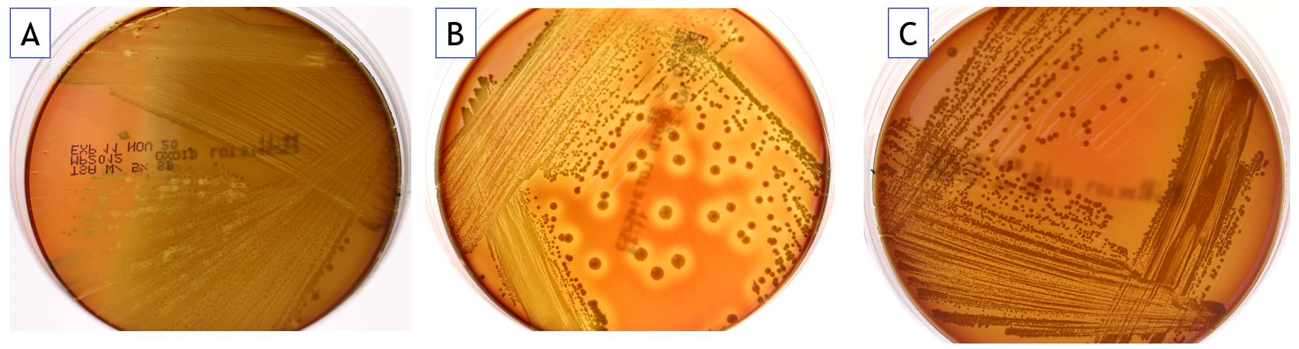
With blood agar plates, the appearance of growth resulting from red blood cell hemolysis (lysing of red blood cells) depends on the bacteria present (Figure 6.4).
Using Controls
When using media, we need to have controls to ensure the media is made properly and the growth conditions give the expected result.
- The positive control is an organism that performs the reaction that the media is testing for.
- The negative control is an organism that does not perform the reaction that the media is testing.
In the media today, one strain will be the positive control, one will be the negative control, and your environmental isolate is the test.
- If the control doesn’t give the expected result, note this. It means either the strain didn’t grow properly (due to contamination most likely) or the media was made improperly. This limits your ability to interpret the test strain.
Media Exercise
Materials
- 1 plate of each: PEA, MSA, MAC, EMB, Blood
- 1 general purpose media (NA, LA, TSA)
- 4 tubes: TSIA
- Strains: Escherichia coli, Proteus vulgaris, Serratia marcesscens, Staphylococcus aureus, S. epidermidis, Bacillus subtilis, Salmonella typhimurium
- Your environmental Isolate
Method
- With a marker, divide the PEA, MSA, MAC, EMB and Blood plates into three.
- Pick a single colony of the stock plate, then using the streak plate technique, label then inoculate the following strains onto each medium (controls are indicated with pos or neg):
- PEA: S. aureus (pos), E. coli (neg), your isolate
- MSA: S. aureus (pos), S. epidermidis (neg), your isolate
- MAC: E. coli (pos), S. typhimurium (neg), your isolate
- EMB: E. coli (pos), S. typhimurium (neg), your isolate
- Blood: B. subtilis (neg), S. aureus (pos), your isolate
- Make a fresh streak plate for your isolate.
- Label the TSIA, then inoculate using the inoculating needle by stabbing into the butt of the tube then streaking across the surface of the slant: E. coli, S. marcescens, P. vulgaris and your isolate.
- Incubate the plates for 24 hours at 37 C.
- Read the test results, noting the colour of the colonies and media in your lab book.
- Take a picture of your plates (use the BSC if you are removing lids), using an ungloved hand on the phone.
- Interpret results using this lab and FOL
- Put your environmental isolate in the fridge to use next week. Either wrap it in parafilm or put it in a Petri plate bag to prevent evaporation.
