Brian’s Health: Tetralogy of Fallot
Electrical Conduction System of the Heart
- Sinoatrial node
- Atrioventricular node
- Bundle of His
- Left bundle branch
- Left posterior fascicle
- Left anterior fascicle
- Left ventricle
- Ventricular septum
- Right ventricle
- Right bundle branch
Chambers & Circulation Through the Heart
- Blood flows from the right atrium to the right ventricle, where it is pumped into the pulmonary circuit.
- The blood in the pulmonary artery branches is low in oxygen but relatively high in carbon dioxide.
- Gas exchange occurs in the pulmonary capillaries (oxygen into the blood, carbon dioxide out), and blood high in oxygen and low in carbon dioxide is returned to the left atrium.
- From here, blood enters the left ventricle, which pumps it into the systemic circuit.
- Following exchange in the systemic capillaries (oxygen and nutrients out of the capillaries and carbon dioxide and wastes in), blood returns to the right atrium and the cycle is repeated.
Facts About Tetralogy of Fallot
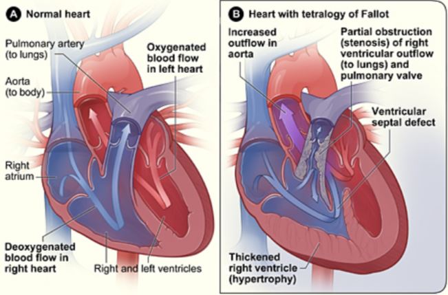
- Birth defect that affects normal blood flow through the heart.
- Happens when the heart does not form correctly as the baby grows & develops in the womb.
- Made up of 4 defects of the heart and its blood vessels.
The 4 Abnormalities
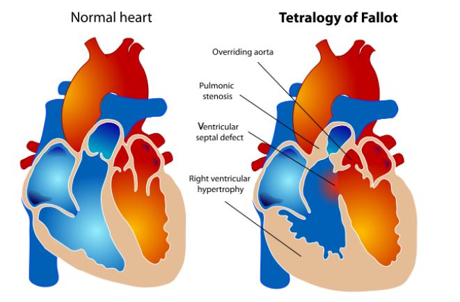
Pulmonary valve stenosis
Pulmonary valve stenosis is a narrowing of the pulmonary valve — the valve that separates the lower right chamber of the heart (right ventricle) from the main blood vessel leading to the lungs (pulmonary artery).
Narrowing (constriction) of the pulmonary valve reduces blood flow to the lungs. The narrowing might also affect the muscle beneath the pulmonary valve. In some severe cases, the pulmonary valve doesn’t form properly (pulmonary atresia) and causes reduced blood flow to the lungs.
Ventricular septal defect
A ventricular septal defect is a hole (defect) in the wall (septum) that separates the two lower chambers of the heart — the left and right ventricles. The hole allows deoxygenated blood in the right ventricle — blood that has circulated through the body and is returning to the lungs to replenish its oxygen supply — to flow into the left ventricle and mix with oxygenated blood fresh from the lungs.
Blood from the left ventricle also flows back to the right ventricle in an inefficient manner. This ability for blood to flow through the ventricular septal defect reduces the supply of oxygenated blood to the body and eventually can weaken the heart.
Overriding aorta
Normally the aorta branches off the left ventricle. In tetralogy of Fallot, the aorta is shifted slightly to the right and lies directly above the ventricular septal defect.
In this position the aorta receives blood from both the right and left ventricles, mixing the oxygen-poor blood from the right ventricle with the oxygen-rich blood from the left ventricle.
Right ventricular hypertrophy
When the heart’s pumping action is overworked, it causes the muscular wall of the right ventricle to thicken. Over time this might cause the heart to stiffen, become weak and eventually fail.
Treatment
- Surgery is the only effective treatment for tetralogy of Fallot.
- Intracardiac repair:
-
- Usually done within the 1st year after birth, involves several repairs
-
- Patch over the ventricular septal defect to close the hole between the ventricles
-
- Repairs or replaces the narrowed pulmonary valve and widens the pulmonary arteries
-
- The right ventricle will go back to normal thickness (doesn’t need to work as hard)
- Temporary surgery:
-
- May be done if premature birth or pulmonary arteries are undeveloped (hypoplastic)
-
- A shunt is inserted between a large artery that branches off the aorta and the pulmonary artery
-
- Intracardiac surgery will be performed when baby is ready and shunt will be removed
After Surgery
- Long-term complications are common:
- Chronic pulmonary regurgitation (right ventricle)
- Other heart valve problems
- Continued leaks after the patch repair, may require a re-repair
- Enlarged right ventricle or left ventricle
- Arrhythmias
- Coronary artery disease
- Aortic root dilation (ascending aorta enlarges)
- Sudden cardiac death
On-going Care
- Lifelong care with a cardiologist trained in treating congenital heart disease
- Routine follow-up appointments which include physical exam, blood tests, echocardiogram, ECG
- Monitor physical activity if there is any pulmonary leakage or obstruction or arrhythmias
- Antibiotics for dental procedures to prevent endocarditis
Coping and Support
- Support groups-provide hope, encouragement and support
- Family physician-provide local resources
- Family & friends-give you a break
Keep a written record of:
- Diagnosis
- Medications
- Surgeries and dates
- Cardiologist’s name and number
