Sensory Systems
Learning Objectives
- Identify the anatomy of the sensory systems and describe the main functions of the sensory systems
- Analyze, translate, and define medical terms and common abbreviations of the sensory systems
- Practice the spelling and pronunciation of sensory systems terminology
- Identify the medical specialties associated with the sensory systems and explore common diseases, disorders, diagnostic tests and procedures related to the sensory systems
Key Word Components
Identify meanings of key word components of the sensory systems:
Prefixes
- bi- (two
- bin- (two)
- a- (absence of, without, no, not, negates meaning)
- an- (absence of, without, no, not negates meaning)
- endo- (within, in)
Combining Forms
- acous/o (hearing)
- audi/o (hearing)
- audit/o (hearing)
- aur/o (ear)
- aur/i (ear)
- blephar/o (eyelid)
- cochle/o (cochlea)
- conjunctiv/o (conjunctiva)
- cor/o (pupil)
- corne/o (cornea)
- core/o (pupil)
- cry/o (cold)
- cyst/o (bladder, sac or cyst)
- dacry/o (tear, tear duct)
- dipl/o (two, double)
- ir/o (iris)
- irid/o (iris)
- is/o (equal)
- kerat/o (cornea)
- labyrith/o (labyrinth, inner ear)
- lacrim/o (tear, tear duct)
- mastoid/o (mastoid bone)
- myring/o (tympanic membrane, eardrum)
- ocul/o (eye)
- ophthalm/o (eye)
- opt/o (vision)
- ossicul/o (ossicle)
- ot/o (ear)
- phac/o (lens)
- phak/o (lens)
- phot/o (light)
- pupill/o (pupil)
- retin/o (retina)
- salping/o (tube)
- scler/o (sclera)
- staped/o (stapes, middle ear)
- ton/o (tension, pressure)
- tympan/o (tympanic membrane, middle ear)
- vestibul/o (vestibule)
Suffixes
- –al (pertaining to)
- -algia (pain)
- -ar (pertaining to)
- -ary (pertaining to)
- -eal (pertaining to)
- -ectomy (excision or surgical removal)
- -gram (record, radiographic image)
- -graphy (process of recording)
- -ia (condition of, diseased or abnormal state)
- -ic (pertaining to)
- -itis (inflammation)
- -logist (specialist or physician who studies and treats)
- -logy (study of)
- -malacia (softening)
- -meter (instrument used to measure)
- -metry (process of measuring)
- -oma (tumor, swelling)
- -opia (vision as it relates to condition)
- -osis (abnormal condition)
- -pathy (disease)
- -pexy (surgical fixation)
- -phobia (abnormal fear, aversion to specific things)
- -plasty (surgical repair)
- -plegia (paralysis)
- -ptosis (prolapse, drooping, sagging)
- -rrhea (flow, discharge)
- -sclerosis (hardening)
- -scope (instrument used to view)
- -scopy (process of viewing)
- -stomy (creation of artificial opening)
- -tomy (incision, cut into)
Sensory Systems Words
Sensory Systems Medical Terms (Text Version)
Practice the following sensory system words by breaking into word parts and pronouncing.
- anisocoria (an-ī-sō-KŌR-ē-ă)
- condition of absence of equal pupil (size)
- aphakia (ă-FĀ-kē-ă)
- condition of no lens
- audiogram (OD-ē-ō-gram)
- graphic record (radiographic image) of hearing
- audiologist (od-ē-OL-ŏ-jĭst)
- specialist who studies and treats the hearing
- audiology (od-ē-OL-ŏ-jē)
- study of the hearing
- audiometer (od-ē-OM-ĕt-ĕr)
- instrument used to measure hearing
- audiometry (od-ē-OM-ĕ-trē)
- measuring hearing
- aural (OR-ăl)
- pertaining to the ear
- binocular (bĭn-ŎK-ū-lăr)
- pertaining to both eyes
- blepharitis (blĕf-ăr-Ī-tĭs)
- inflammation of the eyelid
- blepharoplasty (BLĔF-ă-rō-plăs-tē)
- surgical repair of the eyelid
- blepharoptosis (BLĔF-ă-rōp-TŌ-sĭs)
- condition of drooping of the eyelid
- cochlear (KOK-lē-ăr)
- pertaining to the cochlea
- cochlear implant (KOK-lē-ă IM-plant)
- pertaining to the cochlear implant
- conjunctivitis (kŏn-jŭnk-tĭ-VĪT-ĭs)
- inflammation of the conjunctiva
- corneal (KOR-nē-ă)
- pertaining to the cornea
- cryoretinopexy (krī-ō-RET-in-ō-pek-sē)
- surgical fixation of the retina using extreme cold
- dacrocystitis (dak-rē-ŏ-sis-TĪT-ĭs)
- inflammation of the tear (lacrimal) sac
- dacryocystorhinostomy (dak-rē-ŏ-sis-tŏ-rī-NOS-tŏ-mē)
- creation of an artificial opening between the lacrimal sac and the nose
- diplopia (dip-LŌ-pē-ă)
- condition of double vision
- electrocochleography (ē-lek-trō-kok-lē-OG-ră-fē)
- process of recording the electrical activity in the cochlea
- endophthalmitis (ĕn-dŏf-thăl-MĪ-tĭs)
- inflammation within the eye
- intraocular (in-tră-OK-yŭ-lăr)
- pertaining to within the eye
- iridectomy (ir-ĭ-DEK-tŏ-mē)
- excision of (part of) the iris
- iridoplegia (ir-ĭ-dō-PLĒ-j(ē-)ă, īr)
- paralysis of the iris
- iridotomy (ĭr-ĭ-DŎT-ō-mē)
- incision into the iris
- iritis (ī-RĪT-ĭs)
- inflammation of the iris
- isocoria (ī-sō-KŌ-rē-ă)
- condition of equal pupils
- keratitis (ker-ă-TĪT-ĭs)
- inflammation of the cornea
- keratomalacia (kĕr-ă-tō-mă-LĀ-shē-ă)
- condition of softening of the cornea
- keratometer (kĕr-ă-TŎM-ĕ-ter)
- instrument used to measure (the curvature) of the eye
- keratoplasty (KER-ăt-ō-plas-tē)
- surgical repair of the cornea
- labyrinthectomy (lab-ĭ-rin-THEK-tŏ-mē)
- excision of the inner ear (labyrinth)
- labyrinthitis (lab-ĭ-rin-THĪT-ĭs)
- inflammation of the inner ear (labyrinth)
- lacrimal (LAK-rĭ-măl)
- pertaining to the tear duct
- leukocoria (loo-kō-KŎR-ē-ă)
- condition of white pupil
- mastoidectomy (măs-tŏy-d-ĔK-tō-mē)
- excision of the mastoid bone
- mastoiditis (mas-toyd-ĪT-ĭs)
- inflammation of the mastoid bone
- mastoidotomy (măs-toyd-ŎT-ō-mē)
- incision into the mastoid bone
- myringitis (mĭr-ĭn-JĪ-tĭs)
- inflammation of the tympanic membrane
- myringoplasty (mĭr-ĬN-gō-plăst-ē)
- surgical repair of the tympanic membrane
- myringotomy (mĭr-ĭn-GŎT-ō-mē)
- incision into the tympanic membrane
- nasolacrimal (nā-zō-LAK-rĭ-măl)
- pertaining to the nose and the tear duct
- nasopharyngeal (nā-zō-FAR-in-gēl)
- pertaining to the nose and pharynx (throat)
- oculomycosis (ŏk-ū-lō-mī-KŌ-sĭs)
- abnormal condition of the eye caused by a fungus
- ophthalmalgia (ŏf-thăl-MĂL-jē-ă)
- condition of pain in the eye
- ophthalmic (of-THAL-mik)
- pertaining to the eye
- ophthalmologist (ŏf-thăl-MŎL-ō-jĭst)
- specialist of the eye
- ophthalmology (Ophth) (ŏf-thăl-MŎL-ō-jē)
- study of the eye
- ophthalmopathy (ŏf-thăl-MŎP-ă-thē)
- disease of the eye
- ophthalmoplegia (of-thal-mō-PLĒ-j(ē-)ă)
- paralysis of the eye
- ophthalmoscope (of-THAL-mŏ-skōp)
- instrument used to view the eye
- ophthalmoscopy (of-thal-MOS-kŏ-pē)
- process of viewing the eye
- optic (OP-tik)
- pertaining to vision
- optometry (op-TOM-ĕ-trē)
- measuring vision
- otalgia (ō-TĂL-jē-ă)
- condition of pain in the ear
- otologist ( ō-TŎL-ō-jĭst)
- specialist who studies and treats disorders and diseases of the ear
- otology (ō-TŎL-ō-jē)
- study of the ear
- otomastoiditis (ō-tō-mas-toyd-ĪT-ĭs)
- inflammation of the ear and mastoid bone
- otomycosis (ō-tō-mī-KŌ-sĭs)
- abnormal condition of fungus in the ear
- otopyorrhea (ō-tō-pī-ō-RĒ-ă)
- discharge of pus from the ear
- otorhinolaryngologist (ō-tō-RĪ-nō-lăr-ĭn-GŎL-ō-jĭst)
- specialist or physician who studies and treats diseases and disorders of the ears,
- otorrhea (ō-tō-RĒ-ă)
- discharge from the ear
- otosclerosis (ō-tō-sklē-RŌ-sĭs)
- condition of hardening of the ear
- otoscope(Ō-tō-skōp)
- instrument used to view the ear
- otoscopy (ō-TŎS-kō-pē)
- process of viewing the ear
- phacomalacia (făk-ō-mă-LĀ-shē-ă)
- condition of softening of the lens
- photophobia (fō-tō-FŌ-bē-ă)
- condition of sensitivity to light
- pseudophakia (SOOD-ō-FĀ-kē-a)
- condition of fake lens
- pupillary (PŪ-pĭ-lĕr-ē)
- pertaining to pupil
- pupillometer (pū-pĭl-ŎM-ĕ-tĕr)
- instrument used to measure the pupil
- pupilloscope (pū-pĭl-ŎS-kōp)
- instrument used to view the pupil
- retinal (RĔT-ĭ-năl)
- pertaining to the retina
- retinoblastoma (ret-ĭn-ō-blas-TŌ-mă)
- tumour arising from a developing retinal cell
- retinopathy (ret-ĭn-OP-ă-thē)
- disease of the retina
- retinoscopy (ret-ĭn-OS-kŏ-pē)
- process of viewing the retina
- sclerokeratitis (sklĕr-ō-kĕr-ă-TĪ-tĭs)
- inflammation of the sclera and cornea
- scleromalacia (sklĕ-rō-mā-LĀ-sē-ă)
- softening of the sclera
- sclerotomy (sklĕ-ROT-ŏ-mē)
- incision into the sclera
- stapedectomy (stā-pĕ-DEK-tŏ-mē)
- excision of the stapes
- tonometer (tō-NOM-ĕt-ĕr)
- instrument used to measure pressure (within the eye)
- tonometry (tō-NOM-ĕ-trē)
- process of measuring pressure
- tympanometer (tĭm-pă-NŎM-ĕ-tēr)
- instrument used to measure the middle ear
- tympanometry (tĭm-pă-NŎM-ĕ-trē)
- measurement of the tympanic membrane
- tympanoplasty (tĭm-păn-ō-PLĂS-tē)
- membranesurgical repair of the tympanic
- vestibular (ves-TIB-yŭ-lăr)
- pertaining to the vestibule
- vestibulocochlear (ves-tĭ-būl-ō-KŌ-klē-ar)
- vestibul/o/cochle/ar
- pertaining to the vestibule and cochlea
- xerophthalmia (zer-of-THAL-mē-ă)
- xer/ophthalm/ia
- * Rebel, does not follow the rules*
- condition of dry eye
Activity source: Sensory Systems Medical Terms by Kimberlee Carter, from Building a Medical Terminology Foundation by Kimberlee Carter and Marie Rutherford, licensed under CC BY- 4.0. /Text version added.
Pronouncing and Defining Sensory Systems Medical Terms
Sensory System not easily broken into word parts (Text Version)
- astigmatism (Ast)
- blurry vision due to irregular curvature of the cornea or lens
- Optician
- specialist who fills prescriptions for lenses but cannot prescribe
- anosmia
- condition of being without smell/inability to smell
- stye
- infection of an oil gland of the eyelid (hordeolum)
- amblyopia
- reduced vision in one eye
- associated with strabismus (lazy eye)
- Optometrist
- specialist who diagnoses, treats, and manages diseases and disorders of the eye
- Doctor of Optometry
- visual acuity (VA)
- sharpness or clearness of vision
- cataract
- abnormal progressive disease of lens characterized by lack of transparency or cloudiness
Activity source: Sensory System Terms Not Easily Broken into Word Parts by Kimberlee Carter, from Building a Medical Terminology Foundation by Kimberlee Carter and Marie Rutherford, licensed under CC BY- 4.0. /Text version added.
Pronouncing and Defining Commonly Abbreviated Sensory Systems Terms
Practice pronouncing and defining these commonly abbreviated sensory systems terms:
- AD (right ear)
- AMD (age-related macular degeneration)
- AS (left ear)
- Ast (astigmatism)
- Em (emmetropia)
- IOL (intraocular lens)
- IOP (intraocular pressure)
- LASIK (laser-assisted in situ keratomileusis)
- Ophth (ophthalmology)
- PHACO (phacoemulsification)
- PERRLA (pupils, equal, round, reactive, light, accommodation)
- PRK (photorefractive keratectomy)
- VA (visual acuity)
- VF (visual field)
- AOM (acute otitis media)
- ENT (ears, nose, throat)
- EENT (eyes, ears, nose and throat)
- HOH (hard of hearing)
- OM (otitis media)
Sorting Terms
Sort the terms from the word lists above into the following categories:
- Disease and Disorder (terms describing any deviation from normal structure and function)
- Diagnostic (terms related to process of identifying a disease, condition, or injury from its signs and symptoms)
- Therapeutic (terms related to treatment or curing of diseases)
- Anatomic (terms related to body structure)
Sensory Systems Structures
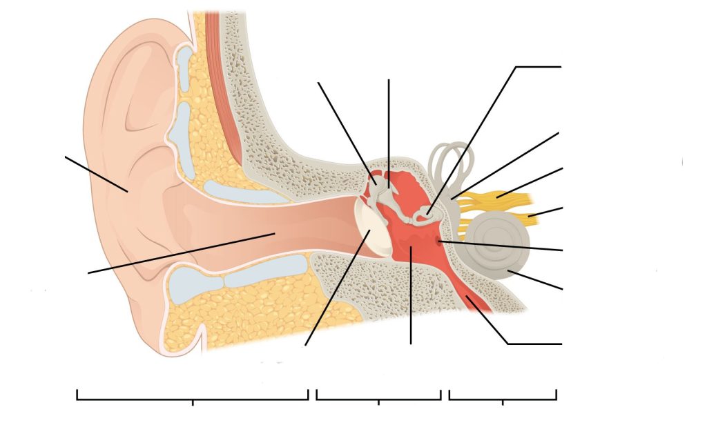
Label the following sensory system ear anatomy:
Sensory System Ear Anatomy labeling activity (Text Version)
Label the diagram with correct words listed below:
- Parieto-occipital sulcus
- Ear canal
- Stapes (attached to oval window)
- Tympanic cavity
- Vestibule
- Cochlear nerve
- Eustachian tube
- Middle ear
- Tympanic membrane
- Malleus
- Incus
- Inner ear
- Round window
- External ear
- Cochlea
- Vestibular nerve

Check your answers [1]
Activity source: Sensory System Ear Anatomy by Gisele tuzon, from Building a Medical Terminology Foundation, illustration from Anatomy and Physiology (OpenStax), licensed under CC BY 4.0./ Text version added.
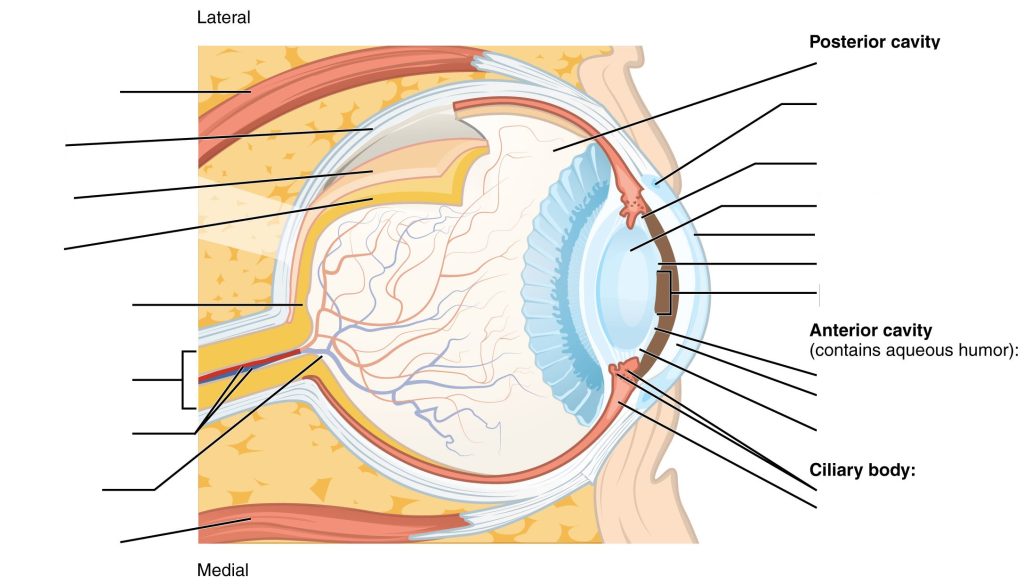
Label the following sensory system eye anatomy:
Sensory System Eye Anatomy (Text Version)
Label the diagram with correct words listed below:
- Fovea centralis
- Suspensory ligaments
- Ciliary muscle
- Retina
- Posterior chamber
- Iris
- Vitreous chamber
- Anterior chamber
- Choroid
- Ciliary process
- Optic disc (blind spot)
- Lens
- Central retinal artery and vein
- Suspensory ligaments
- Lateral rectus muscle
- Optic (II) nerve
- Sclera
- Medial rectus muscle
- Scleral venous sinus (canal of Schlemm)
- Cornea
- Pupil

Sensory System Eye Anatomy Diagram (Text Version)
This diagram shows a lateral and medial view of the eyeball. The major parts are labelled. Labels read from top, clockwise: showing the posterior cavity including the following structures: ______[Blank 1], ______[Blank 2] (canal of Schlemm), ______[Blank 3], _____[Blank 4], ______[Blank 5], _____[Blank 6], and ____[Blank 7]. Next is the anterior cavity (contains aqueous humor), ______[Blank 8], _____[Blank 9], and ______[Blank 10]. The Ciliary body ________[Blank 11] and ________[Blank 12], ______[Blank 13], _____[Blank 14] (blind spot of the eye), ______[Blank 15], _____[Blank 16], _______[Blank 17], ______[Blank 18], ________[Blank 19], and ________[Blank 20].
Check your answers [2]
Activity source: Sensory System Eye Anatomy by Gisele Tuzon, from Building a Medical Terminology Foundation, illustration from Anatomy and Physiology (OpenStax), licensed under CC BY 4.0./ Text version added.
Medical Terms in Context
Place the following medical terms in context to complete the scenario below:
Sensory System – Consultation Report (Text Version)
Use the words below to fill in the consultation report:
- eye
- halos
- acuity
- iris
- dilate
- ophthalmoscope
- cataracts
- subcapsular
- surgery
- intraocular
PATIENT NAME: Betty FOX
AGE: 72
SEX: Female
DOB: October 2
DATE OF CONSULTATION: August 5
CONSULTING PHYSICIAN: Brian Gates, MD, Ophthalmology
REASON FOR CONSULTATION: Cataracts
HISTORY: I saw Mrs. Fox, a 72-year-old, for her regular ______[Blank 1] examination. She has been wearing reading glasses for several years now but has noticed that she has been having trouble reading and has been seeing ______[Blank 2] around lights while driving at night.
PHYSICAL EXAMINATION: A visual ______[Blank 3] test was performed. I used a slit lamp to view the cornea, ______[Blank 4], lens, and the space between the iris and cornea. I detected tiny abnormalities. I administered drops to ______[Blank 5] the pupils to examine the retina. Using an ________[Blank 6], I was able to examine the lenses for signs of ______[Blank 7]. I was able to determine that Mrs. Fox has posterior _________[Blank 8] cataracts in both eyes.
PLAN: I explained to Mrs. Fox that she required cataract _________[Blank 9]. I explained that her clouded lens would be replaced with an _________[Blank 10] lens – a clear artificial lens. She was in agreeance to having the surgery. I told her we would perform the surgery on her right eye first, then in about eight weeks we would do the left eye. Arrangements for her surgery will be made for next month.
_____________________________
Brian Gates, MD, Ophthalmology
Check your answers [3]
Activity source: Sensory System – Consultation Report by Heather Scudder, from Building a Medical Terminology Foundation by Kimberlee Carter and Marie Rutherford, licensed under CC BY- 4.0. / Text version added.
Medical Terms in Context
Place the following medical terms in context to complete the scenario below:
Sensory System – Consultation Report Activity (Text Version)
Use the words below to fill in the consultation report:
- OS
- watering
- antihistamines
- ophthalmalgia
- erythematous
- thyroid
- abnormalities
- masses
- anaesthetic
- puncta
- nasolacrimal
- dacryocystitis
- dacryocystorhinostomy
- medication
PATIENT NAME: Rose MACKENZIE
AGE: 57
SEX: Female
DOB: November 25
DATE OF CONSULTATION: April 16
CONSULTING PHYSICIAN: Ashley Cook MD, Ophthalmology
REASON FOR CONSULTATION: Epiphora in left eye.
HISTORY: Patient is a 57-year-old female who reports epiphora in ________[Blank 1]. Prior to the encounter, she attempted to cure the condition with various ___________[Blank 2]. She states that this has been an ongoing issue for the past 2 years, but the __________[Blank 3] has affected her ability to safely drive over the past 8 months. She denied any persistent _____________[Blank 4], although noted that the surface of the eye was occasionally irritated and _____________[Blank 5] due to rubbing away the tears. She has had no prior eye surgery and no relevant family or personal history of dermatitis or ___________[Blank 6] pathologies.
PHYSICAL EXAMINATION: Patient is alert and oriented x 3, and in no acute distress. Examination of the eye surface revealed no ___________[Blank 7] other than the erythema and tearing. The skin surrounding the eye appeared normal, with no ___________[Blank 8] or swelling.
An irrigation test was then conducted. The eye was treated with ____________[Blank 9] eye drops prior to the test. A syringe filled with saline was inserted into the left _____________[Blank 10] using a hollow wire. The syringe was then pressed to assess the pressure of the left ____________[Blank 11] duct. The fluid did not pass through the nose, indicating inflammation of the duct. No further diagnostic testing was required.
ASSESSMENT: Chronic ____________[Blank 12] of the left nasolacrimal duct.
PLAN: Return for _____________[Blank 13] in 3 months. Patient was instructed to remove tears using tissue instead of her hand to avoid the risk of infection. No _____________[Blank 14] is required in the meantime.
_______________________________
Ashley Cook MD, Ophthalmology
Check your answers [4]
Activity source: Sensory System – Consultation Report Activity by Sheila Bellefeuille & Heather Scudder, licensed under CC BY- 4.0 from “Sensory Systems” in Building a Medical Terminology Foundation by Kimberlee Carter and Marie Rutherford, licensed under CC BY- 4.0. /Text version added.
Test Your Knowledge
Test your knowledge by answering the questions below:
Sensory Systems Glossary Reinforcement activity (Text Version)
- Specialized neurons that respond to changes in temperature are called ____[Blank 1].
- thermoreceptors
- mechanoreceptors
- nociceptors
- Body movement is called _____[Blank 2].
- kinesthesia
- visceral
- proprioception
- Sharpness of vision is called _____[Blank 3].
- visual acuity
- proprioception
- kinesthesia
- Sensory neurons that respond to pain are called _____[Blank 4].
- thermoreceptors
- nociceptors
- glossopharyngeal
- The eardrum is also called _______[Blank 4].
- glossopharyngeal
- mechanoreceptors
- tympanic membrane
Check your answers [5]
Activity source: Sensory Systems Glossary Reinforcement activity by Kimberlee Carter, from Building a Medical Terminology Foundation by Kimberlee Carter and Marie Rutherford, licensed under CC BY- 4.0. /Text version added.
Downloadable Worksheets
View or download & print the PDF or Word format worksheet below:
Worksheet – Sensory Systems Eye/Ear Chapter 15 [Word]
15. Sensory Systems – Definitions Using Word Parts [Word]
15. Sensory Systems – Abbreviations [Word]
Attribution
Except where otherwise noted, this book is adapted from Medical Terminology by Grimm et al. (2022), Nicolet College, CC BY 4.0 International. / A derivative of Building a Medical Terminology Foundation by Carter & Rutherford (2020), and Anatomy and Physiology by Betts, et al., CC BY 4.0, which can be accessed for free at OpenStax Anatomy and Physiology.
-
↵

Check your answers: Sensory System Ear Anatomy labeling activity (Text Version)
text -
↵

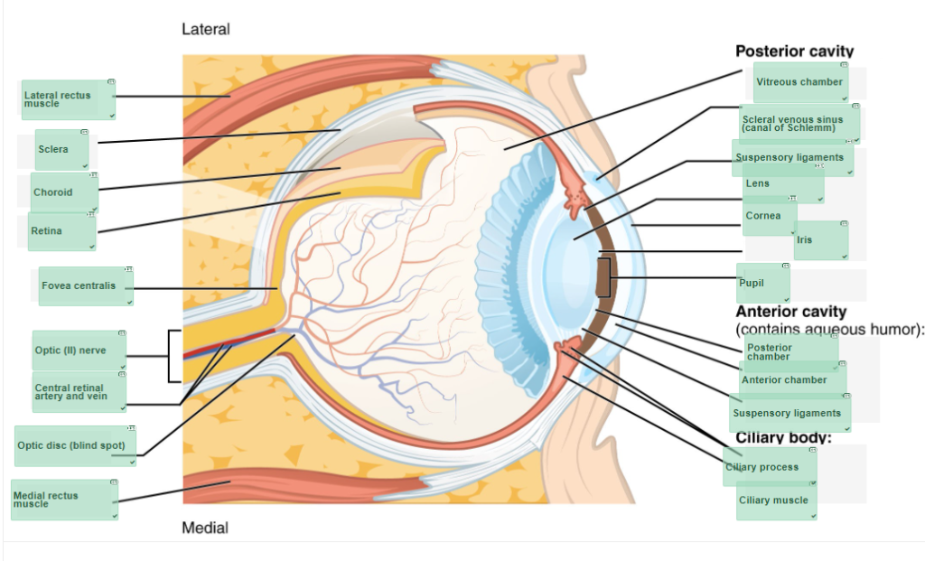
Check your answers: Sensory System Eye Anatomy Diagram (Text Version)
This diagram shows a lateral and medial view of the eyeball. The major parts are labelled. Labels read from top, clockwise: showing the posterior cavity including the following structures: vitreous chamber, scleral venous sinus (canal of Schlemm), suspensory ligaments, lens, cornea, iris, and pupil. Next is the anterior cavity (contains aqueous humor), posterior chamber, anterior chamber, and suspensory ligaments. The Ciliary body ciliary process and ciliary muscle, medial rectus muscle, optic disc (blind spot of the eye), central retinal artery and vein, foveal centralis, optic nerve, retina, choroid, sclera, and lateral rectus muscle. - 1.eye, 2.halos, 3.acuity, 4.iris, 5.dilate, 6.ophthalmoscope, 7.cataracts, 8.subcapsular, 9.surgery, 10.intraocular ↵
- 1.OS, 2.antihistamines, 3.watering, 4.ophthalmalgia, 5.erythematous, 6.thyroid, 7.abnormalities, 8.masses, 9.anaesthetic, 10.puncta, 11.nasolacrimal, 12.dacryocystitis, 13.dacryocystorhinostomy, 14.medication ↵
- 1. thermoreceptors, 2. kinesthesia, 3. Sharpness of vision is called..., 4. Sensory neurons that respond to pain are called..., 5. The ear-drum is also called... ↵

