6.1 – Types of Muscles
 |
6.1. List different types of muscles and briefly explain how they generate locomotion. |
Your last chapter discussed how animals produces energy using different nutrients. The focus of this lecture is on how this energy, in the form of ATP is used to fuel one of the most important physiological responses of animals to their environment, locomotion through muscle contraction.
Types of Muscle Tissue
Muscle cells are excitable cells that are specialized for contraction. They allow for many types of locomotion from small changes in facial expressions to the highly coordinated series of contraction that facilitate walking and even unconscious processes such as respiration and digestion. Muscle tissues differ in their physical appearance, anatomy, location within the body and whether their contraction is controlled consciously or unconsciously. Animals have three types of muscle: skeletal muscle, cardiac muscle, and smooth muscle (Figure 6.2).
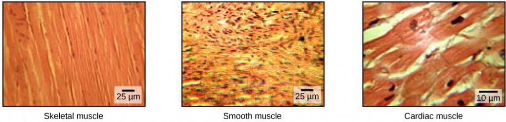
Skeletal muscle tissue forms skeletal muscles, which attach to bones via tendons and contracts to cause locomotion and all other consciously controlled movement. Skeletal muscles make up the fast majority of the animal body, and is also referred to as voluntary muscle as it is consciously controlled. When viewed under a microscope (Figure 6.2), skeletal muscle tissue has a striated appearance. The striations are caused by the regular arrangement of contractile proteins (actin and myosin). Skeletal muscles are also multi nucleated, meaning they have many nuclei in each cell.
Smooth muscle tissue occurs in the walls of hollow organs such as the intestines, stomach, and urinary bladder, and around passages such as the respiratory tract and blood vessels. Smooth muscle has no striations, is not under voluntary control, has only one nucleus per cell, is tapered at both ends, and is called involuntary muscle.
Cardiac muscle tissue is only found in the heart, and cardiac contractions pump blood throughout the body and maintain blood pressure. Like skeletal muscle, cardiac muscle is striated, but unlike skeletal muscle, cardiac muscle cannot be consciously controlled and is called involuntary muscle. It has one nucleus per cell, is branched, and is distinguished by the presence of intercalated disks.
Skeletal Muscle and Muscle Fiber Structure
Skeletal muscle has three layers: Epimysium, Perimysium and Endomysium. The Epimysium surrounds the entire muscle and is on the outermost layer of the muscle. The Perimysium is located beneath the Epimysium and it surrounds the muscle fibers and the Endomysium. The Endomysium is the deepest layer in the muscle and it surrounds each individual myofibril.
Each skeletal muscle fiber is a skeletal muscle cell. These cells are incredibly large, with diameters of up to 100 µm and lengths of up to 30 cm. The plasma membrane of a skeletal muscle fiber is called the sarcolemma. The sarcolemma is the site of action potential conduction, which triggers muscle contraction. Within each muscle fiber are myofibrils—long cylindrical structures that lie parallel to the muscle fiber. Myofibrils run the entire length of the muscle fiber, and because they are only approximately 1.2 µm in diameter, hundreds to thousands can be found inside one muscle fiber. They attach to the sarcolemma at their ends, so that as myofibrils shorten, the entire muscle cell contracts (Figure 6.3).
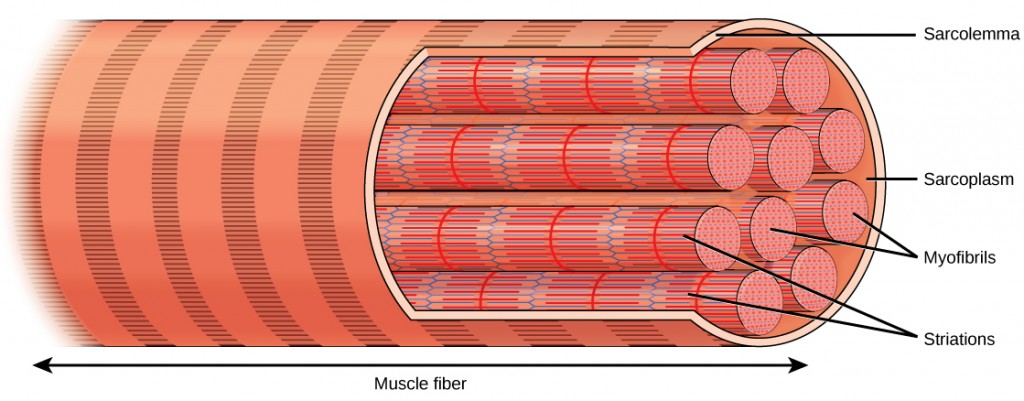
The striated appearance of skeletal muscle tissue is a result of repeating bands of the proteins actin and myosin that are present along the length of myofibrils. Dark A bands and light I bands repeat along myofibrils, and the alignment of myofibrils in the cell causes the entire cell to appear striated or banded.
Each I band has a dense line running vertically through the middle called a Z disc or Z line. The Z discs mark the border of units called sarcomeres, which are the functional units of skeletal muscle. One sarcomere is the space between two consecutive Z discs and contains one entire A band and two halves of an I band, one on either side of the A band. A myofibril is composed of many sarcomeres running along its length, and as the sarcomeres individually contract, the myofibrils and muscle cells shorten (Figure 6.4).

Myofibrils are composed of smaller structures called myofilaments. There are two main types of filaments: thick filaments and thin filaments; each has different compositions and locations. Thick filaments occur only in the A band of a myofibril. Thin filaments attach to a protein in the Z disc called alpha-actinin and occur across the entire length of the I band and partway into the A band. The region at which thick and thin filaments overlap has a dense appearance, as there is little space between the filaments. Thin filaments do not extend all the way into the A bands, leaving a central region of the A band that only contains thick filaments. This central region of the A band looks slightly lighter than the rest of the A band and is called the H zone. The middle of the H zone has a vertical line called the M line, at which accessory proteins hold together thick filaments. Both the Z disc and the M line hold myofilaments in place to maintain the structural arrangement and layering of the myofibril. Myofibrils are connected to each other by intermediate, or desmin, filaments that attach to the Z disc.
Thick and thin filaments are themselves composed of proteins. Thick filaments are composed of the protein myosin. The tail of a myosin molecule connects with other myosin molecules to form the central region of a thick filament near the M line, whereas the heads align on either side of the thick filament where the thin filaments overlap. The primary component of thin filaments is the actin protein. Two other components of the thin filament are tropomyosin and troponin. Actin has binding sites for myosin attachment. Strands of tropomyosin block the binding sites and prevent actin-myosin interactions when the muscles are at rest. Troponin consists of three globular subunits. One subunit binds to tropomyosin, one subunit binds to actin, and one subunit binds Ca2+ ions.
6.1 Concept Questions
True/False:
Multiple Choice:
 |
View this animation showing the organization of muscle fibers. |
In addition to three different types of muscles discussed above, there are also differences in skeletal muscle function. Different skeletal muscles in animals are described as white and red muscle. These different types of skeletal muscles are recruited depending on whether a fast and short versus steady and prolonged locomotion is needed by the animal.
 |
You should read this article about red and white muscles in fish in order to be able to answer the questions at the end of the chapter. |
 |
Based on the article about swimming in fish you read (linked above), answer the following questions.
Question 6.2 Describe the differences between white and red muscle. |
|
Based on the article about swimming in fish you read (linked above), answer the following questions.
Question 6.3 Where is the most of the red muscle found in the fish studied in the above article and what is the functional significance of this muscle location? Hint: review Figures 5 and 6 of the article. |
 |
Question 6.4.
Which is more efficient – red or white muscle? Why? |
