5 CMB 5 – Highway 504
Heidi Daxberger and Phillip Ruscica
GPS Coordinates (44°50’9.89″N, 77°55’10.88″W)
Parking: You can park along the shoulder of the road.
Click here to open the Bedrock Geology legend on the OGS website
At this location you can investigate highly deformed and metamorphosed calc-silicate rocks that show evidence of later intrusion by mafic melts. The road cut outcrop stretches for about 70 metres in a NNW-SSE direction along HW 504 and is here separated in a North and South section. For each section you will be able to view overview and numerous close up images to show the many features of the rocks seen here.
The Digital Outcrops
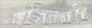
The following model is the northern section of the outcrop of the CMB5 outcrop along HW 504. The model shows the well developed stretching lineation (plunge 24 degrees) on the mica-rich foliation planes. Furthermore, tension gashes and a slight warping of the planes can be seen on many of the foliation planes. The warping (weak folding, axis plunge 70 degrees) is an extension of the boudinage of more competent layers of the calc-silicate rocks that can be seen when standing on top of the outcrop and looking down. However, boudinage can also be seen along the vertical view parallel to the foliation, what indicates a complex three dimensional stretching pattern of the foliated rocks (like a chocolate board). Use hammer (yellow) as scale.
The outcrop image collection below includes close-up images of the described geologic features.
Overview Image 1 and 2 are part of the North Section shown in the google slide show below.
In the following slide show you will find close up images for the North Section.
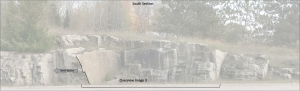
The following model is the centre section (here called south section) of the CMB5 road outcrop along HW 504. In this model you can inspect the nearly vertical foliation that strikes roughly 130 degrees (SE). Furthermore, you can see an intricate pattern of fractures on the nicely exposed foliation plane on the left side. The centre of the model, view parallel to the foliation, shows evidence of shear related passive folding. At the right side of the outcrop model you can see a roughly 20 cm thick metamorphosed mafic intrusions (larger white crystals in mafic matrix) that are concordant to the foliation at the top but cut across the at the bottom.
Overview image 3 below also shows the general trend of steeply dipping NNW-SSE-striking foliation of the calc-silicate rocks.
Virtual Hand Samples (3D Sketchfab Models)
You can investigate the following 3D hand samples by clicking on the respective images.
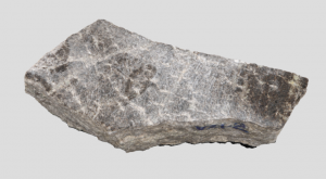
The two virtual 3D models below show examples of hand samples of the calc-silicate rocks found at the CMB5 outcrop (VLS-21-1). The first shows the three dimensional boudinage of more competent, biotite-rich layers of the calc silicate rocks, while the material of the less competent calcite-rich layers flow into the gap between the boudins. 
The second shows the general features of the calc-silicate rocks (VLS-21-1).

The VLS-21-2 shows the general features of the calc-silicate rocks at CMB5 including the mica-rich (brownish) foliation planes.
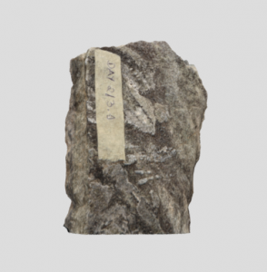
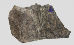
The following two rocks are examples(VLS-21-3) of the mica-rich rocks that are part of the calc-silicate rocks seen at the CMB5 outcrop. The elongated/bladed minerals on the front show a blue hue in hand sample (Kyanite).

The next virtual hand sample (VLS-21-6) shows part of the metamorphosed mafic intrusions including the contact to the surrounding calc-silicate rocks. The larger minerals in the mafic rocks may represent Scapolite.
Description of Thin Sections
In the following section you can take a look at thin section imagery of sSamples take at CMB 5 which correlate to the samples shown above where available. The slide show below features the whole section scans of the provided thin sections. Further below you will find videos of full 360 degree rotations at specific close up locations of each thin section.
The following videos show full 360 rotations at a higher magnification of each thin section.
Scanning Electron Microscope
Sample VLS-21-4
Sample VLS-21-6 Point 2
Sample VLS-21-6 Point 10000
Sample VLS-21-6 Point 20000
Sample VLS-21-1
Check out the methodologies used in this book here: Appendix: Methods!