Want to create or adapt books like this? Learn more about how Pressbooks supports open publishing practices.
37 Case 1-2010: A 68-year-old male with breathlessness
Lower respiratory tract infection and rapid expansion of an abdominal aortic aneurysm: a case report. Journal of Medical Case Reports,2010, 4(1). doi: 10.1186/1752-1947-4-333
Naylor, S., Gamie, Z., Vohra, R., Puppala, S., Kent, P., & Scott, D.
Case Summary 1
A 68-year-old Caucasian male was admitted with a contaminated lower respiratory tract infection (LRTI), increasing back pain and epigastric discomfort. This patient’s medical history includes consistent cigarette smoking for 20 years, a coronary artery bypass graft for ischemic heart disease, and an existing aortic aneurysm. Clinical examinations showed tender epigastrium and left bronchopneumonia. A computed tomography (CT) aortogram, CT thorax, and coronal CT angiogram were used to further investigate this patient’s condition and diagnosis.
Learning Objectives
Investigating the clinical history of the patient and selecting appropriate examinations to identify this respiratory infection.
Correlating the patient’s symptoms and the case study clinical examinations to narrow down the possible diagnosis.
Familiarizing and defining new medical terminology associated with this patient’s condition.
Extrapolating key lifestyle factors that have contributed to the respiratory infection and preventative measures that can be put in place to ensure the future health of the patient.
Clinical History 1
Age: 68 years old
Sex: Male
Ethnicity: Caucasian
Medical History 1
Has been previously diagnosed with infra-renal AAA which had been monitored for six-months.
Maximum diameter was 4.9 cm, grown to 5.2 cm over one year.
Most recent ultrasound scan was conducted two months before admission.
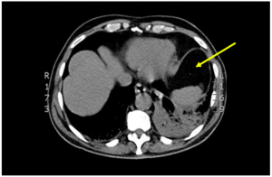
Extensive lower lobe consolidation and collapse with hilar lymphadenopathy was observed (figure 2, yellow arrow).
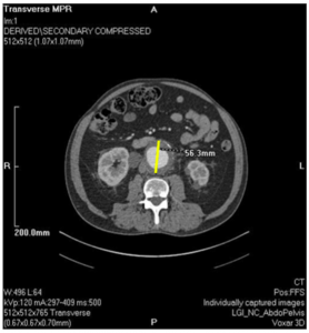
Figure 1: CT aortogram revealing the 5.6 cm (yellow line) anteroposterior diameter AAA.1
Figure 2: CT thorax shows the lower lobe consolidation and collapse in the left lung (yellow arrow) with extensive hilar lymphadenopathy.1
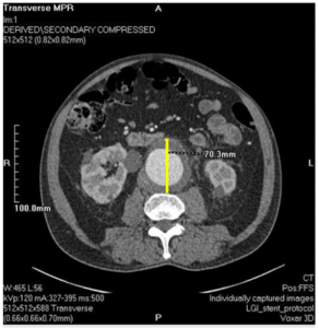
Due to symptoms of hypotension 48-hours after admission, a repeat CT aortogram was conducted revealing the increased size of the AAA to 7.0 cm (figure 3, yellow line). This would also reveal retroperitoneal fat stranding (figure 4, yellow line).
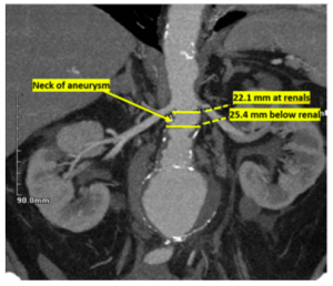
No significant angulation (changes in angle formation) was shown in the neck of the aneurysm. The juxta-renal diameter (aneurysms adjacent to renal artery origins) had been 22.1 mm before increasing to 25.4 mm in its infra-renal (below the renal artery origins) segment (figure 4).
These repeat CT scans revealed significant stenosis (narrowing) of the left common iliac artery.
Figure 3: CT aortogram shows a rapid increased 7.0 cm (yellow line) size of the AAA in the anteroposterior diameter. This also shows sign of impending rupture and beak in left lateral aortic thrombus.1Figure 4: Coronal CT angiogram image shows 7.0 cm aneurysm. The diameter of the neck of aneurysm at the renal arteries were 22.1 mm and the renal arteries below were 25.4 mm.1
Question & Answers Leading to Diagnosis:
Question 1:Considering the patient’s previous medical history and current symptoms, what diagnosis could we expect?
Question 2:From the patient’s respiratory symptoms, lab investigations and CT scans, what is the confirmatory diagnosis?
Question 3:The CT scans have shown AAA expansion and lower lobe consolidation, what biomarkers could be used to support this diagnosis? How can both diagnoses be correlated?
** For answers please check the next chapter.
Medical terminology/Abbreviations:
Abdominal aortic aneurysm (AAA) – The enlargement of the lower area of the major vessel which supplies blood to the body.10
Chemokines – A family of chemoattractant cytokines which are secreted by cells in response to the body’s immune system.11
Computed tomography (CT) aortogram – A technique using CT scanning and an injection of contrast material into the blood vessels to examine and diagnose cardiovascular diseases. 12
Computed tomography (CT) thorax – An imaging test to examine organs and chest using X-ray and computer technology. 13
Coronal computed tomography (CT) angiogram – A technique using CT scanning and an injection of contrast material into the blood vessels to evaluate structure and patency of arteries suppling lower limbs and abdomen with blood. 14
Coronary artery bypass graft – Surgery redirecting blood around a blocked artery of the heart. 15
Cytokines – Group of glycoproteins, peptides and proteins secreted by cells in response to the immune system, they regulate and mediate immunity. 16
Epigastric – Upper central region of the abdomen. 17
Growth factor – Substance required for growth stimulation in living cells. 18
Hypoxic – Condition where the areas of the body or the body does not have an adequate oxygen supply at the tissue level. 19
Ischemic heart disease (or coronary heart/artery disease) – Disease with the heart is getting an inadequate supply of blood and oxygen due to narrowing of arteries. 20
Lower respiratory tract infection (LRTI) or pneumonia – Infection that involves the lungs abscess and acute bronchitis. 21
Pro-inflammatory biomarkers – Regulatory proteins that can be used to detect inflammation. 22
Sepsis – Condition where the body’s response to an existing infection begins to damage it’s own tissues, this can be a potentially life-threatening. 23
Stenosis – Narrowing of the diameter of bodily passages. 24
References
Naylor, S., Gamie, Z., Vohra, R., Puppala, S., Kent, P., & Scott, D. (2010). Lower respiratory tract infection and rapid expansion of an abdominal aortic aneurysm: a case report. Journal Of Medical Case Reports, 4(1). doi: 10.1186/1752-1947-4-333
Middleton, R. K., Lloyd, G. M., Bown, M. J., Cooper, N. J., London, N. J., & Sayers, R. D. (2007). The pro-inflammatory and chemotactic cytokine microenvironment of the abdominal aortic aneurysm wall: a protein array study. Journal of vascular surgery, 45(3), 574–580. https://doi.org/10.1016/j.jvs.2006.11.020
Wallinder, J., Bergqvist, D., & Henriksson, A. E. (2009). Proinflammatory and anti-inflammatory cytokine balance in patients with abdominal aortic aneurysm and the impact of aneurysm size. Vascular and endovascular surgery, 43(3), 258–261. https://doi.org/10.1177/1538574408324617
Gallagher, P. M., Lowe, G., Fitzgerald, T., Bella, A., Greene, C. M., McElvaney, N. G., & O’Neill, S. J. (2003). Association of IL-10 polymorphism with severity of illness in community acquired pneumonia. Thorax, 58(2), 154–156. https://doi.org/10.1136/thorax.58.2.154
Monaco, M., Di Tommaso, L., Oliviero, U., Iannelli, G., & Stassano, P. (2008). A rapidly expanding descending thoracic aortic aneurysm: an unusual complication. Journal of cardiac surgery, 23(3), 260–261. https://doi.org/10.1111/j.1540-8191.2007.00525.x
Mory, M., Hansmann, J., Allenberg, J. R., & Böckler, D. (2007). Images in vascular medicine. Rapid expansion of an inflammatory abdominal aortic aneurysm. Vascular medicine (London, England), 12(4), 381–382. https://doi.org/10.1177/1358863X07083276
Oxford Learner’s Dictionaries | Find definitions, translations, and grammar explanations at Oxford Learner’s Dictionaries. (2021). Retrieved 27 April 2021, from https://www.oxfordlearnersdictionaries.com/
Samuel J., Franklin C. (2008) Hypoxemia and Hypoxia. In: Myers J.A., Millikan K.W., Saclarides T.J. (eds) Common Surgical Diseases. Springer, New York, NY. https://doi.org/10.1007/978-0-387-75246-4_97
Institute of Medicine (US) Committee on Social Security Cardiovascular Disability Criteria. Cardiovascular Disability: Updating the Social Security Listings. Washington (DC): National Academies Press (US); 2010. 7, Ischemic Heart Disease. Available from: https://www.ncbi.nlm.nih.gov/books/NBK209964/
Therapeutic Guidelines Limited. (2014). Therapeutic guidelines. West Melbourne, Victoria.
van den Berg, R., Jongbloed, E., de Schepper, E., Bierma-Zeinstra, S., Koes, B., & Luijsterburg, P. (2018). The association between pro-inflammatory biomarkers and nonspecific low back pain: a systematic review. The Spine Journal, 18(11), 2140-2151. doi: 10.1016/j.spinee.2018.06.349