8.2 – Procedures Related to Obstetrics
In Vitro Fertilization (IVF)
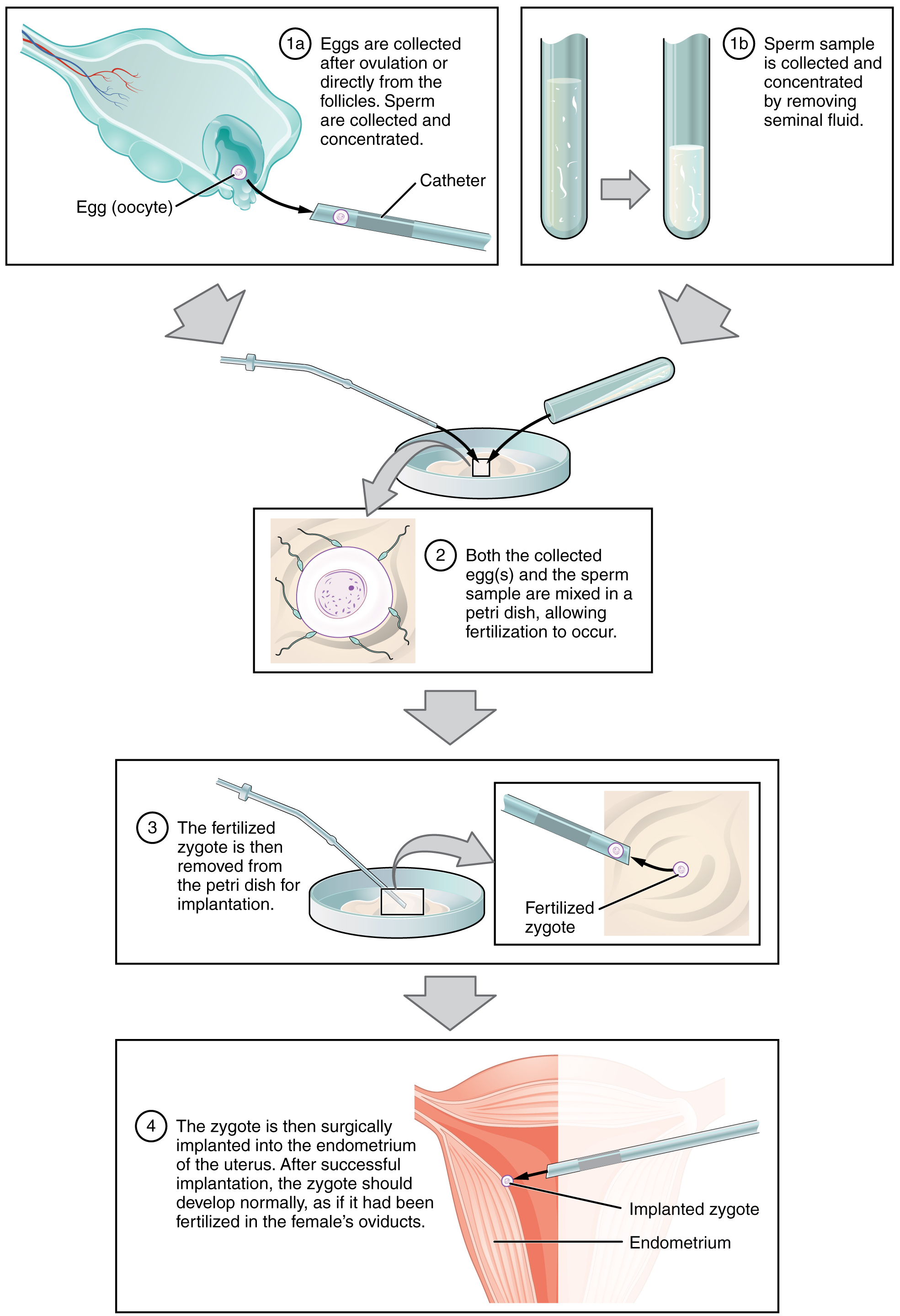
IVF, which stands for in vitro fertilization, is an assisted reproductive technology. In vitro, which in Latin translates to in glass, refers to a procedure that takes place outside of the body. There are many different indications for IVF. For example, a woman may produce normal eggs, but the eggs cannot reach the uterus because the uterine tubes are blocked or otherwise compromised. A man may have a low sperm count, low sperm motility, sperm with an unusually high percentage of morphological abnormalities, or sperm that are incapable of penetrating the zona pellucida of an egg. Figure 8.2 illustrates the steps involved in IVF.
Did You Know?
Prenatal Screening and Diagnostic Testing
Approximately 4% of Canadian babies are born with a congenital anomaly. The most common anomalies include structural heart defects, cleft lip/palate, or anomalies like Down syndrome. Prenatal testing may include blood work, ultrasound, chorionic villus sampling (CVS) and amniocentesis (Genetics Education Canada Knowledge Organization, 2019). To learn more about prenatal screening tests, visit GECKO’s Guide to Understanding Prenatal Screening Tests [PDF].
Image Descriptions
Figure 8.2 image description: This multi-part figure shows the different steps of in vitro fertilization. The top panel shows how the oocytes and the sperm are collected and prepared (text reads: 1a) eggs are collected after ovulation or directly from the follicles. Sperm are collected and concentrated. 1b) Sperm sample is collected and concentrated by removing seminal fluid). The next panel shows the sperm and oocytes being mixed in a petri dish (text labels read: 2) both the collected eggs and the sperm sample are mixed in a petri dish, allowing fertilization to occur). The panel below that shows the fertilized zygote being prepared for implantation (text labels read: 3a) the fertilized zygote is then removed from the petri dish for implantation. 3b) fertilized zygote). The last panel shows the fertilized zygote being implanted into the uterus (text label reads: 4) The zygote is then surgically implanted into the endometrium of the uterus. After successful implantation, the zygote should develop normally, as if it had been fertilized in the female’s oviducts). [Return to Figure 8.2].
Attribution
Except where otherwise noted, this chapter is adapted from “Obstetrics” in Building a Medical Terminology Foundation by Kimberlee Carter and Marie Rutherford, licensed under CC BY 4.0. / A derivative of Betts et al., which can be accessed for free from Anatomy and Physiology (OpenStax). Adaptations: dividing Obstetrics chapter content into sub-chapters.
assisted reproductive technology
A small piece of placenta is taken and tested to determine potential for birth differences
surgical puncture (using a needle) to remove amniotic fluid for sampling.
