6.3 – Physiology (Function) of the Male Reproductive System
Spermatogenesis
Spermatogenesis occurs in the seminiferous tubules that form the bulk of each testis. The process begins at puberty, after which time sperm are produced constantly throughout a man’s life. One production cycle takes approximately 64 days. One production cycle is considered from spermatogonia through to formed sperm. A new cycle starts approximately every 16 days, although this timing is not synchronous across the seminiferous tubules.
Did You Know?
Sperm
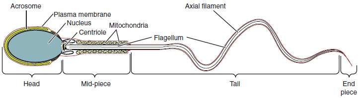
Sperm are smaller than most cells in the body; in fact, the volume of a sperm cell is 85,000 times smaller than that of the female gamete. Approximately 100 to 300 million sperm are produced each day, whereas women typically ovulate only one oocyte per month. As is true for most cells in the body, the structure of sperm cells speaks to their function. Sperm have a distinctive head, mid-piece, and tail region (see Figure 6.2).
Sperm Transport
To fertilize an egg, sperm must be moved from the seminiferous tubules in the testes, through the epididymis, and—later during ejaculation—along the length of the penis and out into the female reproductive tract. It takes an average of 12 days for sperm to move through the coils of the epididymis, with the shortest recorded transit time in humans being one day.
Epididymis
Sperm enter the head of the epididymis and are moved by the contraction of smooth muscles lining the epididymal tubes. As the sperm mature they acquire the ability to move under their own power. Once inside the female reproductive tract, they will use this ability to move independently toward the unfertilized egg. The more mature sperm are then stored in the tail of the epididymis until ejaculation occurs.
Ducts
During ejaculation, sperm exit the tail of the epididymis and are pushed by smooth muscle contraction to the vas deferens (also called the ductus deferens). The vas deferens is a thick, muscular tube that is bundled together inside the scrotum with connective tissue, blood vessels, and nerves into a structure called the spermatic cord. From each epididymis, each vas deferens extends through the inguinal canal in the abdominal wall and continues to a region called the ampulla. The sperm is mixed with fluid from the paired seminal vesicles and moves into its associated ejaculatory duct. The ejaculatory ducts transport the seminal fluid to the prostate gland.
Prostate Gland
The prostate gland secretes an alkaline, milky fluid to the passing seminal fluid (referred to as semen) to first coagulate and then decoagulate the semen following ejaculation. The temporary thickening of semen helps retain it within the female reproductive tract and once decoagulated the sperm can pass farther into the female reproductive tract.
Bulbourethral Glands
Bulbourethral glands release a thick, salty fluid that lubricates the end of the urethra and vagina, and helps to clean urine residues from the penile urethra.
Check Your Knowledge of the Male Reproductive System
Concept Check
- Write or draw out the components of the pathway that sperm takes from beginning until the end.
- Consider fertility challenges that may be experienced if a large number of defective sperm are produced.
Anatomy Labeling Activity
Anatomy Labeling Activity (Text Version)
Label the following diagram correctly with words:
- Ureter
- Seminal Vesicle
- Pubic Bone
- Anus
- Prostate Gland
- Urethra
- Urinary Bladder
- Penis
- Testis
- Epididymis
- Vas Deferens
- Rectum
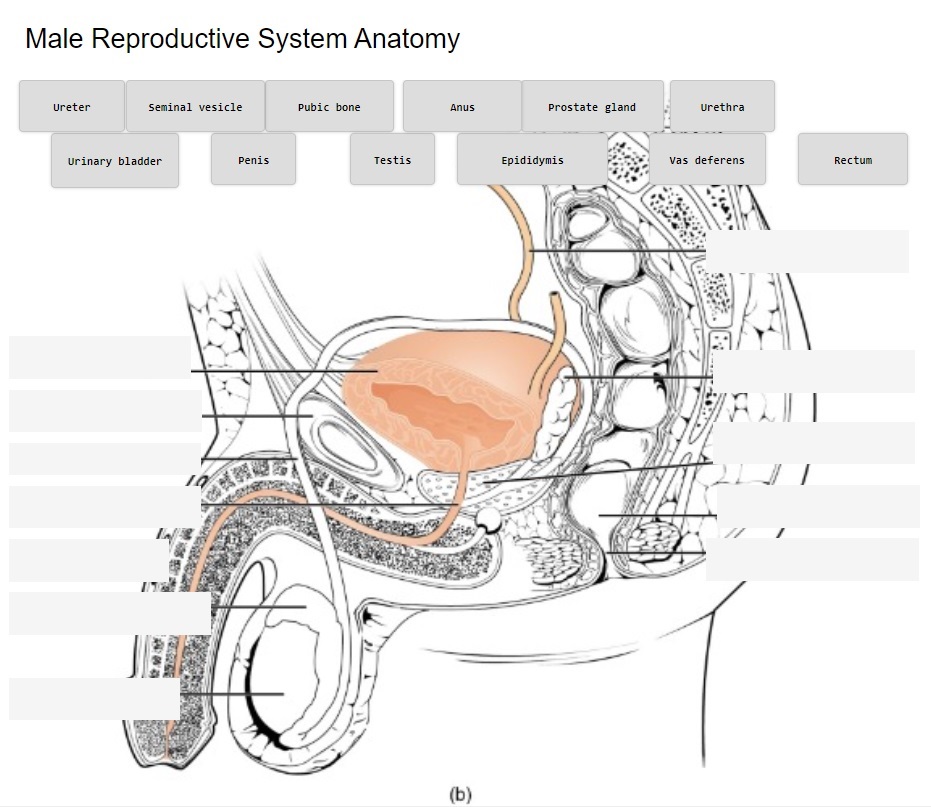
Anatomy Labeling Activity Diagram (Text Version)
Detailed anatomical diagram of the male reproductive system from a lateral viewpoint. The diagram highlights the location of key components from top of the diagram is the _______[Blank 1]. Then from right to left is the _________[Blank 2] followed by the ________[Blank 3] which are a pair of glands that secrete fluid making up a substantial portion of seminal fluid. The _______[Blank 4] is one of the three bones making up the pelvis. The _______[Blank 5] is a carrying vessel that transports sperm from the testes to the urethra. Located at the base of the bladder is ________[Blank 6], this gland secretes nourishing fluid for sperm and becomes a component of semen. The ______[Blank 7] extends from the urinary bladder and carries the semen towards the penis. The _______[Blank 8], the external male sex organ used to inseminate a female during reproduction. The ______[Blank 9] is the straight portion of the lower large intestines, and the _______[Blank 10] expels fecal matter. Located under the penis is the ______[Blank 11] and extending from the testes is a cordlike structure known as the ________[Blank 12].
Check your answers: [1]
Activity source: Male Reproductive System Anatomy by Gisele Tuzon, from Building a Medical Terminology Foundation by Kimberlee Carter and Marie Rutherford, licensed under CC BY 4.0./ Text version added.
Male Reproductive Terms Not Easily Broken into Word Parts
Male Reproductive System terms not easily broken down into word parts (Text Version)
- hydrocele
- fluid-filled sac around the testicle
- varicocele
- enlarged veins of the spermatic cord
- ablation
- destruction of abnormal or excessive tissue by eroding, vaporizing or melting
- circumcision
- surgical removal of the prepuce (foreskin)
- enucleation
- excision of a whole organ or mass without cutting into it
- hydrocelectomy
- surgical removal of a fluid-filled sac around the testicle causing scrotal swelling (hydrocele)
- coitus
- sexual intercourse between male and female
- condom
- sheath (cover) for penis worn during coitus to prevent conception and spread of sexually transmitted infection
- infertility
- inability to achieve pregnancy
- sterility
- a condition of being unable to conceive or reproduce the species
Activity Source: Male Reproductive System terms not easily broken down into word parts by Kimberlee Carter, from Building a Medical Terminology Foundation by Kimberlee Carter and Marie Rutherford, licensed under CC BY 4.0./ Text version added.
Common Male Reproductive System Abbreviations
Common Male Reproductive System Abbreviations
- AIDS (acquired immunodeficiency syndrome)
- BPH (benign prostatic hyperplasia, benign prostatic hypertrophy)
- Bx (biopsy)
- CT (chlamydia)
- DRE (digital rectal examination)
- ED (erectile dysfunction)
- FTA-ABS (florescent treponemal antibody absorption test)
- GC (gonococcus)
- GU (genitourinary)
- HIV (human immunodeficiency virus)
- HoLEP (holmium laser enucleation of the prostate gland)
- HPV (human papillomavirus)
- HSV-2 (herpes simplex virus 2)
- LUTS (lower urinary tract symptoms)
- NGU (nongonococcal urethritis)
- PSA (prostate-specific antigen)
- PVP (photoselective vaporization of the prostate gland)
- RP (radical prostatectomy)
- STD (sexually transmitted disease)
- STI (sexually transmitted infection)
- TRUS (transrectal ultrasound)
- TSE (testicular self-examination)
- TUIP (transurethral incision of the prostate gland)
- TUMT (transurethral microwave thermotherapy)
- TURP (transurethral resection of the prostate gland)
- VD (venereal disease)
- VDRL (Venereal Disease Research Laboratory)
Activity source: Male Reproductive System Common Abbreviations by Kimberlee Carter, from Building a Medical Terminology Foundation by Kimberlee Carter and Marie Rutherford licensed under CC BY 4.0./ Text version.
Image Descriptions
Figure 6.2 image description: This diagram shows the structure of sperm; the major parts are labeled (from left to right): head section (acrosome, plasma membrane, nucleus), mid-piece (centriole, mitochondria, flagellum), tail (flagellum, axial filament), end piece (end piece). [Return to Figure 6.2].
Attribution
Except where otherwise noted, this chapter is adapted from “Male Reproductive System” in Building a Medical Terminology Foundation by Kimberlee Carter and Marie Rutherford licensed under CC BY 4.0. / A derivative of Betts et al., which can be accessed for free from Anatomy and Physiology (OpenStax). Adaptations: dividing Male Reproductive System chapter content into sub-chapters.
-
↵
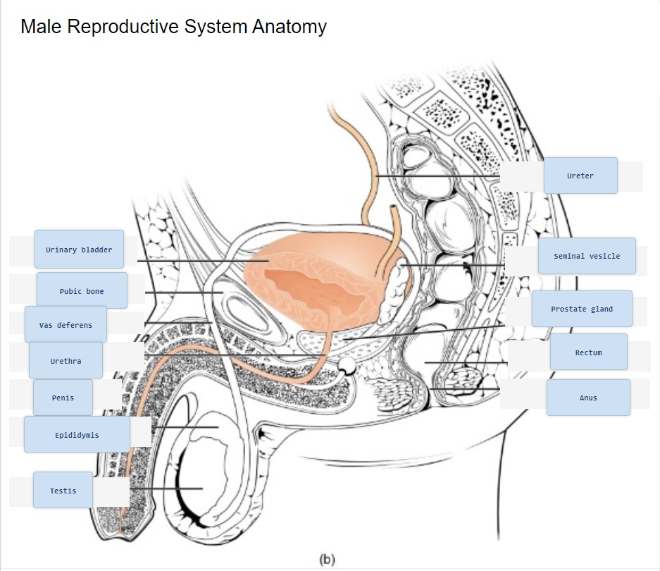
Check your answer: Anatomy Labeling Activity Diagram (Text Version)
Detailed anatomical diagram of the male reproductive system from a lateral viewpoint. The diagram highlights the location of key components from top of the diagram is the ureter. Then from right to left is the urinary bladder followed by the seminal vesicles which are a pair of glands that secrete fluid making up a substantial portion of seminal fluid. The pubic bone is one of the three bones making up the pelvis. The vas deferens is a carrying vessel that transports sperm from the testes to the urethra. Located at the base of the bladder is prostate gland, this gland secretes nourishing fluid for sperm and becomes a component of semen. The urethra extends from the urinary bladder and carries the semen towards the penis. The penis, the external male sex organ used to inseminate a female during reproduction. The rectum is the straight portion of the lower large intestines, and the anus expels fecal matter. Located under the penis is the testes and extending from the testes is a cordlike structure known as the epididymis.
Process of producing sperm
diploid precursor cells that become sperm (singular = spermatogonium)
tube structures within the testes where spermatogenesis occurs
male gamete (spermatozoon)
female gamete
(plural = epididymides) coiled tubular structure in which sperm start to mature and are stored until ejaculation
Pertaining to the epididymis
(also, ductus deferens) duct that transports sperm from the epididymis through the spermatic cord and into the ejaculatory duct; also referred as the vas deferens
doughnut-shaped gland at the base of the bladder surrounding the urethra and contributing fluid to semen during ejaculation
(also, Cowper’s glands) glands that secrete a lubricating mucus that cleans and lubricates the urethra prior to and during ejaculation
