2.1 – Levels of Organization
Learning Objectives
- Connect medical language learning to the context of anatomy and physiology
- Introduce the basic architecture and levels of organization of the human body
- Evaluate the anatomical position, regional terms, directional terms, body planes, and body quadrants for anatomical positioning
- Describe body cavities and the functions of associated membranes
As you memorize the language components of medical terminology, it is important to support that learning within the context of anatomy and physiology. Proceeding through the body system chapters, you will learn word parts, whole medical terms, and common abbreviations. It is important to put into context where in the body the medical term is referencing, and then consider how it works within the body.
Anatomy focuses on structure and physiology focuses on function. Much of the study of physiology centers on the body’s tendency toward homeostasis.
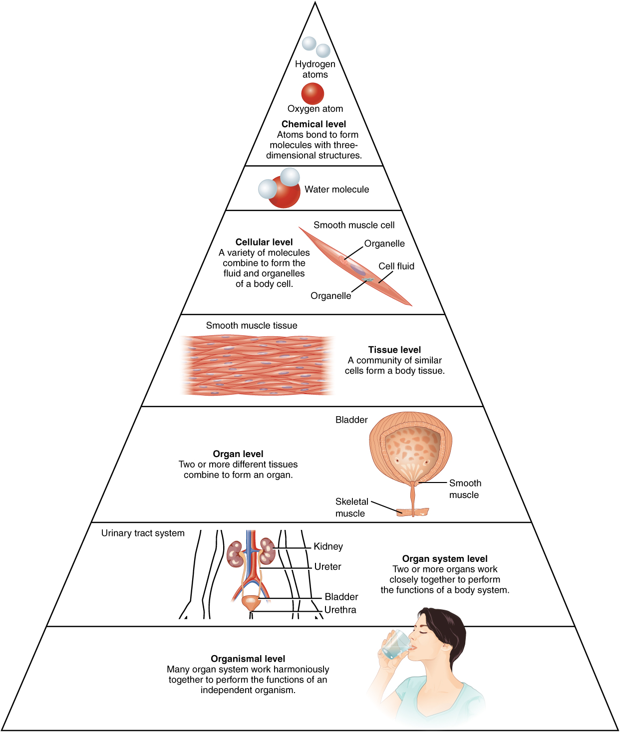
Consider the structures of the body in terms of fundamental levels of organization that increase in complexity: subatomic particles, atoms, molecules, organelles, cells, tissues, organs, organ systems, organisms, and biosphere (Figure 2.1).
The Levels of Organization
All matter in the universe is composed of one or more unique pure substances called elements; familiar examples are hydrogen, oxygen, carbon, nitrogen, calcium, and iron.
- The smallest unit of any of these pure substances (elements) is an atom.
- Atoms are made up of subatomic particles such as the proton, electron, and neutron.
- Two or more atoms combine to form a molecule, such as the water molecules, proteins, and sugars found in living things.
- Molecules are the chemical building blocks of all body structures.
- A cell is the smallest independently functioning unit of a living organism.
- Even bacteria, which are extremely small, independently-living organisms, have a cellular structure. Each bacterium is a single cell. All living structures of human anatomy contain cells, and almost all functions of human physiology are performed in cells or are initiated by cells
- A human cell typically consists of flexible membranes that enclose cytoplasm, a water-based cellular fluid, together with a variety of tiny functioning units called organelles. In humans, as in all organisms, cells perform all functions of life.
- A tissue is a group of many similar cells (though sometimes composed of a few related types) that work together to perform a specific function.
- An organ is an anatomically distinct structure of the body composed of two or more tissue types. Each organ performs one or more specific physiological functions.
An organ system is a group of organs that work together to perform major functions or meet the physiological needs of the body.
Did You Know?
- Organs are very collaborative and work with multiple body systems.
- For example, the heart (cardiovascular system) and lungs (respiratory system) work together to deliver oxygen throughout the body and remove carbon dioxide from the body.
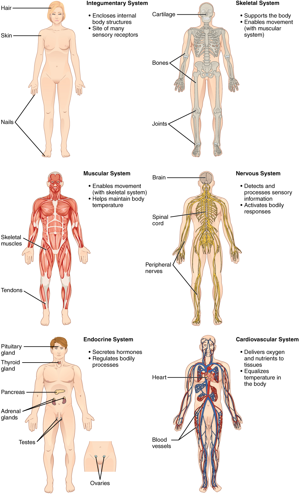
Consider the breakdown into eleven distinct organ systems in the human body (Figure 2.2 and Figure 2.3). Assigning organs to organ systems can be imprecise, since organs that “belong” to one system can also have functions integral to another system. In fact, most organs contribute to more than one system.
The organism level is the highest level of organization. An organism is a living being that has a cellular structure and that can independently perform all physiologic functions necessary for life. In multicellular organisms, including humans, all cells, tissues, organs, and organ systems of the body work together to maintain the life and health of the organism.
Image Descriptions
Figure 2.1 image description: This illustration shows biological organization as a pyramid. The chemical level is at the apex of the pyramid where atoms bond to form molecules with three dimensional structures. An example is shown with two white hydrogen atoms bonding to a red oxygen atom to create water. The next level down on the pyramid is the cellular level, as illustrated with a long, tapered, smooth muscle cell. At this level, a variety of molecules combine to form the interior fluid and organelles of a body cell. The next level down is the tissue level. A community of similar cells forms body tissue. The example given here is a section of smooth muscle tissue, which contains many smooth muscle cells closely bound side by side. The next level down is the organ level, as illustrated with the bladder and urethra. The bladder contains smooth muscle while the urethra contains skeletal muscle. These are both examples of muscle tissues. The next level down is the organ system level, as illustrated by the entire urinary system containing the kidney, ureters, bladder and urethra. At this level, two or more organs work closely together to perform the functions of a body system. At the base of the pyramid is the organismal level illustrated with a woman drinking water. At this level, many organ systems work harmoniously together to perform the functions of an independent organism. [Return to Figure 2.1].
Figure 2.2 image description: This illustration shows eight silhouettes of a human female, each showing the components of a different organ system. The integumentary system encloses internal body structures and is the site of many sensory receptors. The integumentary system includes the hair, skin, and nails. The skeletal system supports the body and, along with the muscular system, enables movement. The skeletal system includes cartilage, such as that at the tip of the nose, as well as the bones and joints. The muscular system enables movement, along with the skeletal system, but also helps to maintain body temperature. The muscular system includes skeletal muscles, as well as tendons that connect skeletal muscles to bones. The nervous system detects and processes sensory information and activates bodily responses. The nervous system includes the brain, spinal cord, and peripheral nerves, such as those located in the limbs. The endocrine system secretes hormones and regulates bodily processes. The endocrine system includes the pituitary gland in the brain, the thyroid gland in the throat, the pancreas in the abdomen, the adrenal glands on top of the kidneys, and the testes in the scrotum of males as well as the ovaries in the pelvic region of females. The cardiovascular system delivers oxygen and nutrients to the tissues as well as equalizes temperature in the body. The cardiovascular system includes the heart and blood vessels.[Return to Figure 2.2].
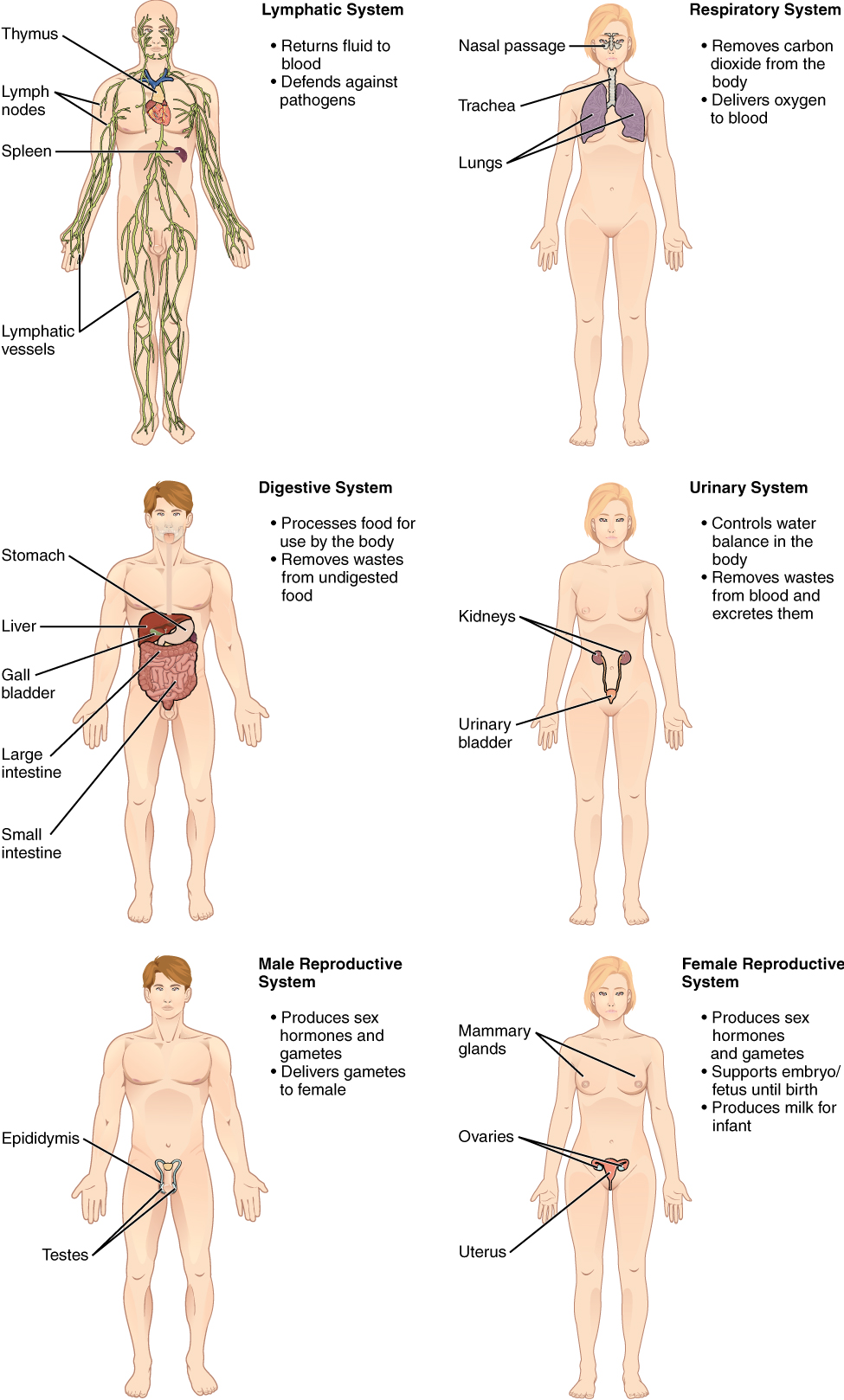
Figure 2.3 image description: The lymphatic system returns fluid to the blood and defends against pathogens. The lymphatic system includes the thymus in the chest, the spleen in the abdomen, the lymphatic vessels that spread throughout the body, and the lymph nodes distributed along the lymphatic vessels. The respiratory system removes carbon dioxide from the body and delivers oxygen to the blood. The respiratory system includes the nasal passages, the trachea, and the lungs. The digestive system processes food for use by the body and removes wastes from undigested food. The digestive system includes the stomach, the liver, the gall bladder (connected to the liver), the large intestine, and the small intestine. The urinary system controls water balance in the body and removes and excretes waste from the blood. The urinary system includes the kidneys and the urinary bladder. The reproductive system of males and females produce sex hormones and gametes. The male reproductive system is specialized to deliver gametes to the female while the female reproductive system is specialized to support the embryo and fetus until birth and produce milk for the infant after birth. The male reproductive system includes the two testes within the scrotum as well as the epididymis which wraps around each testis. The female reproductive system includes the mammary glands within the breasts and the ovaries and uterus within the pelvic cavity. [Return to Figure 2.3]
Attribution
Except where otherwise noted, this chapter is adapted from “Medical Language Within the Context of Anatomy and Physiology” in Building a Medical Terminology Foundation by Kimberlee Carter and Marie Rutherford licensed under CC BY 4.0. / A derivative of Betts et al., which can be accessed for free from Anatomy and Physiology (OpenStax). Adaptations: dividing Medical Language: Within the Context of Anatomy and Physiology chapter content into sub-chapters.
biological process that results in stable equilibrium
