13.4 – Skeletal Diseases, Disorders and Diagnostic Testing
Common Diseases and Disorders
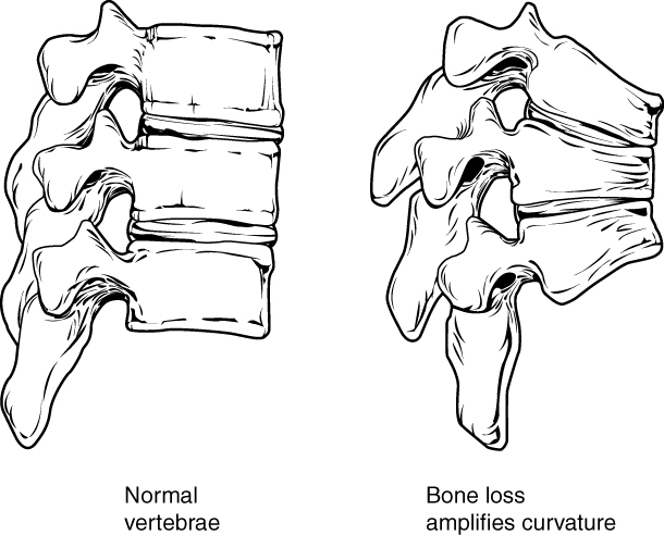
Osteoporosis
Health Canada (2018) describes osteoporosis as bone loss that causes bones to become weak and thin over time. This weakness can lead to fractures from simple movements and occurs often in the wrist, shoulder, spine, and hip. To learn more about osteoporosis, please visit the Osteoporosis Health Canada website [New Tab].
Arthritis
Arthritis often presents as edema, arthralgia, and ankylosis (Centers for Disease Control and Prevention, 2019). Common types of arthritis are osteoarthritis (OA), rheumatoid arthritis (RA), fibromyalgia, gout, and lupus. To learn more about arthritis, visit the CDC’s Arthritis Basics webpage [New Tab].
Osteoarthritis
Osteoarthritis is the most common form of arthritis and, according to the Arthritis Society, affects Canadians more than the combination of all other types of arthritis. The breakdown of cartilage and bone occurs over time when joints are exposed to heavy workloads either through occupation, obesity, and/or prior injury to a joint. Common symptoms are pain, stiffness, and aching that worsens over time. While there is no cure, symptoms can be managed through exercise, medications and, in severe cases, joint replacements (Arthritis Society, 2020).
Rheumatoid arthritis
The CDC describes rheumatoid arthritis (RA) as an autoimmune and inflammatory disease. Autoimmune diseases are disorders in which the immune system overreacts and begins to attack itself. In the case of RA, inflammation of the joint tissues of the hands, wrists and knees is painful and debilitating. Treatments may include immunosuppressive drugs and anti-inflammatory drugs (Betts et al., 2013). RA can also affect other tissues throughout the body and cause problems in organs such as the lungs, heart, and eyes. RA can affect children, and in this case, it is referred to as juvenile rheumatoid arthritis (Centers for Disease Control and Prevention, 2022).
Gout
According to the Arthritis Society, gout is an inflammatory arthritis caused when the immune system attacks the crystals that form when uric acid builds up in a joint. Gout has periods of exacerbation and remission and is commonly treated through lifestyle changes and medication. While any joint can be affected, it is common in the lower extremities and most often in the big toe (Choy & MacMullan, 2017). To learn more about the causes and treatments, please visit the Arthritis Society’s web page about gout [New Tab].
Myasthenia Gravis
The National Institute of Neurological Disorders and Strokes describes myasthenia gravis as a “chronic autoimmune neuromuscular disorder that causes weakness in the skeletal muscle” (Office of Communications and Public Liaison, 2020). To learn more, visit the National Institute of Neurological Disorders and Stroke’s page on Myasthenia Gravis [New Tab].
Fibromyalgia
Fibromyalgia is a challenging disease to diagnose, since symptoms manifest differently and are similar to other diseases. Symptoms may include chronic fatigue, gastrointestinal problems, headaches, and increased pain sensitivity. Historically, fibromyalgia was often misdiagnosed or dismissed as not real. According to The National Institute of Arthritis and Musculoskeletal and Skin Disease, there is agreement on the definition and treatment for fibromyalgia, but it is recommended to find a specialist who is familiar with fibromyalgia (National Institute of Arthritis and Musculoskeletal and Skin Diseases, 2021). To learn more about the diagnosis and treatment for fibromyalgia, please visit the National Institute of Arthritis and Musculoskeletal and Skin Diseases’ fibromyalgia web page [New Tab].
Osteomyelitis
Osteomyelitis is a bone infection caused when staphylococcus bacteria travels through the blood stream from an infection in one part of the body to the bone. Staphylococcus bacteria is found on the skin and it can transfer to the bone through a wound and/or surgical contamination. The risk increases as people age or if their immune system is compromised (Mayo Clinic Staff, 2022). To learn more about the causes, symptoms and treatments for osteomyelitis, please visit the Mayo Clinic’s osteomyelitis web page [New Tab].
Disorders of the Curvature of the Spine
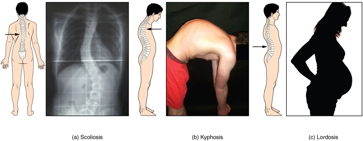
Developmental anomalies, pathological changes, or obesity can enhance the normal vertebral column curves, resulting in the development of abnormal or excessive curvatures (see Figure 13.10). Disorders associated with the curvature of the spine include:
- Kyphosis: Also referred to as humpback, is an excessive posterior curvature of the thoracic region. This can develop when osteoporosis causes weakening and erosion of the anterior portions of the upper thoracic vertebrae, resulting in their gradual collapse (see Figure 13.11).
- Lordosis: Also referred to as swayback, is an excessive anterior curvature of the lumbar region and is most commonly associated with obesity or late pregnancy. The accumulation of body weight in the abdominal region results in an anterior shift in the line of gravity that carries the weight of the body. This causes an anterior tilt of the pelvis and a pronounced enhancement of the lumbar curve.
- Scoliosis: An abnormal lateral curvature accompanied by twisting of the vertebral column. Scoliosis is the most common vertebral abnormality among girls. The cause is usually unknown, but it may result from weakness of the back muscles, defects such as differential growth rates in the right and left sides of the vertebral column, or differences in the length of the lower limbs. When present, scoliosis tends to get worse during adolescent growth spurts. Although most individuals do not require treatment, a back brace may be recommended for growing children. In extreme cases, surgery may be required (Betts et al., 2013).
Fractures
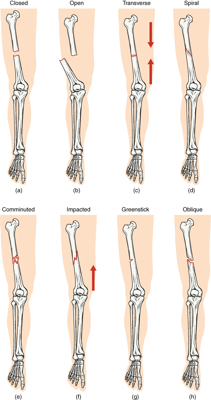
A fracture is a broken bone. It will heal whether or not a physician resets it in its anatomical position. If the bone is not reset correctly, the healing process will keep the bone in its deformed position. Crepitation or crepitus is the creaking or popping sound that is heard when fractured bones move against each other Fractures are classified by their complexity, location, and other features (see Figure 13.12). Some fractures may be described using more than one term because it may have features of more than one type (e.g., an open transverse fracture) (Betts et al., 2013; Cleveland Clinic, 2022).
Types of fractures include:
- Closed or simple: bones are broken but does not protrude the skin
- Open or compound: bones are broken and pierce through the skin
- Transverse; bone is broken straight across
- Spiral: bone has twisted apart
- Comminuted: bones are broken and crushed into pieces
- Greenstick: bones are partially broken; occurs mainly in children
- Oblique: bones are broken at an angle
- Coles: bones are broken and occur at the wrist or distal radius
- Stress: small crack in a bone
Bone Cancer
There are three types of primary bone cancers: osteosarcoma, Ewing Sarcoma, and chondrosarcoma. These are considered primary cancers because they originate in the bones. Osteosarcoma and Ewing Sarcoma are cancers found in children, teenagers, and young adults. Ewing Sarcoma is considered to be the more aggressive of the two cancers since it tends to metastasize quickly. Osteosarcoma is the most common type of bone cancer and it begins in the tissues of growing bones. Chondrosarcoma is a slow-growing bone cancer that affects adults and rarely metastasizes (Government of Canada, 2013). To learn more, visit the Public Health Agency of Canada’s web page on bone cancer [New Tab].
Diagnostic Procedures
Common diagnostic procedures related specifically to the skeletal system include x-rays, bone mineral density testing, and arthroscopy.
- X-rays are common diagnostic tests used to confirm or rule out fractures and broken bones. The radiation dose is low so it is considered a safe diagnostic test (Ontario Association of Radiologist, 2020).
- Dual x-ray absorptionmetry (BMD), also called a bone mineral density test, is a test to determine osteoporosis by measuring the amount of bone mineral in a particular amount of bone (Radiology Info, 2022).
- Arthroscopy is a common procedure performed by orthopedic surgeons to view the inside of a joint to diagnose and/or repair joint problems. The patient is given a local anesthetic and the surgeon inserts an arthroscope through an incision in the skin. Depending on what the surgeon finds, a repair of the joint may take place during the procedure (Mayo Clinic Staff, 2022).
Medical Specialties Related to the Skeletal System
Orthopedic Surgeon
Orthopedic Surgeons are medical doctors who complete an additional 5-years of specialized training in the prevention, diagnosis, treatment and surgery of disorders and diseases related to the musculoskeletal systems (Canadian Medical Association, 2019). For more details, please visit the Canadian Medical Association’s page on Orthopedic Surgery [PDF].
Rheumatologist
Rheumatologists are medical doctors who have additional training as internists with a sub-specialty in rheumatology. Many rheumatology disorders have an underlying autoimmune disorders. Consequently, rheumatologists are interested in autoimmune disorders and their impact on multiple body systems including the musculoskeletal systems (Canadian Medical Association, 2019). For more details, please follow the link to the Canadian Medical Association’s page on Rheumatology [PDF].
Doctor of Chiropractic (DC)/Chiropractor
A Doctor of Chiropractic (DC) is regulated and licensed by each province in Canada. Chiropractors have seven years of university education, a supervised internship, and national examinations. Chiropractors are trained in the prevention, assessment and treatment of the spine, muscular system and nervous system. Chiropractors focus on spinal adjustments, nutrition, and preventing injury without the use of pharmaceuticals or surgical procedures (Cleveland Clinic, 2022). To learn more, visit the Cleveland Clinic’s page on Chiropractic adjustment [New Tab].
Physiotherapist
A physiotherapist in Canada has a Master’s degree in physiotherapy and has successfully completed a national Physiotherapy Competency Examination (PCE). Physiotherapists use an evidenced-based approach when assessing and designing treatment plans for their clients. Treatments may include exercises, massage, joint manipulation, and occupational retraining (Canadian Physiotherapy Association, n.d.). To learn more, please visit the Canadian Physiotherapy Association website [New Tab].
Image Descriptions
Figure 13.10 image description: This image shows the changes to the abnormal curves of the vertebral columns in different diseases. The left panel shows the change in the curve of the vertebral column in scoliosis, the middle panel shows the change in the curve of the vertebral column in kyphosis, and the right panel shows the change in the curve of the vertebral column in lordosis. [Return to Figure 13.10].
Figure 13.11 image description: This figure shows the changes to the spine in osteoporosis. The left panel shows the structure of normal vertebrae and the right panel shows the curved vertebrae in osteoporosis. [Return to Figure 13.11].
Figure 13.12 image description: In this illustration, each type of fracture is shown on the right femur from an anterior view. In the closed fracture, the femur is broken in the middle of the shaft with the upper and lower halves of the bone completely separated. However, the two halves of the bones are still aligned in that the broken edges are still facing each other. In an open fracture, the femur is broken in the middle of the shaft with the upper and lower halves of the bone completely separated. Unlike the closed fracture, in the open fracture, the two bone halves are misaligned. The lower half is turned laterally and it has protruded through the skin of the thigh. The broken ends no longer line up with each other. In a transverse fracture, the bone has a crack entirely through its width, however, the broken ends are not separated. The crack is perpendicular to the long axis of the bone. Arrows indicate that this is usually caused by compression of the bone in a superior-inferior direction. A spiral fracture travels diagonally through the diameter of the bone. In a comminuted fracture, the bone has several connecting cracks at its middle. It is possible that the bone could splinter into several small pieces at the site of the comminuted fracture. In an impacted fracture, the crack zig zags throughout the width of the bone like a lightning bolt. An arrow indicates that these are usually caused by an impact that pushes the femur up into the body. A greenstick fracture is a small crack that does not extend through the entire width of the bone. The oblique fracture shown here is travelling diagonally through the shaft of the femur at about a thirty degree angle. [Return to Figure 13.12].
Attribution
Except where otherwise noted, this chapter is adapted from “Skeletal System” in Building a Medical Terminology Foundation by Kimberlee Carter and Marie Rutherford, licensed under CC BY 4.0. / A derivative of Betts et al., which can be accessed for free from Anatomy and Physiology (OpenStax). Adaptations: dividing Skeletal System chapter content into sub-chapters.
abnormal condition of bones that are porous.
Inflammation of the joints
swelling
painful joint(s)
abnormal condition of stiffness
Inflammation of bones and joints
pain in the fibrous tissues of muscles
increase in severity of a problem
grave or serious muscle weakness
a condition that lasts a long time with periods of remission and exacerbation
Production of cells that can mobilize and establish tumors in other organs of the body
Instrument that contains a small camera on a tube with a light. This is a tool used to view the inside of a joint.
