16.4 – Nervous Systems Diseases, Disorders and Diagnostic Testing
Neurodegenerative Diseases – Alzheimer’s Disease, Parkinson’s Disease, Amyotrophic Lateral Sclerosis (ALS), Multiple sclerosis (MS)
A class of disorders that affect the nervous system are the neurodegenerative diseases: Alzheimer’s disease, Parkinson’s disease, Huntington’s disease, amyotrophic lateral sclerosis (ALS), Creutzfeld–Jacob disease, multiple sclerosis (MS), and other disorders that are the result of nervous tissue degeneration. In diseases like Alzheimer’s, Parkinson’s, or ALS, neurons die; in diseases like MS, myelin is affected. Some of these disorders affect motor function and others present with dementia. Some are the result of genetics, such as Huntington’s disease, or the result of autoimmunity, such as MS; others are not entirely understood, such as Alzheimer’s and Parkinson’s diseases.
Several diseases can result from the demyelination of axons. The causes of these diseases are not the same; some have genetic causes, some are caused by pathogens, and others are the result of autoimmune disorders. Though the causes are varied, the results are largely similar. The myelin insulation of axons is compromised, making electrical signaling slower (Betts et al., 2013).
Multiple sclerosis (MS) is one such disease. It is an example of an autoimmune disease. The antibodies produced by lymphocytes (a type of white blood cell) mark myelin as something that should not be in the body. This causes inflammation and the destruction of the myelin in the central nervous system. As the insulation around the axons is destroyed by the disease, scarring becomes obvious (Betts et al., 2013).
Guillain-Barre (pronounced gee-YAN bah-RAY) syndrome is an example of a demyelinating disease of the peripheral nervous system. It is also the result of an autoimmune reaction, but the inflammation is in peripheral nerves. Sensory symptoms or motor deficits are common, and autonomic failures can lead to changes in the heart rhythm or a drop in blood pressure, especially when standing, which causes dizziness (Betts et al., 2013).
Other Nerve Disorders
Infection, trauma, and congenital disorders can all lead to significant signs, as identified through the neurological exam. It is important to differentiate between an acute event, such as stroke, and a chronic or global condition, such as blunt force trauma. Responses seen in the neurological exam can help. A loss of language function observed in all its aspects is more likely a global event as opposed to a discrete loss of one function, such as not being able to say certain types of words. A concern, however, is that a specific function—such as controlling the muscles of speech—may mask other language functions. The various subtests within the mental status exam can address these finer points and help clarify the underlying cause of the neurological loss (Betts et al., 2013).
Stroke
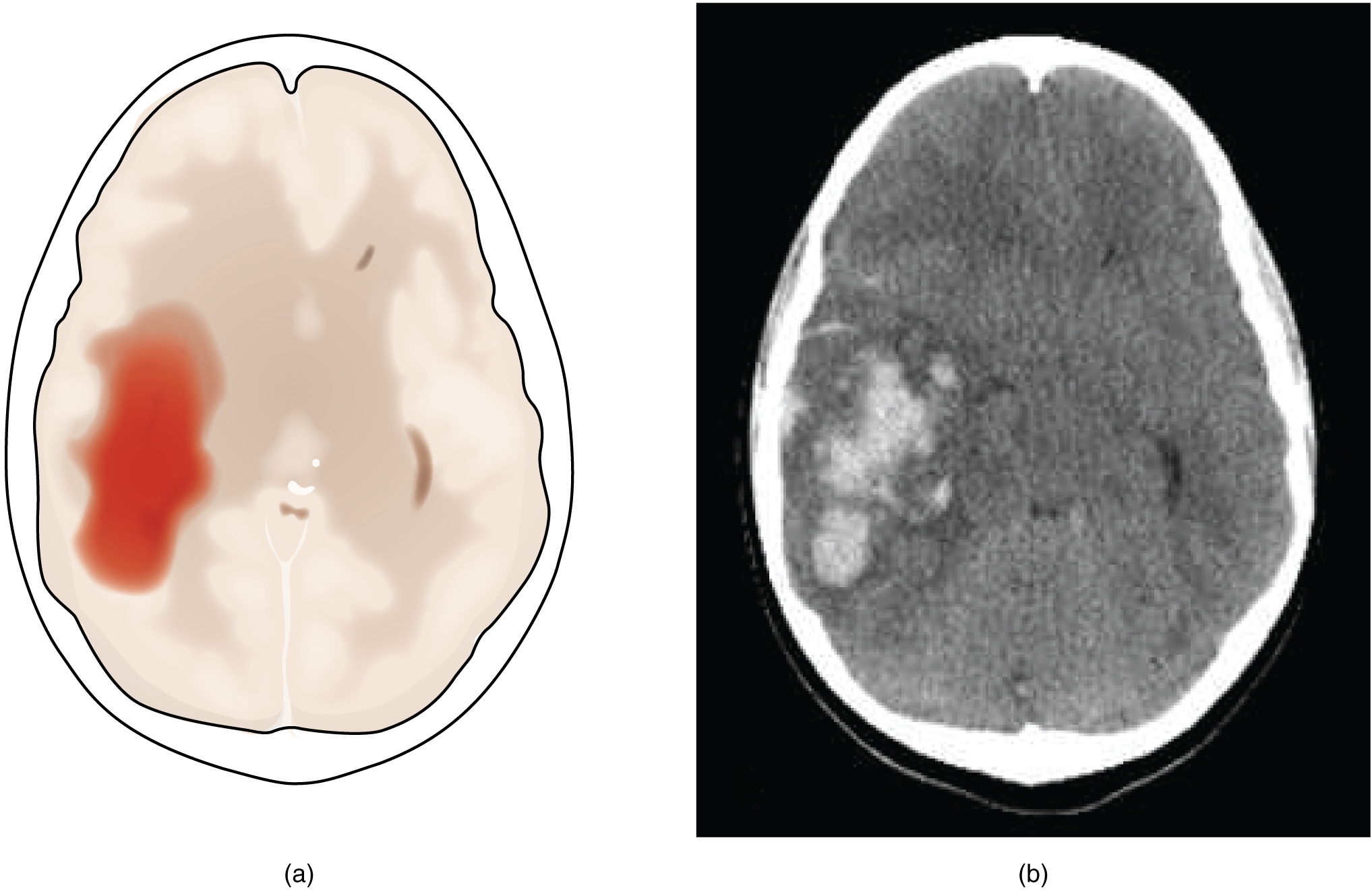
Damage to the nervous system can be limited to individual structures or can be distributed across broad areas of the brain and spinal cord. Localized, limited injury to the nervous system is most often the result of circulatory problems. The loss of blood flow to part of the brain is known as a stroke, or a cerebrovascular accident (CVA). There are two main types of stroke, depending on how the blood supply is compromised: ischemic and hemorrhagic. An ischemic stroke is the loss of blood flow to an area because vessels are blocked or narrowed. This is often caused by an embolus, which may be a blood clot or fat deposit. Ischemia may also be the result of thickening of the blood vessel wall, or a drop in blood volume in the brain known as hypovolemia. A hemorrhagic stroke is bleeding into the brain because of a damaged blood vessel. Accumulated blood fills a region of the cranial vault and presses against the tissue in the brain (see Figure 16.15) (Betts et al., 2013).
Cerebral Palsy
Cerebral Palsy (CP) is caused by an interruption to the normal development of a person’s brain leading to weakness with muscles. Depending on the area of the brain that is affected, signs and symptoms will vary in the type and severity between individuals. Balance and coordination are often challenging due the inability to control muscles (Centers for Disease Control and Prevention, 2024, Ontario Federation for Cerebral Palsy, 2018). To learn more about cerebral palsy, please visit the Centers for Disease Control and Prevention’s web page on cerebral palsy [New Tab].
Traumatic Brain Injury (TBI)
According to the Minister of Health, approximately 20,000 people in Canada are hospitalized (each year) for traumatic brain injures. Brain injuries range from moderate to severe and include concussions. TBI can be caused by falls, automobile accidents, sports, assaults, and strokes. Investment has been made to educate people on how to prevent TBIs with a focus on concussions from sports (Taylor, 2019).
Neurological System Medical Terms in Use
Neurological System
Neurological System – History and Physical Examination (Text version)
Use the words below to fill in the history and physical examination form:
- festination
- depression
- fatigue
- postural
- rigidity
- cognition
- downgoing
- q.i.d
PATIENT NAME: Susan LOGAN
AGE: 62
SEX: Female
DOB: March 24
DATE OF ASSESSMENT: December 10
ADMITTING PHYSICIAN: Martin Lewis, MD, Neurology
DIAGNOSIS: Parkinson disease
HISTORY: This pleasant and co-operative 62-year-old woman has advanced parkinsonism which presents for more than 10 years. It is affecting her daily living to a great degree. She has minor difficulty with ADLs noted in difficulty dressing and meal preparation. She has had frequent falls occasionally related to freezing or to _______[Blank 1] but also occurring without any apparent cause. She has marked hesitancy on changing direction and unsteadiness after exertion and develops _______[Blank 2]. She has a minor problem with sialorrhea, eating, and swallowing. She can maintain basic personal hygiene without any difficulty. However, showering or bathing requires assistance. She has had some symptoms of anxiety and ________[Blank 3] along with her Parkinson disease.
PHYSICAL EXAMINATION: On neurologic exam, she did have mild-to-moderate impairment in _________[Blank 4] and short-time memory, although she is alert and oriented x3. She has pronounced tremor, worse in the left arm than the right. She has marked ________[Blank 5] in the upper left extremity; mild in the right. She has marked reduction of movement with long delays in initiating movement and frequent freezing. She has a moderately-flexed posture and cannot straighten to command. She has __________[Blank 6] instability. Her speech is mildly dysarthric. She has paucity of spontaneous facial expression. She has an unsteady and erratic gait characterized by shuffling strides with festination in propulsion. She can arise from a chair with difficulty only after multiple attempts. Deep tendon reflexes (DTRs) are symmetrical, and toes are __________[Blank 7]. Cranial nerves are intact and unremarkable.
TREATMENT AND PLAN: She has been on Sinemet 25/100 t.i.d. for the last 7 years or so. She will be going on vacation soon, and I do not wish to start a second antiparkinsonian medication while she away from medical supervision. However, I have asked her to increase her Sinemet dose to _______[Blank 8]. We will see how she does with Sinemet and plan to add bromocriptine 1 mg per day when she returns.
FOLLOW UP: The patient has been scheduled for follow up in 3 weeks, upon her return from vacation. Her treatment regimen will be adjusted at that time.
________________________
Martin Lewis, MD, Neurology
Check your answers: [1]
Activity source: Neurological System – History and Physical Examination by Sheila Bellefeuille and Heather Scudder, from Building a Medical Terminology Foundation by Kimberlee Carter and Marie Rutherford, licensed under CC BY- 4.0. /Text version added.
Neurological System – Consultation Report
Neurological System – Consultation Report (Text version)
Use the word below to fill in the consultation report:
- cognition
- dementia
- hypertension
- downgoing
- neurological
- symptomatic
- MRI
- stroke
- blurred
NEUROLOGICAL SYSTEM – CONSULTATION REPORT
PATIENT NAME: Robert BROWN
AGE: 74
SEX: Male
DOB: July 5
DATE OF CONSULTATION: April 15
CONSULTING PHYSICIAN: Martin Lewis, MD, Neurology
REASON FOR CONSULTATION: Assessment of cognitive changes and testing.
HISTORY: The patient presented a few days ago with a marked change in __________[Blank 1] identified by his family members and care staff. The reports describe two episodes of the patient presenting a somewhat confused state, instability with a “holding of the temples” and a report of blurring vision. The patient was also observed holding on to walls and furniture to walk around. This seems to have been two transient episodes and has not recurred since. Prior to that, he had maintained excellent cognitive abilities with full lingual ability, no signs of aphasia, __________[Blank 2] or loss of consciousness. The cognitive decline noticed was not of gradual onset but rather an acute change within hours to a day. The time span is unclear as the patient lives alone and there was a time lapse between a family visit and the arrival of a personal care assistant.
The patient is a good historian to questioning and does admit to some recent occasional headaches and ___________[Blank 3] vision. These are new to him as he reports never having “had a headache” in his “whole life”. He reports that the blurring is not constant but only seems to occur when he turns his head to right or left suddenly. There is a “tilting sensation” like he will fall but this clears when he brings his head back to center. He has no history of epilepsy or seizure disorders. No history of TM or ear trauma.
PHYSICAL EXAMINATION: HEENT: Head is normocephalic. EYES: PERRLA. EARS: Auditory exam reveals intact TMs bilaterally. No erythema. The nose and throat exam is unremarkable. NECK: JVD appears normal. VITAL SIGNS: Blood pressure is 132/86 with no previous history of ____________[Blank 4]. Pulse is 83 and resp. 22 but the patient does admit to feeling anxious during the assessment. Temperature 37C.
NEURO: Orientation and language are normal. Extremity strength testing show some minimal weakness in the right upper. Reflexes are normal. Toes are ___________[Blank 5] bilaterally. Has difficulty with heel-and toe-walk and is unable to tandem walk. The gait is alternately normal and minimally spastic.
IMPRESSION: What appears to be a transient or acute cognitive change with altered awareness, headache and cephalo-positional blurring of vision. There is some ____________[Blank 6] change, although minimal and not clinically diagnostic, as evidenced by the slight changes in gait during testing but it does not remain consistently. This is puzzling.
PLAN: It is still not clear to me what these spells are. Some of the neurological possibilities to be considered are TIA, __________[Blank 7], brain and spinal cord tumors, inflammation, infection, vascular irregularities, and some neurodegenerative disorders. I have ordered a stat cerebral _________[Blank 8], electroencephalogram (EEG) and blood levels for CBC, chem panel. However, I feel we should also rule out the more common possibilities of pseudo-seizure, vertigo, and inner ear anomalies and am in the process of making these appropriate bookings.
I have booked a follow up with this patient in 10 days to review the results. He and his family have been advised to contact me immediately if he has another “spell” or to present to the ER where we can complete testing when the patient is ____________[Blank 9].
Thank you for this most interesting referral. I will be in touch after I have reviewed the patient.
_________________________
Martin Lewis, MD, Neurology
Check your answers: [2]
Activity source: NEUROLOGICAL SYSTEM – CONSULTATION REPORT by Sheila Bellefeuille and Heather Scudder, from Building a Medical Terminology Foundation by Kimberlee Carter and Marie Rutherford, licensed under CC BY- 4.0. /Text version added.
Neurological System- Follow Up Report
Neurological System – Follow Up Report (Text version)
Use the words below to fill in the follow-up report:
- electroencephalogram
- balance
- pathology
- vertigo
- coordination
- white matter
- hemorrhagic
- wasting
- calcifications
- mass
- somnolence
- symptomatic
PATIENT NAME: Randy NORTON
AGE: 74
SEX: Male
DOB: October 14
DATE OF ASSESSMENT: January 18
ASSESSING PHYSICIAN: Martin Lewis, MD, Neurology
REASON FOR ASSESSMENT: Follow up assessment of cognitive changes and testing.
HISTORY: This 74-year-old patient was seen in consultation 10 days ago for assessment of cognitive changes. He underwent prescribed testing in the forms of cerebral MRI, _____________[Blank 1] (EEG) and blood was drawn for CBC and chem panel. The patient was seen by our local ENT for inner ear and __________[Blank 2] testing. Test results showed normal hearing. No evidence of an inner ear __________[Blank 3] that might have contributed to the __________[Blank 4] or lack of balance and _____________[Blank 5] reported as part of the presenting symptoms.
TEST RESULTS: Cerebral MRI reveals a few T2 hyperintensities in the ____________[Blank 6] in the left temporal lobe. The right hemisphere shows some diffuse ___________[Blank 7] and some occipital wasting. There are multiple, small dark ____________[Blank 8] areas and a few areas indicative of ischemia.
EEG: This showed some depressive effect indicative of an encephalopathy. The patient did not sleep during the exam but did show some signs of ___________[Blank 9]. CBC and chem panels were normal.
IMPRESSIONS: This patient present with testing result that may be warning for Alzheimer wasting and also, some localized hemorrhagic events that have since stopped. This type of “leakage” is often not ____________[Blank 10], and I do not feel that they are connected to his presenting complaints. I see no signs of tumor or __________[Blank 11] formation nor infectious process.
On repeat verbal assessment, the patient reports he has not experienced any more of the spells. We will follow him closely in regards to the ____________[Blank 1]2 with a repeat MRI and perhaps a CT also in 3 months for results comparison to see whether the wasting has advanced or receded.
Thank you for asking me in on this most interesting case.
_________________________
Martin Lewis, MD, Neurology
Check your answers: [3]
Activity source: Neurological System – Follow Up Report by Sheila Bellefeuille and Heather Scudder, from Building a Medical Terminology Foundation by Kimberlee Carter and Marie Rutherford, licensed under CC BY- 4.0. /Text version added.
Medical Specialties
Primary Specialist – Neurologist
Neurologists are medical doctors who complete an additional 5 years of specialized training in the prevention, diagnosis, and treatment of disorders and conditions related to the brain, spinal cord, nerves and muscles (Canadian Medical Association, 2019). For more details, please follow the link to the Canadian Medical Association’s page on Neurology profile [PDF].
Procedures Related to the Nervous System
Lumbar Puncture
A neurologist may order this procedure to test cerebrospinal fluid (CSF). This procedure is recommended if they believe symptoms are caused by a problem in the nervous system that can be detected in the cerebrospinal fluid. The procedure involves inserting a needle into the spine after numbing it and taking a sample of cerebrospinal fluid (Canadian Cancer Society, n.d.).
Tensilon Test
This procedure can help a neurologist diagnose myasthenia gravis. In this test, the doctor injects with a medicine called Tensilon, then they observe how it affects muscle movements (Bergen, 2018). For more information, visit Healthline’s Tensilon Test web page [New Tab].
Electromyography (EMG)
An EMG measures electrical activity between your brain or spinal cord to a peripheral nerve. This nerve is found in your arms and legs, and is responsible for muscle control during times of movement and rest. EMGs can help your neurologist diagnose spinal cord disease as well as general muscle or nerve dysfunction (Moores & Cirino, 2018).
Electroencephalogram (EEG)
With electrodes applied to your scalp, an EEG measures electrical activity in the brain. It’s used to help diagnose conditions of the brain, including inflammation, tumors, and injuries, as well as seizures and psychiatric disorders.
Image Descriptions
Figure 16.15 image description: The left panel of this figure shows an image of the brain with a region in red. This red region indicates a hemorrhage associated with a stroke. The right panel shows a hemorrhage as it might appear on a CT scan. [Return to Figure 16.15].
Attribution
Except where otherwise noted, this chapter is adapted from “Nervous System” in Building a Medical Terminology Foundation by Kimberlee Carter and Marie Rutherford, licensed under CC BY 4.0. / A derivative of Betts et al., which can be accessed for free from Anatomy and Physiology (OpenStax). Adaptations: dividing Nervous System chapter content into sub-chapters.
- 1.festination, 2.fatigue, 3.depression, 4.cognition, 5.rigidity, 6.postural, 7.downgoing, 8.q.i.d, ↵
- 1.cognition, 2.dementia, 3.blurred, 4.hypertension, 5.downgoing, 6.neurological, 7.stroke, 8.MRI, 9.symptomatic ↵
- 1.electroencephalogram, 2.balance, 3.pathology, 4.vertigo, 5.coordination, 6.white matter, 7.calcifications, 8.hemorrhagic, 9.somnolence, 10.symptomatic, 11.mass, 12.wasting ↵
