14.2 – Anatomy (Structures) of the Muscular System
Muscle is one of the four primary tissue types of the body, and it is made up of specialized cells called fibers. The body contains three types of muscle tissue: skeletal muscle, cardiac muscle, and smooth muscle (see Figure 14.1). All three muscle tissues have some properties in common; they all exhibit a quality called excitability, as their plasma membranes can change their electrical states (from polarized to depolarized) and send an electrical wave called an action potential along the entire length of the membrane. Fascia is fibrous connective tissue that encloses muscles.
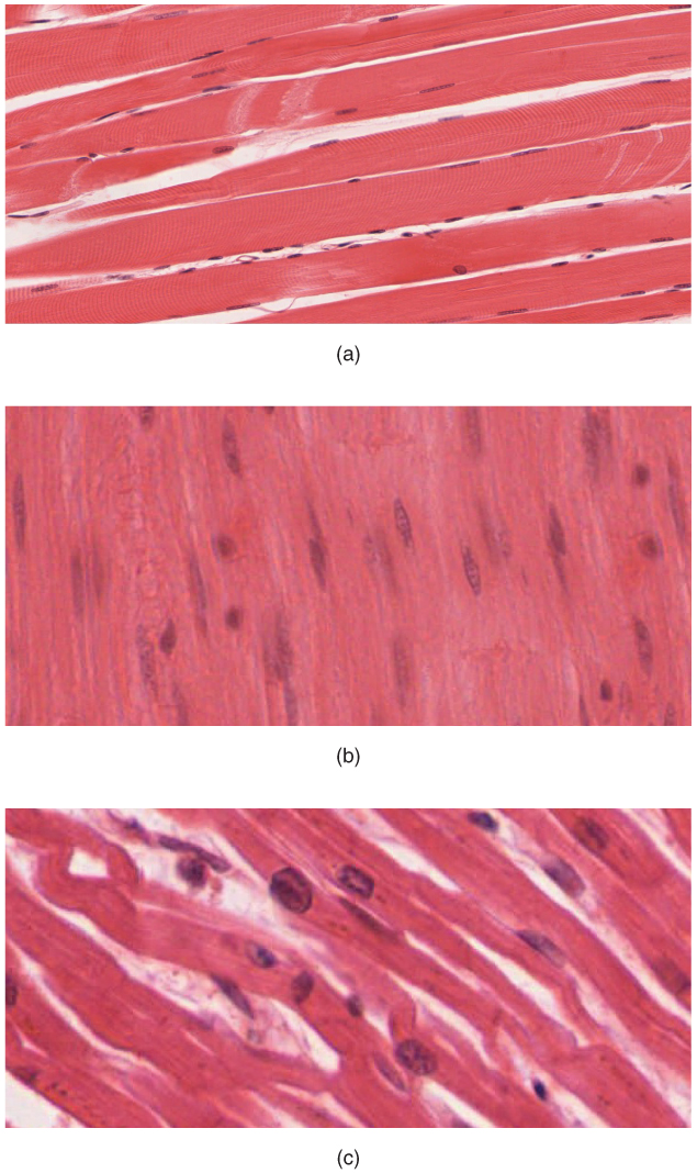
Three Types of Muscle Tissues
- Skeletal: closely associated with the skeletal system. Also known as striated muscles and are responsible for voluntary muscle movement – such as swallowing, etc.
- Smooth: mainly associated with the walls of the internal organs. Also known as visceral muscles and are responsible for involuntary muscle movement – such as breathing, etc.
- Cardiac: heart muscle or myocardium. Its appearance is similar to a skeletal muscle and is responsible for the pumping of blood. It gives the heartbeat.
Did You Know?
The gluteus maximus is the largest muscle and the heart is the hardest working muscle.
Skeletal Muscle
Skeletal muscles act not only to produce movement but also to stop movement, such as resisting gravity to maintain posture. Small, constant adjustments of the skeletal muscles are needed to hold a body upright or balanced in any position. Muscles also prevent excess movement of the bones and joints, maintaining skeletal stability and preventing skeletal structure damage or deformation.
Skeletal muscles are located throughout the body at the openings of internal tracts to control the movement of various substances. These muscles allow functions, such as swallowing, urination, and defecation, under voluntary control. Skeletal muscles also protect internal organs (particularly abdominal and pelvic organs) by acting as an external barrier or shield to external trauma and by supporting the weight of the organs.
Skeletal muscles contribute to the maintenance of homeostasis in the body by generating heat. This heat is very noticeable during exercise when sustained muscle movement causes body temperature to rise, and in cases of extreme cold, when shivering produces random skeletal muscle contractions to generate heat.
Smooth Muscle
Smooth muscle, so named because the cells do not have striations, is present in the walls of hollow organs like the urinary bladder, uterus, stomach, intestines, in the walls of passageways, such as the arteries and veins of the circulatory system, and the tracts of the respiratory, urinary, and reproductive systems. Smooth muscle is also present in the eyes, where it functions to change the size of the iris and alter the shape of the lens, and in the skin, where it causes hair to stand erect in response to cold temperature or fear.
Cardiac Muscle
Cardiac muscle tissue is only found in the heart. Highly coordinated contractions of cardiac muscle pump blood into the vessels of the circulatory system. Similar to skeletal muscle, cardiac muscle is striated and organized into sarcomeres, possessing the same banding organization as skeletal muscle (see Figure 14.1). Cardiac muscle fibers cells also are extensively branched and are connected to one another at their ends by intercalated discs. An intercalated disc allows the cardiac muscle cells to contract in a wave-like pattern so that the heart can work as a pump.
Concept Check
- Compare and contrast the 3 types of muscles tissues.
- Where in the body do you find each of the muscle types?
Attribution
Except where otherwise noted, this chapter is adapted from “Muscular System” in Building a Medical Terminology Foundation by Kimberlee Carter and Marie Rutherford, licensed under CC BY 4.0. / A derivative of Betts et al., which can be accessed for free from Anatomy and Physiology (OpenStax). Adaptations: dividing Muscular System chapter content into sub-chapters.
Also known as striated muscles. Skeletal muscles are responsible for voluntary muscle movement.
Is the heart muscle also known as the myocardium. Its appearance is similar to skeletal muscle. It pumps blood and gives the heart beat.
Also known as visceral muscles. Smooth muscle is mainly associated with the walls of internal organs. Smooth muscles are responsible for involuntary muscle movement.
biological process that results in stable equilibrium
