3.2 – Anatomy (Structures) of the Integumentary System
The skin and its accessory structures make up the integumentary system, which provides the body with overall protection. The skin is made of multiple layers of cells and tissues, which are held to underlying structures by connective tissue. The deeper layer of skin is well vascularized. It also has numerous sensory, and autonomic and sympathetic nerve fibers ensuring communication to and from the brain.
Layers of the skin
The skin is composed of three main layers:
- The epidermis
- The dermis
- Beneath the dermis lies the hypodermis
Concept Check
- On the diagram above, find the two layers of the skin; epidermis and dermis.
- The literal breakdown for hypodermis is below the dermis. On the diagram above, where can you locate it?
- Can you find a hair follicle, hair root, and hair shaft?
- Keep reading to find out what the arrector pili muscle does when you are frightened.
Epidermis
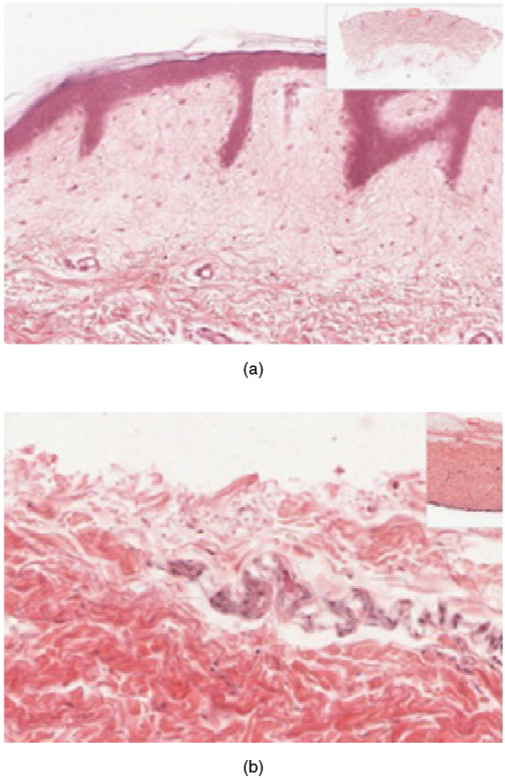
- Thin skin has four layers of cells. From deep to superficial, these layers are the stratum basale, stratum spinosum, stratum granulosum, and stratum corneum. Most of the skin can be classified as thin skin.
- Thick skin is found only on the palms of the hands and the soles of the feet. It has a fifth layer, called the stratum lucidum, located between the stratum corneum and the stratum granulosum (see Figure 3.2).
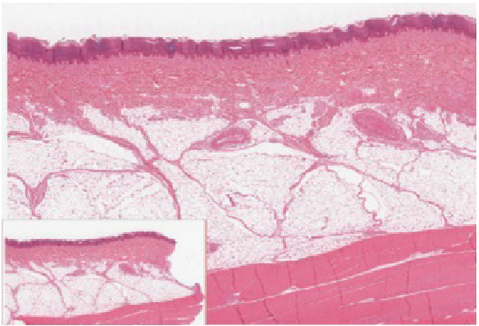
The cells in all of the layers except the stratum basale are called keratinocytes. Keratin is an intracellular fibrous protein that gives hair, nails, and skin their hardness and water-resistant properties. The keratinocytes in the stratum corneum are dead and regularly slough away, being replaced by cells from the deeper layers (see Figure 3.3).
Dermis
The dermis contains blood and lymph vessels, nerves, and other structures, such as hair follicles and sweat glands. The dermis is made of two layers (papillary layer and reticular layer) of connective tissue that compose an interconnected mesh of elastin and collagenous fibers, produced by fibroblasts (see Figure 3.4).
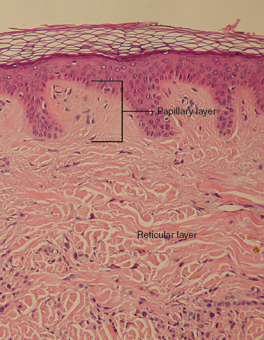
Papillary Layer
The papillary layer is made of loose, areolar connective tissue, which means the collagen and elastin fibers of this layer form a loose mesh. This superficial layer of the dermis projects into the stratum basale of the epidermis to form finger-like dermal papillae (see Figure 3.4). Within the papillary layer are fibroblasts, a small number adipocytes, and an abundance of small blood vessels. In addition, the papillary layer contains phagocytes, that help fight bacteria or other infections that have breached the skin. This layer also contains lymphatic capillaries, nerve fibers, and Meissner corpuscles.
Reticular Layer
Underlying the papillary layer is the much thicker reticular layer, composed of dense, irregular connective tissue. This layer is well vascularized and has a rich sensory and sympathetic nerve supply. The reticular layer appears reticulated due to a tight meshwork of fibers. Elastin fibers provide some elasticity to the skin, enabling movement. Collagen fibers provide structure and tensile strength, with strands of collagen extending into both the papillary layer and the hypodermis. In addition, collagen binds water to keep the skin hydrated. Collagen injections and Retin-A creams help restore skin turgor by either introducing collagen externally or stimulating blood flow and repair of the dermis, respectively.
Hypodermis
The hypodermis serves to connect the skin to the underlying fascia of the bones and muscles. It is not strictly a part of the skin, although the border between the hypodermis and dermis can be difficult to distinguish. The hypodermis consists of well-vascularized, loose, areolar connective tissue and adipose tissue, which functions as a mode of fat storage and provides insulation and cushioning for the integument.
Layers of the Skin
Practice labeling the layers of the skin.
Layers of the Skin (Text Version)
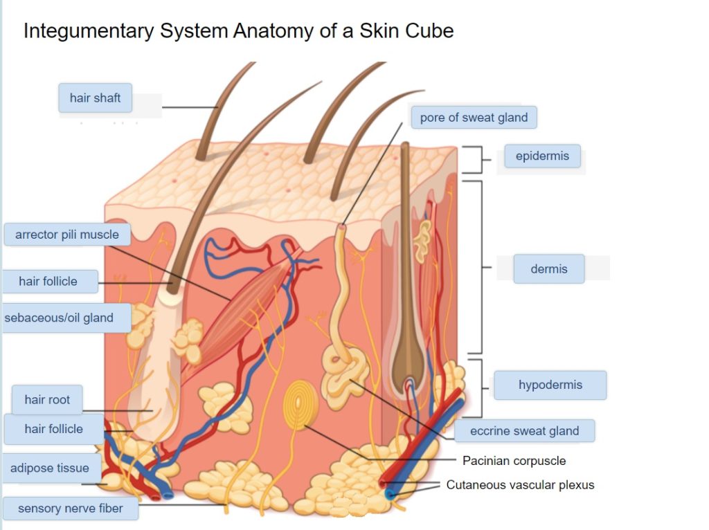
Label the diagram with correct words listed below:
- Hair Root
- Hair Follicle
- Eccrine Sweat Gland
- Hair shaft
- Adipose tissue
- Hypodermis
- Hair follicle
- dermis
- epidermis
- arrector pili muscle
- pore of sweat gland
- sensory nerve fiber
- sebaceous/oil gland
Layers of the Skin Diagram (Text Version)
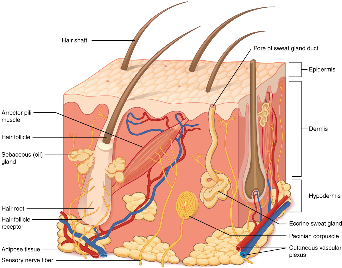
This illustration shows a cross section of skin tissue. The outermost layer is called the _______[Blank 1] and occupies one fifth of the cross section. Several hairs are emerging from the surface. The epidermis dives around one of the hairs, forming a ________[Blank 2]. The ________[Blank 3] is located above the hair follicle. Surrounding the base of the hair follicle is the ________[Blank 4] which lubricates the _________ [Blank 5]. Extending the surface of the skin is the ___________[Blank 6]. The middle layer is called the _________[Blank 7], which occupies four fifths of the cross section. The dermis contains an __________[Blank 8] that causes contraction of the hair follicle making the hair stand on end such as when someone experiences goosebumps. The dermis also contains an __________[Blank 9], composed of a bunch of tubules. One tubule travels up from the bunch, through the epidermis, opening onto the surface a __________[Blank 10]. There are two string-like nerves travelling vertically through the dermis. The right nerve is attached to a Pacinian corpuscle, which is a yellow structure consisting of concentric ovals like an onion. The lowest level of the skin, the _________[Blank 11], contains __________[Blank 12], arteries, and veins. Blood vessels travel from the hypodermis and connect to hair follicles and erector pili muscle in the dermis. ____________[Blank 13] located in the hypodermis supports the interpretation of touch.
Check your answers: [1]
Activity source: Layers of the Skin by Kimberlee Carter from Building a Medical Terminology Foundation, illustration from Anatomy and Physiology (OpenStax), licensed under CC BY 4.0./ Text version added.
Image Descriptions
Figure 3.1 image description: This illustration shows a cross section of skin tissue. The outermost layer is called the epidermis, and occupies one fifth of the cross section. Several hairs are emerging from the surface. The epidermis dives around one of the hairs, forming a follicle. The middle layer is called the dermis, which occupies four fifths of the cross section. The dermis contains an erector pilli muscle connected to one of the follicles. The dermis also contains an eccrine sweat gland, composed of a bunch of tubules. One tubule travels up from the bunch, through the epidermis, opening onto the surface a pore. There are two string-like nerves travelling vertically through the dermis. The right nerve is attached to a Pacinian corpuscle, which is a yellow structure consisting of concentric ovals similar to an onion. The lowest level of the skin, the hypodermis, contains fatty tissue, arteries, and veins. Blood vessels travel from the hypodermis and connect to hair follicles and erector pilli muscle in the dermis. [Return to Figure 3.1].
Figure 3.2 image description: Part A is a micrograph showing a cross section of thin skin. The topmost layer is a thin, translucent layer with irregular texture and areas where cells are sloughing off. The deepest layer is dark purple and extends into the third layer with finger like projections. The third light purple layer contains thin bands of fibers and small, dark cells. The fourth, and deepest layer, is darker than the third layer, but is still light purple. It contains thick fiber bands that are loosely packed. Part B is a magnified view of the epidermis of thick skin. It shows the topmost layer is five times thicker than the topmost layer of thin skin. The topmost layer of thick skin is also denser and less translucent than the topmost layer of thin skin. [Return to Figure 3.2].
Figure 3.3 image description: The outer layer of cells in this micrograph is the thinnest layer and stained deep purple due to full keratinization of dead cells. The next layer occupies one quarter of the micrograph, is lightly stained, and is a dense collection of cells. The third layer from the top is mostly white, with lightly stained, loosely-packed strands radiating in random directions. The bottom-most layer is densely-packed, with thick bands of highly organized muscle tissue that are darkly stained. [Return to Figure 3.3].
Figure 3.4 image description: This micrograph shows layers of skin in a cross section. The papillary layer of the dermis extends between the downward fingers of the darkly stained epidermis. The papillary layer appears finer than the reticular layer, consisting of smaller, densely-packed fibers. The reticular layer is three times thicker than the papillary layer and contains larger, thicker fibers. The fibers seem more loosely packed than those of the papillary layer, with some separated by empty spaces. Both layers of the dermis contain cells with darkly stained nuclei. [Return to Figure 3.4].
Attribution
Except where otherwise noted, this chapter is adapted from “Integumentary System” In Building a Medical Terminology Foundation by Kimberlee Carter and Marie Rutherford licensed under CC BY 4.0. / A derivative of Betts et al., which can be accessed for free from Anatomy and Physiology (OpenStax). Adaptations: dividing Integumentary System chapter content into sub-chapters.
-
↵
Check your answers: Layers of the Skin Diagram (Text Version)
This illustration shows a cross section of skin tissue. The outermost layer is called the epidermis and occupies one fifth of the cross section. Several hairs are emerging from the surface. The epidermis dives around one of the hairs, forming a hair follicle. The hair root is located above the hair follicle. Surrounding the base of the hair follicle is the sebaceous/oil gland which lubricates the hair follicle. Extending the surface of the skin is the hair shaft. The middle layer is called the dermis, which occupies four fifths of the cross section. The dermis contains an arrector pili muscle that causes contraction of the hair follicle making the hair stand on end such as when someone experiences goosebumps. The dermis also contains an eccrine sweat gland, composed of a bunch of tubules. One tubule travels up from the bunch, through the epidermis, opening onto the surface a pore of sweat gland. There are two string-like nerves travelling vertically through the dermis. The right nerve is attached to a Pacinian corpuscle, which is a yellow structure consisting of concentric ovals like an onion. The lowest level of the skin, the hypodermis, contains adipose tissue, arteries, and veins. Blood vessels travel from the hypodermis and connect to hair follicles and erector pili muscle in the dermis. Sensory nerve fibers located in the hypodermis supports the interpretation of touch.
has numerous blood vessels
unconsciously regulates
flight or fight response
outer layer of skin, made of closely packed epithelial cells
The layer that is made of dense, irregular connective tissue that houses blood vessels, hair follicles, sweat glands, and other structures
Literally means below the dermis. The layer of the skin below the dermis that is composed mainly of loose connective and fatty tissues
deepest layer of the epidermal
cells that manufacture and store the protein keratin
fat cells
Cells that engulf and absorb bacteria and cell particles
Tactile corpuscles that responds to light and touch, touch receptors.
net like
fibrous tissue
fat
