9.2 – Anatomy of the Heart
Location
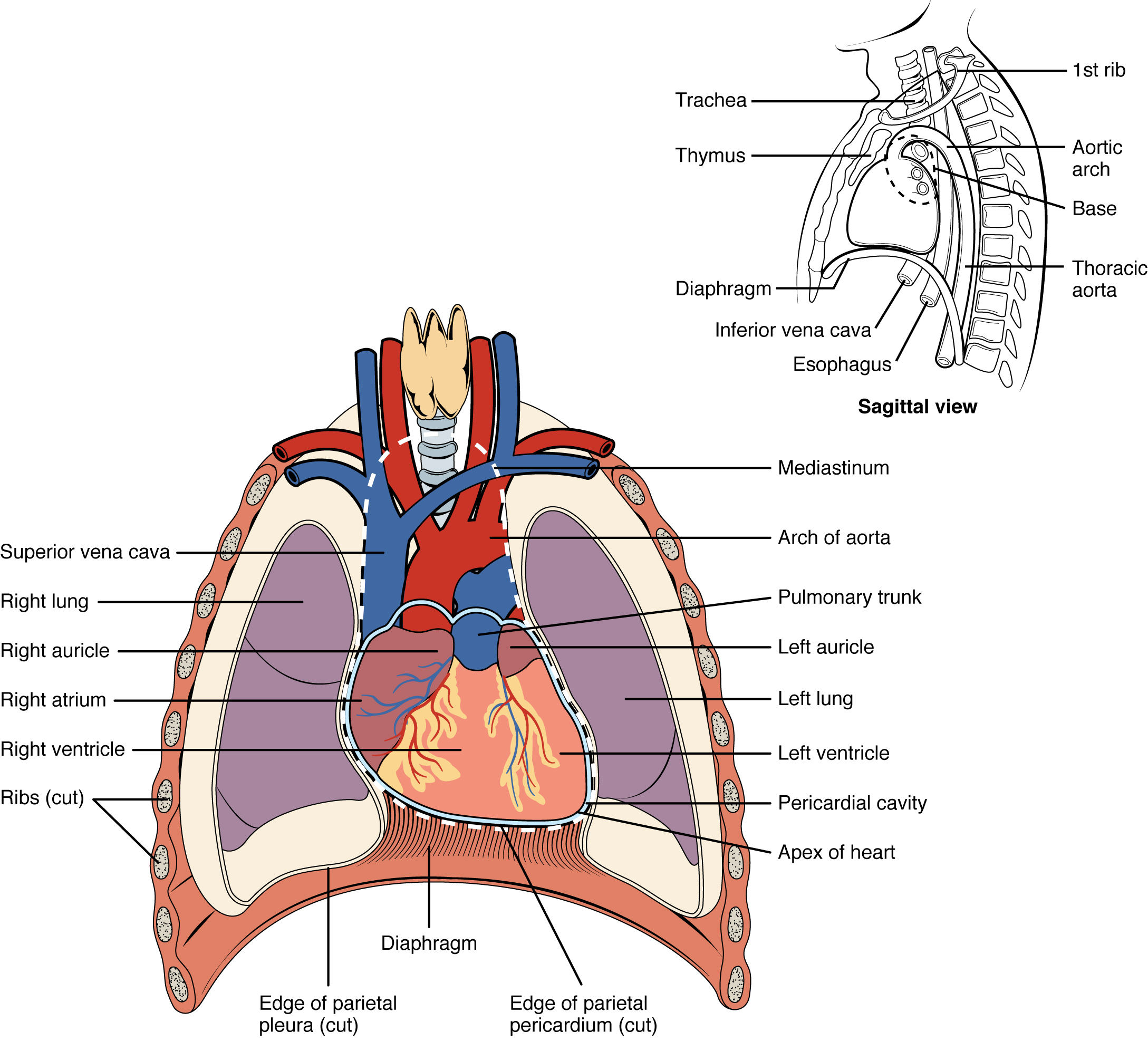
The human heart is located within the thoracic cavity, between the lungs in the space known as the mediastinum. Figure 9.1 shows the position of the heart within the thoracic cavity. Within the mediastinum, the heart is separated from the other mediastinal structures by a tough membrane known as the pericardium, or pericardial sac, and sits in its own space called the pericardial cavity. The great vessels, which carry blood to and from the heart, are attached to the superior surface of the heart, which is called the base. The base of the heart is located at the level of the third costal cartilage. The inferior tip of the heart is called the apex. The apex lies just to the left of the sternum between the junction of the fourth and fifth ribs.
Concept Check 1
- On the diagram below (Figure 9.1), locate the mediastinum, the pericardial cavity, the base of the heart and the apex of the heart.
- Locate the largest vein in the body superior vena cava.
Membranes and Layers of the Heart Walls
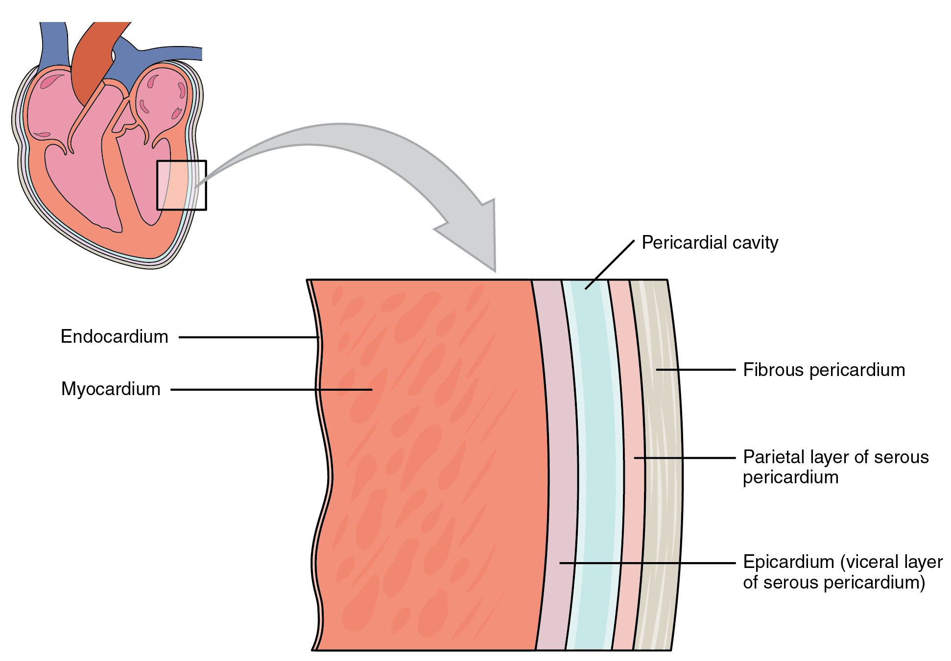
The heart and the roots of the great vessels are surrounded by a membrane known as the pericardium or pericardial sac (Figure 9.2). The pericardium consists of two distinct sub layers:
- The sturdy outer fibrous pericardium is made of tough, dense connective tissue that protects the heart and holds it in position.
- The inner serous pericardium is separated by the pericardial cavity and contains pericardial fluid. It consists of two layers:
- the outer parietal pericardium, which is fused to the fibrous pericardium.
- the inner visceral pericardium, or epicardium, which is fused to the heart and forms the outer layer of the heart wall.
The walls of the heart consist of three layers:
- The outer epicardium, which is another name for the visceral pericardium mentioned above.
- The thick, middle myocardium, which is made of muscle tissue and gives the heart its ability to contract.
- The inner endocardium, which lines the heart chambers and is the main component of the heart valves.
Concept Check 2
- Look at Figure 9.2 below, and name the layers of the heart wall and surrounding membranes, starting with the innermost layer.
- As shown on the diagram, suggest why the myocardium layer is thicker than the endocardium layer.
Internal Structures of the Heart
The heart consists of four chambers:
- The upper chambers are the right and left atria (singular: atrium).
- The lower chambers are the right and left ventricles (singular: ventricle).
The interventricular septum is a muscular wall that separates the right and left ventricles. The interatrial septum separates the right and left atria.
The atrium and ventricle on each side of the heart are separated by an atrioventricular (AV) valve:
- The right AV valve, or tricuspid valve, separates the right atrium and right ventricle.
- The left AV valve, or bicuspid valve, separates the left ventricle and the left atrium. This valve is also called the mitral valve.
There are also two semilunar valves:
- The pulmonary valve separates the right ventricle from the pulmonary trunk.
- The aortic valve separates the left ventricle from the aorta (De Saix, et al., 2013).
Anatomy Labeling Activity
Cardiovascular System: The Heart Anatomy (Text Version)
Label the diagram with correct words listed below:
- Aortic valve
- Mitral (bicuspid) valve
- Aorta
- Pulmonary trunk
- Tricuspid valve
- Interventricular septum
- Inferior vena cava
- Left pulmonary artery
- Right atrium
- Left pulmonary veins
- Left atrium
- Right ventricle
- Right pulmonary veins
- Right pulmonary artery
- Pulmonary valve
- left ventricle
- Superior vena cava
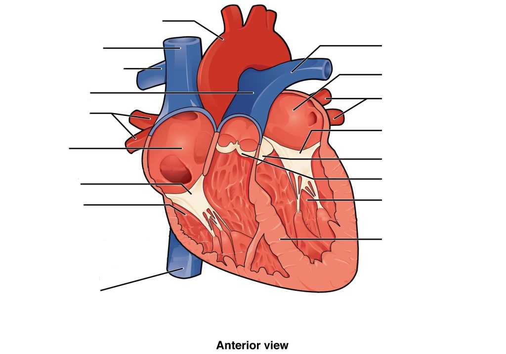
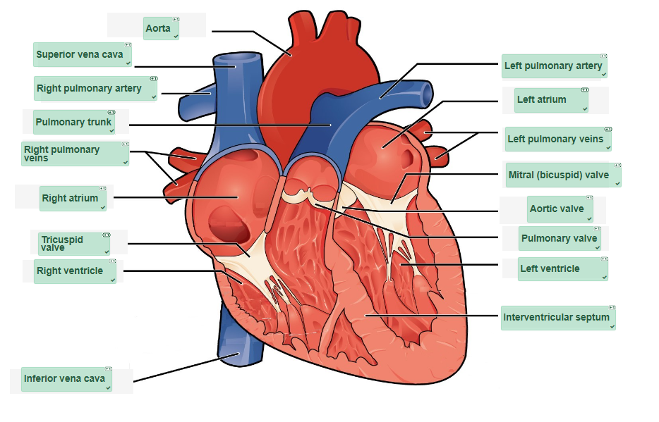
Cardiovascular System: The Heart Anatomy Diagram (Text Version)
This diagram shows the heart with an anterior view. The view shows from (from top, clockwise): the largest artery in the body known as the _______[Blank 1]. The _______[Blank 2] is shown which is the only vein in the body to carry oxygenated blood. The heart is divided into four chambers the_______[Blank 3] is one of the four chambers of the heart it is in the upper left portion of the heart. The _______[Blank 4] also know as the bicuspid valve contains to cusps or flaps and is positioned between the left atrium and lower left ventricle. The _______[Blank 5] is a structure located between the aorta and ______[Blank 6] of the heart which is the left lower chamber of the heart. The ________[Blank 7] is a thick wall of tissue divided the right side of the heart from the left. The _______[Blank 8] lies between the right atrium and pulmonary artery. The ______[Blank 9] is a large vein that carries deoxygenated blood to the heart. The ______[Blank 10] is the lower right chamber of the heart. The ________[Blank 11] lies between the right ventricle and the _______[Blank 12] which is the upper right chamber of the heart. The ________[Blank 13] transfer oxygenated blood from the lungs to the heart. The __________[Blank 14] is part of the _________[Blank 15] and transfers deoxygenated blood to the lungs. The _______[Blank 16] a large vein that returns deoxygenated blood from systemic circulation to the right atrium of the heart.
Check your answers: [1]
Activity source: Cardiovascular System: The Heart Anatomy by Gisele Tuzon, from Building a Medical Terminology Foundation, illustration from Anatomy and Physiology (OpenStax), licensed under CC BY 4.0./ Text version added.
Image Descriptions
Figure 9.1 image description: This diagram shows the location of the heart in the thorax (sagittal and anterior views). The sagittal view labels read (from top, clockwise): first rib, aortic arch, thoracic arch, esophagus, inferior vena cava, diaphragm, thymus, trachea. The anterior view labels read (from top, clockwise): mediastinum, arch of aorta, pulmonary trunk, left auricle, left lung, left ventricle, pericardial cavity, apex of heart, edge of parietal pericardium, diaphragm, edge of parietal pleura, ribs, right ventricle, right atrium, right auricle, right lung, superior vena cava. [Return to Figure 9.1].
Figure 9.2 image description: This image shows a magnified view of the structure of the heart wall. Labels read (from top, clockwise): pericardial cavity, fibrous pericardium, parietal layer of serous pericardium, epicardium (visceral layer of serous pericardium), myocardium, endocardium. [Return to Figure 9.2].
Attribution
Except where otherwise noted, this chapter is adapted from “Cardiovascular System – Heart” in Building a Medical Terminology Foundation by Kimberlee Carter and Marie Rutherford, licensed under CC BY 4.0. / A derivative of Betts et al., which can be accessed for free from Anatomy and Physiology (OpenStax). Adaptations: dividing Cardiovascular System – Heart chapter content into sub-chapters.
-
↵
Check your answers: Cardiovascular System: The Heart Anatomy Diagram (Text Version)
This diagram shows the heart with an anterior view. The view shows from (from top, clockwise): the largest artery in the body known as the aorta. The left pulmonary vein is shown which is the only vein in the body to carry oxygenated blood. The heart is divided into four chambers the left atrium is one of the four chambers of the heart it is in the upper left portion of the heart. The mitral valve also know as the bicuspid valve contains to cusps or flaps and is positioned between the left atrium and lower left ventricle. The aortic valve is a structure located between the aorta and left ventricle of the heart which is the left lower chamber of the heart. The interventricular septum is a thick wall of tissue divided the right side of the heart from the left. The pulmonary valve lies between the right atrium and pulmonary artery. The inferior vena cava is a large vein that carries deoxygenated blood to the heart. The right ventricle is the lower right chamber of the heart. The tricuspid valve lies between the right ventricle and the right atrium which is the upper right chamber of the heart. The right pulmonary veins transfer oxygenated blood from the lungs to the heart. The pulmonary trunk is part of the right pulmonary artery and transfers deoxygenated blood to the lungs. The superior vena cava a large vein that returns deoxygenated blood from systemic circulation to the right atrium of the heart.
The great vessels include the superior vena cava, inferior vena cava, aorta and pulmonary trunk.
The part of each great vessel (aorta, pulmonary trunk, inferior vena cava, superior vena cava) that connects to the base of the heart
You may recall that serous membranes throughout the body are folded back on themselves, which results in a double-layered membrane separated by serous fluid. The serous membrane surrounding the lungs is called pleura, The serous membrane surrounding the abdominopelvic organs is called peritoneum.
