13.2 – Anatomy (Structures) of the Skeletal System
The skeletal system includes all of the bones, cartilages, and ligaments of the body that support and give shape to the body and body structures. The skeleton consists of the bones of the body. For adults, there are 206 bones in the skeleton. Younger individuals have higher numbers of bones because some bones fuse together during childhood and adolescence to form an adult bone. The primary functions of the skeleton are to provide a rigid, internal structure that can support the weight of the body against the force of gravity, and to provide a structure upon which muscles can act to produce movements of the body.
In addition to providing for support and movements of the body, the skeleton has protective and storage functions. It protects the internal organs, including the brain, spinal cord, heart, lungs, and pelvic organs. The bones of the skeleton serve as the primary storage site for important minerals such as calcium and phosphate. The bone marrow found within bones stores fat and houses the blood-cell producing tissue of the body.
The skeleton is subdivided into two major divisions: the axial and appendicular.
The Axial Skeleton
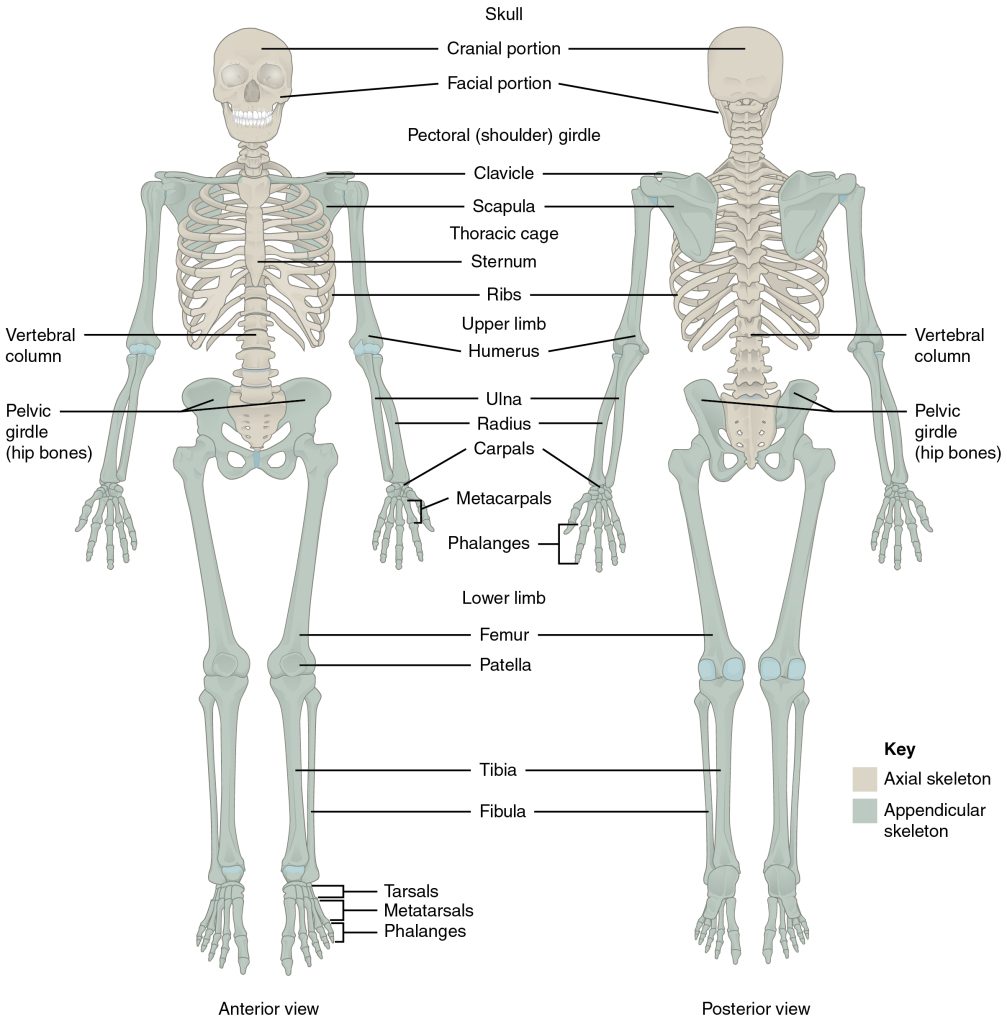
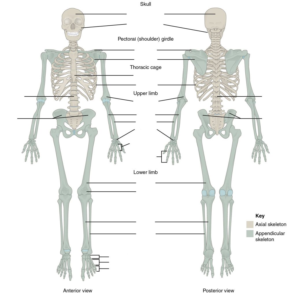
The axial skeleton forms the vertical, central axis of the body and includes all bones of the head, neck, chest, and back (see Figure 13.1). It serves to protect the brain, spinal cord, heart, and lungs. It also serves as the attachment site for muscles that move the head, neck, and back and for muscles that act across the shoulder and hip joints to move their corresponding limbs.
The axial skeleton of the adult consists of 80 bones, including the skull, the vertebral column, and the thoracic cage. The skull is formed by 22 bones. Also associated with the head are an additional seven bones, including the hyoid bone and the ear ossicles (three small bones found in each middle ear). The vertebral column consists of 24 bones, each called a vertebra, plus the sacrum and coccyx. The thoracic cage includes the 12 pairs of ribs, and the sternum, the flattened bone of the anterior chest.
Did You Know 1?
The axial skeleton has 80 bones and includes bones of the skull (and face), vertebral column, and thoracic cage.
The cranium or skull supports the face and protects the brain. It is subdivided into the bones of the skull and the bones of the face.
Bones of the Skull
- Frontal – forms the forehead
- Parietal – the upper lateral sides of the cranium
- Occipital – the posterior skull and base of the cranial cavity
- Temporal – the lower lateral sides of the cranium
- Sphenoid -the ‘keystone’ bone that forms part of the base of skull and eye sockets
- Ethmoid – forms part of the nose and orbit and base of the cranium
- Auditory ossicles – the small bones of the middle ear
- External auditory meatus – the external opening of the ear and temporal bone
Bones of the Face
- Zygomatic – the cheekbone
- Maxillary – the upper jaw and hard palate
- Palatine – the lateral walls of the nose
- Lacrimal – the walls of the orbit
- Inferior conchae – the lower lateral wall of the nasal cavity
- Vomer – the separates the left and right nasal cavity
- Mandible – the lower jaw bone (The only movable bone of the skull)
- Hyoid – the bone located between the mandible and larynx, not connected to other bones
Bones of the Vertebral Column
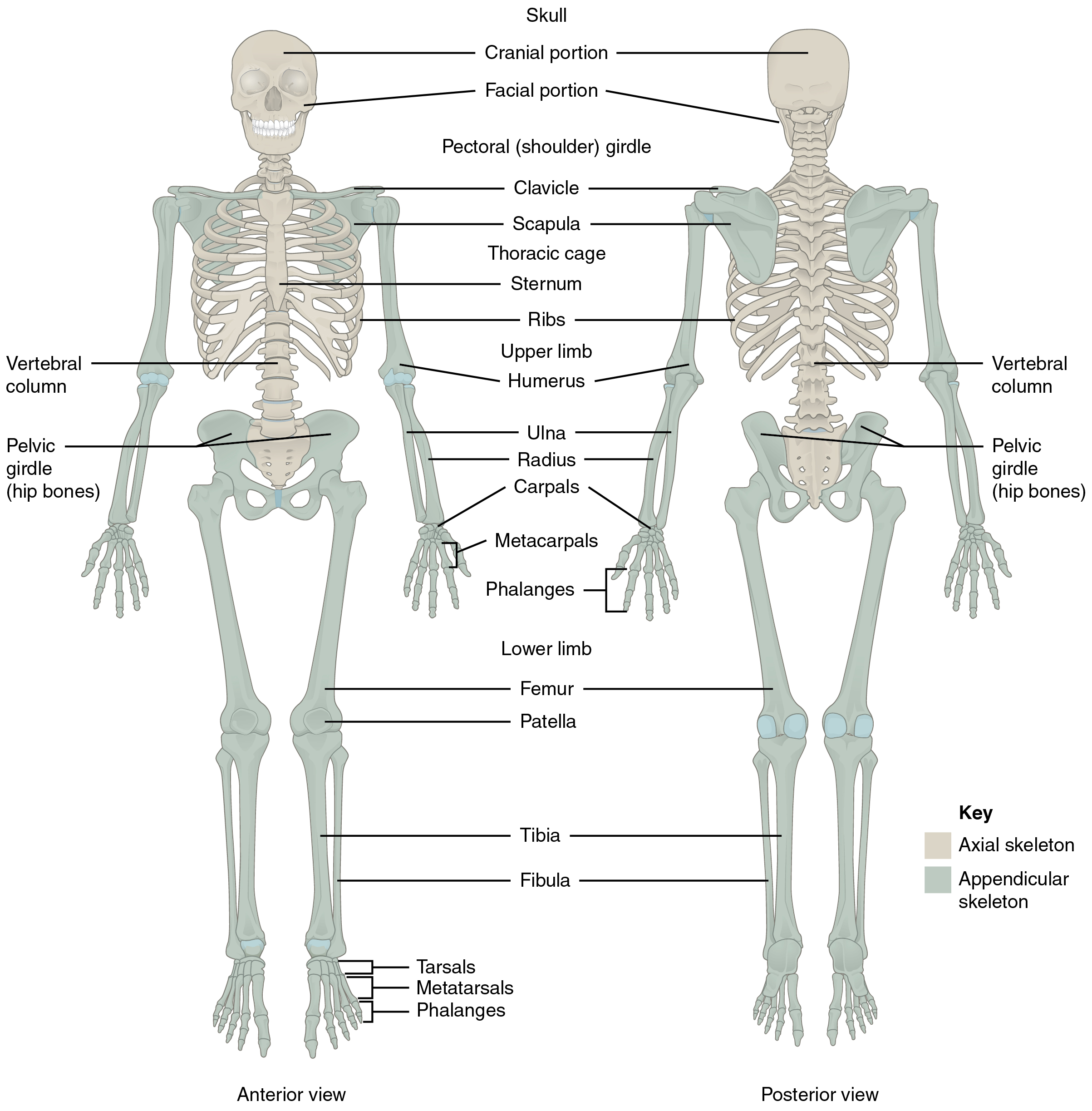
The vertebral column is also known as the spinal column or spine (see Figure 13.2). It consists of a sequence of vertebrae (singular = vertebra), each of which is separated and united by an intervertebral disc. Together, the vertebrae and intervertebral discs form the vertebral column. It is a flexible column that supports the head, neck, and body and allows for their movements. It also protects the spinal cord, which passes down the back through openings in the vertebrae.
Types of Vertebrae
- Cervical: C1 to C7 – the first 7 vertebrae in the neck region
- Thoracic: T1 to T12 – the next 12 vertebrae that form the outward curvature of the spine
- Lumbar: L1 to L5 – the next 5 vertebrae that form the inner curvature of spine
- Sacrum: the triangular-shaped bone at the base of the spine
- Coccyx: the tailbone
Bones of the Thoracic Cavity
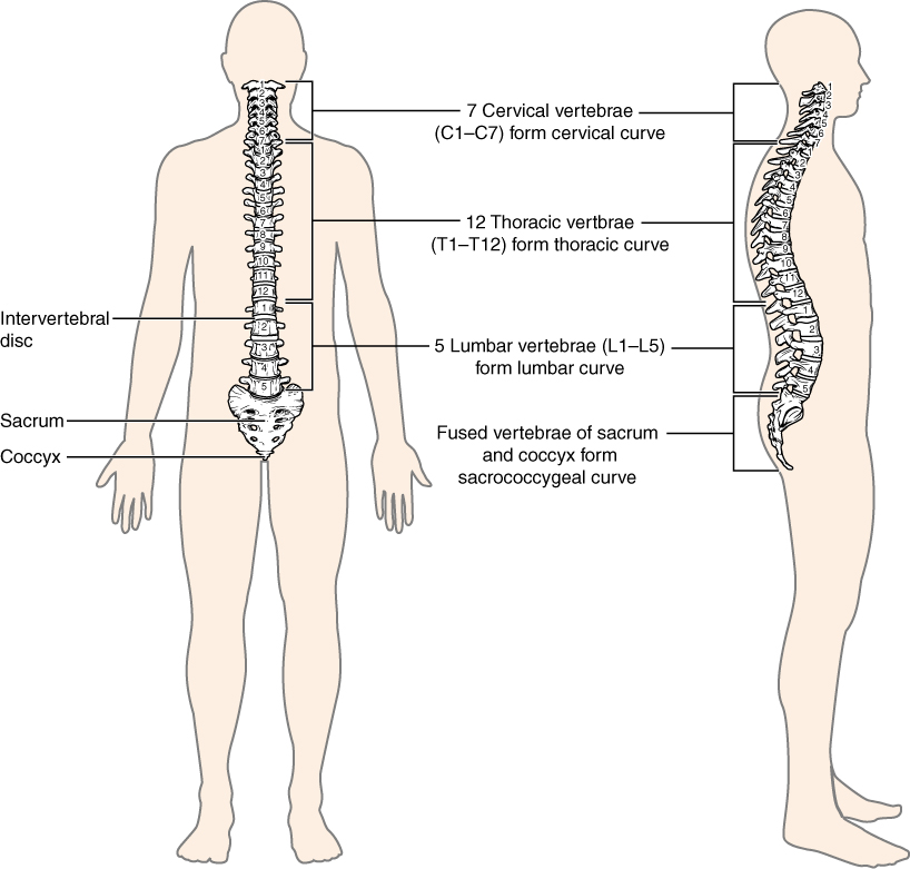
The thoracic cage (rib cage) forms the thorax (chest) portion of the body. It consists of 12 pairs of ribs with their costal cartilages and the sternum (see Figure 13.3). The ribs are anchored posteriorly to the 12 thoracic vertebrae (T1–T12). The thoracic cage protects the heart and lungs.
Ribs
There are 12 sets of ribs and can be divided as such:
- 7 true ribs as they are attached to the front of the sternum
- 3 false ribs as they are attached to the cartilage that joins the sternum
- 2 floating ribs as they are not attached to the front of the sternum
Sternum
The sternum, also known as the breast bone, is divided into 3 parts:
- manubrium – the upper portion of the breastbone
- body – the middle portion of the breastbone
- xiphoid process – the lower portion of the breastbone and is made up of cartilage
Concept Check 2
Answer the following questions:
- What is the medical term for the upper jaw bone and for the lower jaw bone?
- What medical term is used for the bones of the inner ear?
- How many bones make up the cervical region of the vertebral column?
The Appendicular Skeleton
The appendicular skeleton includes all bones of the upper and lower limbs, plus the bones that attach each limb to the axial skeleton. There are 126 bones in the appendicular skeleton of an adult.
Did You Know 2?
The appendicular skeleton has 126 bones. It is divided into the bones of the upper limbs and lower limb s that attach each limb to skeleton (Betts et al., 2013).
Bones of the Pectoral Girdle
- Scapula: the shoulder blades
- Clavicle: the collar bones. It connects the sternum to the scapula
- Acromion: the extension that forms the bony point of the shoulder
Bones of the Upper Limbs
The bones of the upper limbs include the bones of the arms, wrists, and hands.
Bones of the Arm
- Humerus: the bone in upper arm
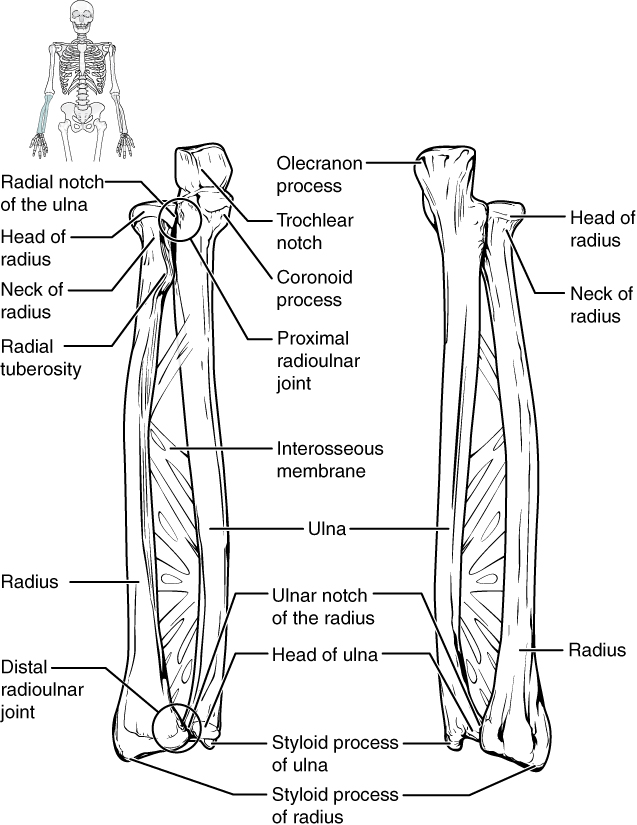
- Radius – the bone that runs thumb-side of the forearm
- Ulna – the bone that runs on the side of the little finger of the forearm
Bones of the Wrist and Hand
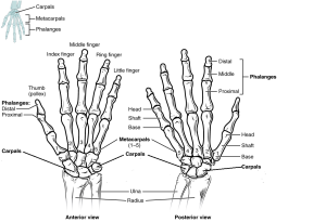
- Carpals – the wrist bones
- Metacarpals – the bones in the palm of hand
- Phalanges – the finger and toe bones
Each phalanx has three bones: the distal, medial, and proximal. The exception is the thumb and big toe which has two bones: distal and proximal (see Fig 16.5 below). There are 30 bones in each upper limb. Can you count them on your limb?
Bones of the Pelvic Region
The bones of the pelvic region protect the reproductive, urinary, and excretory organs.
- Pelvic girdle – the hip or coxal bone. It is formed by the fusion of three bones during adolescence
- Illium – the largest part of the hip bone
- Ischium – the lower portion of the pelvic girdle
- Pubis – the anterior portion of the pelvic girdle
- Pelvis – consists of four bones: the left and right hip bones as well as the sacrum and coccyx
- Acetabulum – the large socket in the pelvic bones that holds the head of the femur
The shape of the pelvic girdle is different for males than females. In the male, it is a funnel shape. In the female it is shaped like a basin to accommodate for the fetus during pregnancy.
Did You know 3?
The femur is the longest and strongest bone of the body, and accounts for approximately one-quarter of a person’s total height (Betts et al., 2013).
Bones of the Lower Limbs
The bones of the lower limb include bones of the leg and the feet.
Bones of the Leg
- Femur – the thigh bone and is also referred to as the upper leg bone. It is the longest and strongest bone in the human body
- Patella – the kneecap
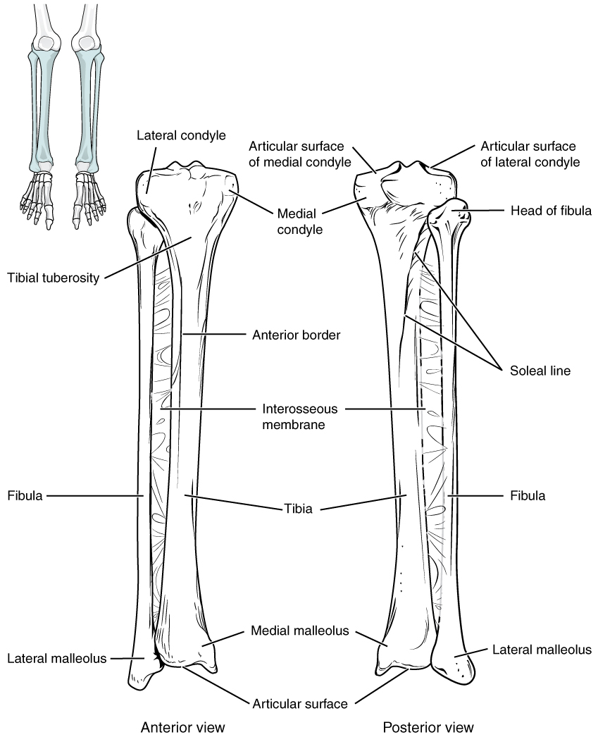
- Tibia – the shin bone. It is a medial bone and the main weight-bearing bone of the lower leg
- Fibula – the smaller of the lower leg bone (see Figure 13.6)
Bones of the Ankles and Feet
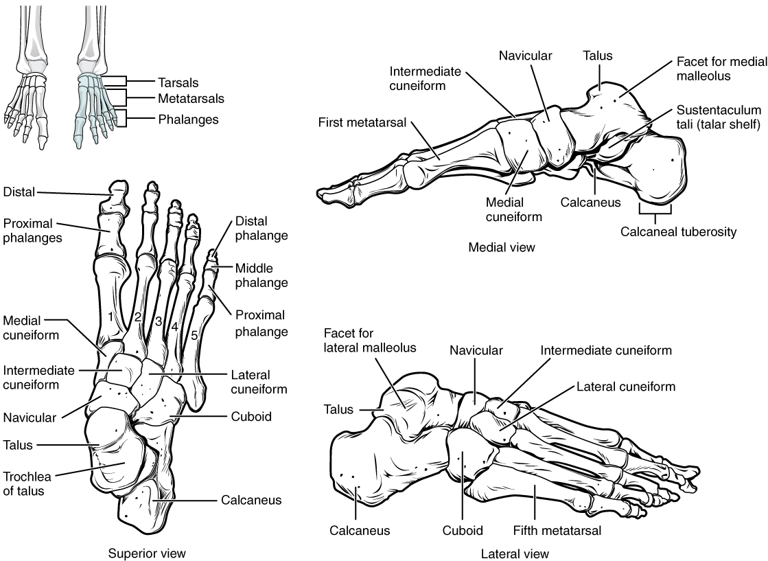
- Tarsals – the ankle bones (7 total)
- Malleous – the bony protrusions of the ankle bones
- Talus – the superior ankle bones
- Calcaneous – the heel bones
- Metatarsals – the foot bones
- Phalanges – the bones of the toes (see Figure 13.7)
Check Your Knowledge of the Skeletal System
Concept Check 1
Answer the following questions:
- Is the humerus the same as the funny bone?
- What is the medical term for the kneecap?
Musculoskeletal System-Skeleton Anatomy
Musculoskeletal System-Skeleton Anatomy (Text Version)
Label the diagram with the correct words listed below:
- Fibula
- Clavicle
- Femur
- Cranial portion
- Radius
- Facial portion
- Vertebral column
- Pelvic girdle
- Vertebral column
- Tibia
- Ulna
- Phalanges
- Ribs
- Tarsals
- Phalanges
- Pelvic girdle
- Scapula
- Metatarsals
- Sternum
- Metacarpals
- Patella
- Humerus
- Carpals
Check your answers [1]
Activity source: Musculoskeletal System-Skeleton Anatomy by Gisele Tuzon, from Building a Medical Terminology Foundation, illustration from Anatomy and Physiology (OpenStax), licensed under CC BY 4.0./ Text version added.
Image Descriptions
Figure 13.1 image description: This diagram shows the human skeleton and identifies the major bones. The left panel shows the anterior view (from the front) and the right panel shows the posterior view (from the back). Labels read (from the top of skull): skull (cranial portion, facial portion), pectoral shoulder girdle, clavicle, scapula, thoracic cage (sternum, ribs), upper limb (humerus, ulna, radius, carpals, metacarpals, phalanges), vertebral column, pelvic girdle (hip bones), lower limb (femur, patella, tibia, fibula, tarsals, metatarsals, phalanges). [Return to Figure 13.1].
Figure 13.2 image description: This image shows the structure of the vertebral column. The left panel shows the front view of the vertebral column. Labels and the right panel shows the side view of the vertebral column. Labels read (from top): 7 cervical vertebrae (C1-C7) form cervical curve, 12 thoracic vertebrae (T1-T12) form thoracic curve, intervertebral disc, 5 lumbar vertebrae (L1-L5) form lumbar curve, Fused vertebrae of sacrum and coccyx form sacrococcygeal curve, sacrum, coccyx. [Return to Figure 13.2].
Figure 13.3 image description: This figure shows the skeletal structure of the rib cage. The left panel shows the anterior view of the sternum. Labels read (from top): clavicular notch, jugular notch, manubrium, sternal angle, body, xiphoid process. The right panel shows the anterior panel of the sternum including the entire rib cage. Labels read (from top): jugular notch, clavicular notch, clavicle, sternum (manubrium, body, xyphoid process), scapula, sternal angle, costal cartilages, intercostal space. Ribs are numbered 1-12 from the top. [Return to Figure 13.3].
Figure 13.4 image description: This diagram labels the bones of the lower arm (excluding the hands). Labels read (from top): olecranon process, head of radius, radial notch of the ulna, trochlear notch, coronoid process, radial tuberosity, proximal radioulnar joint, neck of radius, radius, interosseous membrane, ulna, ulnar notch of the radius, head of the ulna, distal radioulnar joint, styloid process of ulna, styloid process of radius. [Return to Figure 13.4].
Figure 13.5 image description: This diagram shows an anterior and posterior view of the hands with corresponding labels. Anterior view labels read (from top): middle finger, ring finger, index finger, little finger, thumb, phalanges (distal, proximal), metacarpals, carpals, ulna, radius. Posterior view labels read (from top): Phalanges (distal, middle, proximal), head shaft and base of proximal phalange, head shaft and base of metatarsal, metatarsals 1-5, carpals, ulna, radius. [Return to Figure 13.5].
Figure 13.6 image description: This image shows the structure of the tibia and the fibula. The left panel shows the anterior view. Labels read (from top): lateral condyle, medial condyle, tibial tuberosity, anterior border, interosseous membrane, fibula, tibia, medial malleolus, lateral malleolus, articular surface. The right panel shows the posterior view. Labels read (from top): articular surface of medial and lateral condyles, medial condyle, head of fibula, soleal line, interosseous membrane, tibia, fibula, medial malleolus, lateral malleolus, articular surface. [Return to Figure 13.6].
Figure 13.7 image description: This figure shows the bones of the foot. The left panel shows the superior view. Labels read (from toes): distal, proximal phalanges, distal phalange, middle phalange, proximal phalange, medial cuneiform, intermediate and lateral cuneiforms, navicular, cuboid, talus, trochlea of talus, calcaneus. The top right panel shows the medial view. Labels read (from left to right starting at toe): first metatarsal, medial cuneiform, intermediate cuneiform, navicular, talus, calcaneus, facet for medial malleolus, sustentaculum tali (talar shelf), calcaneal tuberosity. The bottom right panel shows the lateral view. Labels read (from left at the heel to right): calcaneus, talus, facet for lateral malleolus, cuboid, navicular, intermediate and lateral cuneiforms, fifth metatarsal. [Return to Figure 13.7].
Attribution
Except where otherwise noted, this chapter is adapted from “Skeletal System” in Building a Medical Terminology Foundation by Kimberlee Carter and Marie Rutherford, licensed under CC BY 4.0. / A derivative of Betts et al., which can be accessed for free from Anatomy and Physiology (OpenStax). Adaptations: dividing Skeletal System chapter content into sub-chapters.