11.2 – Anatomy & Physiology of the Lymphatic System
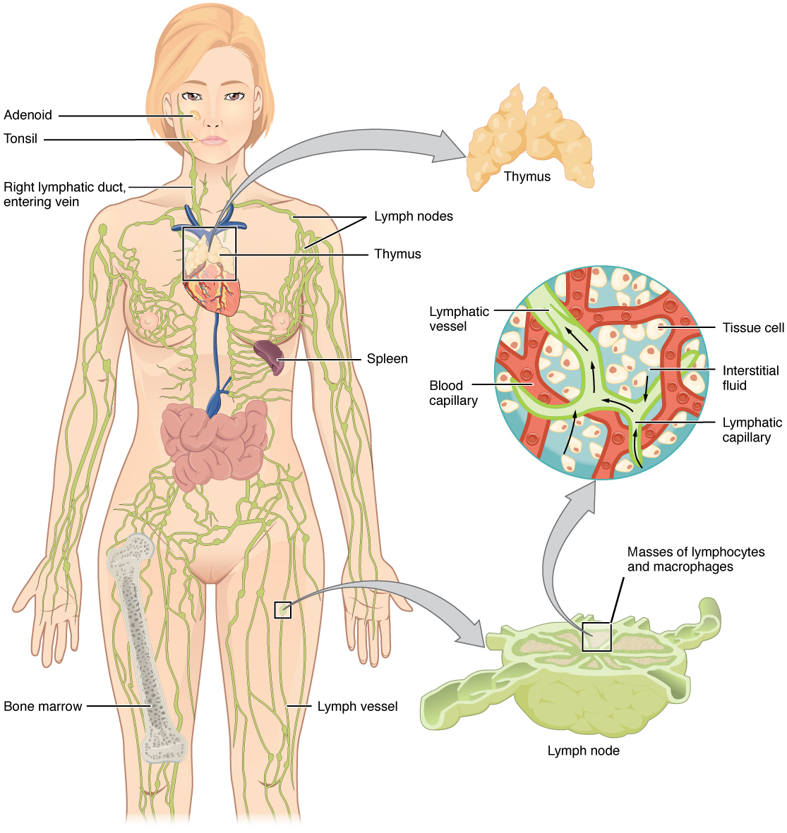
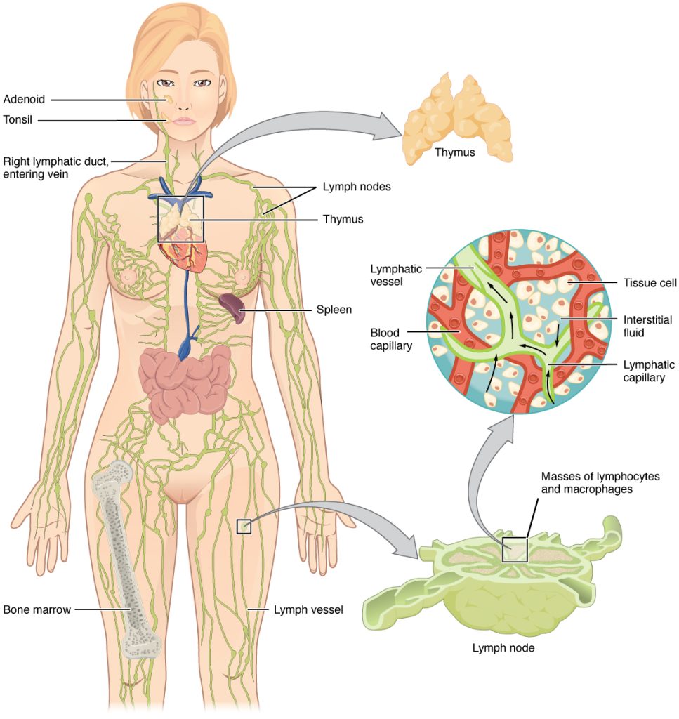
The lymphatic vessels begin as open-ended capillaries, which feed into larger and larger lymphatic vessels, and eventually empty into the bloodstream. Along the way, the lymph travels through the lymph nodes, which are commonly found near the groin, armpits, neck, chest, and abdomen. Humans have about 500–600 lymph nodes throughout the body (see Figure 11.1). Several organs and tissues that participate in immunity are also part of the lymphatic system.
Lymphatic Capillaries
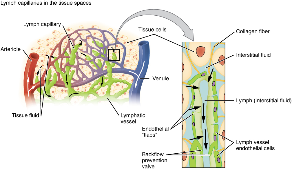
An important function of the lymphatic system is to return the fluid (lymph) to the blood. Lymph may be thought of as recycled blood plasma. Blood pressure causes leakage of fluid from the blood capillaries, resulting in the accumulation of fluid in the interstitial space. In humans, 20 liters of plasma is released into the interstitial space of the tissues each day due to capillary leakage. The blood vessels reabsorb 17 liters of this interstitial fluid, leaving three litres in the tissues for the lymphatic system to transport back to the circulation. If the lymphatic system is damaged in some way, such as by being blocked by cancer cells or destroyed by injury, interstitial fluid accumulates in the tissue spaces, causing a condition called lymphedema.
Did You Know 1?
Lymphatic capillaries, also called the terminal lymphatics, are vessels where interstitial fluid enters the lymphatic system to become lymph. Located in almost every tissue in the body, these vessels are interlaced among the arterioles and venules of the circulatory system in the soft connective tissues of the body. See Figure 11.2. Exceptions are the central nervous system, bone marrow, bones, teeth, and the cornea of the eye, which do not contain lymph vessels.
Larger Lymphatic Vessels, Trunks, and Ducts
The lymphatic capillaries empty into larger lymphatic vessels, which are similar to veins in terms of their three-tunic structure and the presence of valves. These one-way valves are located fairly close to one another, and each one causes a bulge in the lymphatic vessel, giving the vessels a beaded appearance (see Figure 11.2).
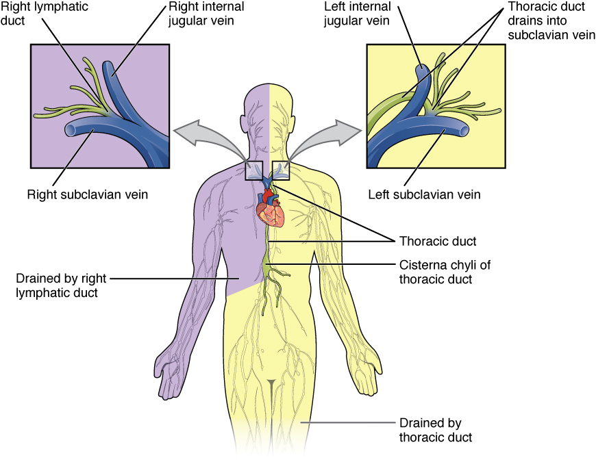
In general, superficial lymphatics follow the same routes as veins, whereas deep lymphatic vessels of the viscera generally follow the paths of arteries. The superficial and deep lymphatics eventually merge to form larger lymphatic structures known as lymphatic trunks. On the right side of the body, the right sides of the head, thorax, and right upper limb trunks drain lymph fluid into the right subclavian vein via the right lymphatic duct (see Figure 11.3). On the left side of the body, the trunks from the remaining portions of the body drain into the larger thoracic duct, which drains into the left subclavian vein. The thoracic duct itself begins just beneath the diaphragm in the cisterna chyli.
Primary Lymphoid Organs
The primary lymphoid organs are the bone marrow and thymus gland. The lymphoid organs are where lymphocytes mature, proliferate, and are selected, which enables them to attack pathogens without harming the cells of the body.
- Bone Marrow
- Recall that all blood cells, including lymphocytes, are formed in the red bone marrow. The B cell undergoes nearly all of its development in the red bone marrow, whereas the immature T cell, called a thymocyte, leaves the bone marrow and matures largely in the thymus gland.
- Thymus
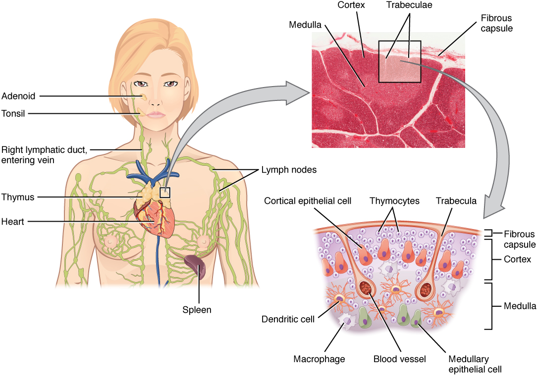
- The thymus gland, where T cells mature, is a bilobed organ found in the space between the sternum and the aorta of the heart (see Figure 11.4). Connective tissue holds the lobes closely together but also separates them and forms a capsule.
- The loss of immune function with age is called immunosenescence. One major cause of age-related immune deficiencies is thymic involution.
- The shrinking of the thymus gland begins at birth at a rate of about three percent tissue loss per year. This shrinking continues until 35–45 years of age, then the rate declines to about one percent loss per year for the rest of one’s life. At that pace, the total loss of thymic epithelial tissue and thymocytes would occur at about 120 years of age. So, in theory, 120 years could be the maximum life span.
Concept Check
- Do you remember what the suffix “-oid” means?
- Can you explain the term lymphoid?
Did You Know 2?
The thymus gland produces a hormone called thymosin and is therefore also considered to be part of the endocrine system.
Secondary Lymphoid Organs
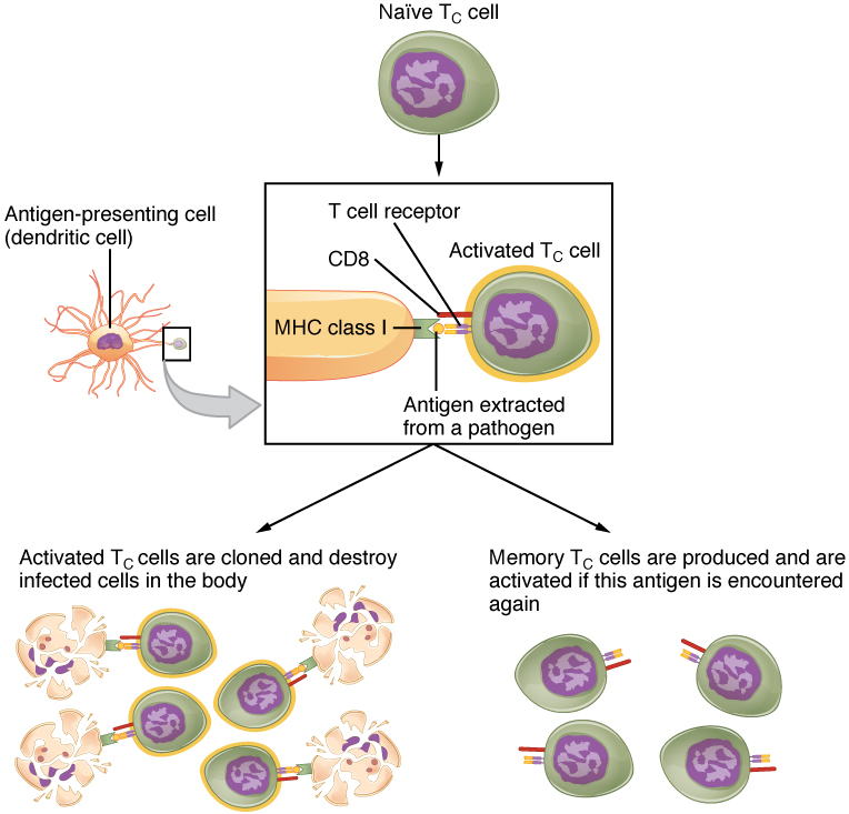
Lymphocytes develop and mature in the primary lymphoid organs, but they mount immune responses from the secondary lymphoid organs, which include the lymph nodes, spleen, and lymphoid nodules. A naïve lymphocyte is one that has left the primary organ, where it learned to function immunologically, and entered a secondary lymphoid organ where it waits to encounter an antigen against which it will mount a response (see Figure 11.5).
Lymph Nodes
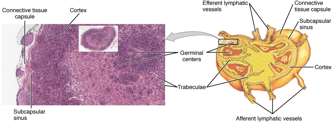
Lymph nodes function to remove debris and pathogens from the lymph, and are thus sometimes referred to as the “filters of the lymph” (see Figure 11.6). Any bacteria that infect the interstitial fluid are taken up by the lymphatic capillaries and transported to a regional lymph node. Dendritic cells and macrophages within this organ internalize and kill many of the pathogens that pass through, thereby removing them from the body. The lymph node is also the site of adaptive immune responses mediated by T cells, B cells, and accessory cells of the adaptive immune system.
Did You Know 3?
Spleen
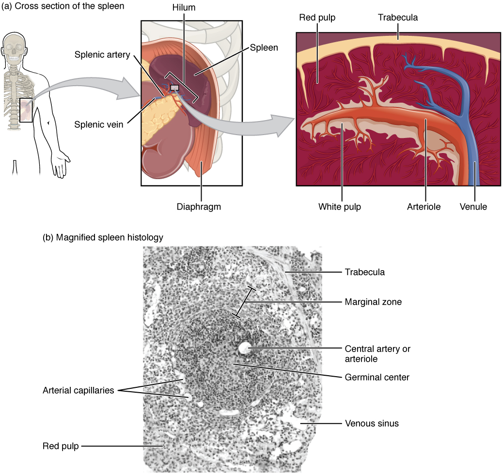
The spleen is a vascular organ that is somewhat fragile due to the absence of a capsule. It is about 12 cm long and is attached to the lateral border of the stomach. The spleen is sometimes called the “filter of the blood” because of its extensive vascularization and the presence of macrophages and dendritic cells that remove microbes and other materials from the blood, including dying red blood cells. The spleen also functions as the location of immune responses to blood-borne pathogens.
Lymphoid Nodules
The other lymphoid tissues, the lymphoid nodules, consist of a dense cluster of lymphocytes without a surrounding fibrous capsule. These nodules are located in the respiratory and digestive tracts, areas routinely exposed to environmental pathogens.
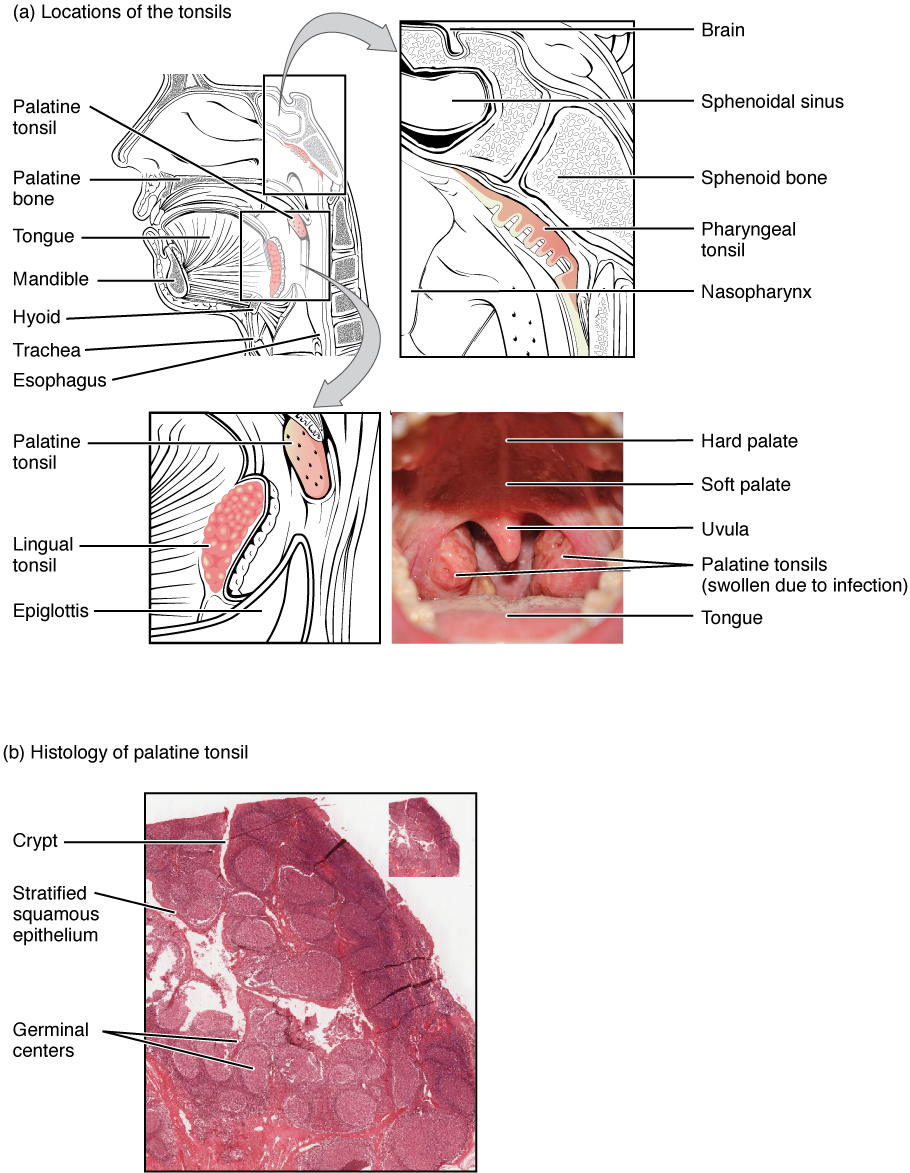
Tonsils are lymphoid nodules located along the inner surface of the pharynx and are important in developing immunity to oral pathogens (see Figure 11.8). The tonsil located at the back of the throat, the pharyngeal tonsil, is sometimes referred to as the adenoid when swollen. Such swelling is an indication of an active immune response to infection. Tonsils have deep grooves called crypts, which accumulate all sorts of materials taken into the body through eating and breathing and actually “encourage” pathogens to penetrate deep into the tonsillar tissues where they are eliminated. A major function of tonsils is to help children’s bodies recognize, destroy, and develop immunity to common environmental pathogens so that they will be protected in their later lives. Tonsils are often removed in children who have recurring throat infections since swollen palatine tonsils can interfere with breathing and/or swallowing.
Concept Check
Tonsils are named after their locations.
- Look at the figure above and determine which anatomical structure is closely associated with each set of tonsils and was therefore used to name the tonsils; for example, the lingual tonsils are named after the tongue (lingula).
- Can you tell which structures were used to name the palatine tonsils and the pharyngeal tonsils?
Bronchus-associated lymphoid tissue (BALT) consists of lymphoid follicular structures with an overlying epithelial layer found along the bifurcations of the bronchi and between bronchi and arteries. These tissues, in addition to the tonsils, are effective against inhaled pathogens.
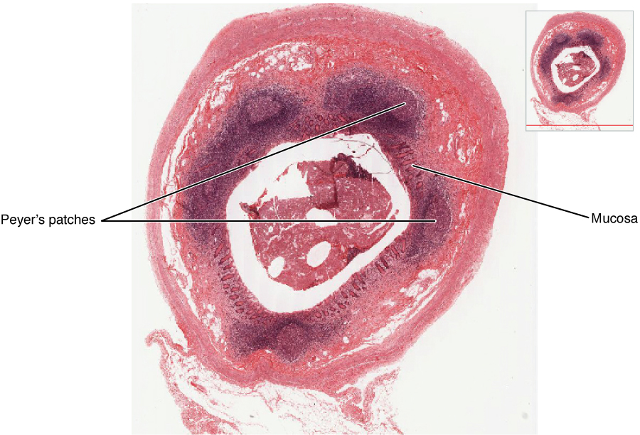
Mucosa-associated lymphoid tissue (MALT) consists of an aggregate of lymphoid follicles directly associated with mucous membrane. MALT makes up dome-shaped structures found underlying the mucosa of the gastrointestinal tract, breast tissue, lungs, and eyes. Peyer’s patches, a type of MALT in the small intestine, are especially important for immune responses against ingested substances (see Figure 11.9). Peyer’s patches contain specialized cells that sample material from the intestinal lumen and transport it to nearby follicles so that adaptive immune responses to potential pathogens can be mounted.
Check Your Knowledge of the Lymphatic System
Anatomy Labeling Activity
Lymphatic System Anatomy (Text Version)
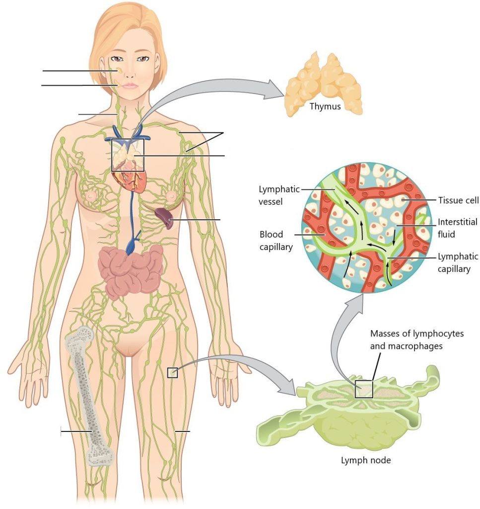
Label the diagram with correct words listed below:
- Adenoid
- Lymph nodes
- Tonsil
- Thymus
- Bone marrow
- Spleen
- Right lymphatic duct, entering vein
- Lymph vessel
Lymphatic System Anatomy Diagram (Text Version)
The diagram shows a female human body standing upright, and the entire lymphatic system is shown and labeled (clockwise from top): The _______[Blank 1] is a small gland located in the centre of the chest and it is responsible for supporting the immune function by producing T-cells a type of white blood cell which fights infections and diseases. A collection of oval shaped structures known as _______[Blank 2] serve as filtration units. The _____[Blank 3] is an organ located under the left part of the diaphragm and is responsible for blood filtration. Thin-walled tube known as a ______[Blank 4] carry lymph tissue throughout the body. The ______[Blank 5] is a primary site for T-cell activity in the lymphatic system. The ___________[Blank 6] receives lymph fluid from the right side of the head, neck, and thorax, as it drains the venous system. Located at the back of the throat is a fleshy structure known as the ________[Blank 7] and serves as the first line of defence against inhaled harmful substances. Located in the nasopharyngeal region is the _________[Blank 8] which also filter and trap harmful substances from entering the body. The right panel shows magnified images of the thymus and the lymph node. Labels read (clockwise from top): tissue cell, interstitial fluid, lymphatic capillary, blood capillary, lymphatic vessel. Label of lymph node reads masses of lymphocytes and macrophages.
Check your answers [1]
Activity source: Lymphatic System Anatomy by Gisele Tuzon, from Building a Medical Terminology Foundation, illustration from Anatomy and Physiology (OpenStax), licensed under CC BY 4.0./ Text version added.
Image Descriptions
Figure 11.1 image description: The left panel shows a female human body, and the entire lymphatic system is shown Labels read (clockwise from top): thymus, lymph nodes, thymus, spleen, lymph vessel, bone marrow, right lymphatic duct, entering vein, tonsil, adenoid. The right panel shows magnified images of the thymus and the lymph node. Labels read (clockwise from top): tissue cell, interstitial fluid, lymphatic capillary, blood capillary, lymphatic vessel. Label of lymph node reads masses of lymphocytes and macrophages. [Return to Figure 11.1].
Figure 11.2 image description: This image shows the lymph capillaries in the tissue spaces. Labels read (clockwise, from top): lymph capillary, tissue cells, venule, lymphatic vessel, tissue fluid, arteriole). It also shows a magnified image shows the interstitial fluid and the lymph vessels. Labels read (clockwise, from top): collagen fiber, interstitial fluid, lymph, lymph vessel endothelial cells, backflow prevention valve, endothelial flaps. [Return to Figure 11.2].
Figure 11.3 image description: This figure shows the lymphatic trunks and the duct system in the human body. Labels read (clockwise from top) thoracic duct, cisterna chyli of thoracic duct, drained by thoracic duct, drained by right lymphatic duct. Callouts to the left and right show the magnified views of the left and right jugular vein respectively. Labels read (right lymphatic duct): right internal jugular vein, right subclavian vein, right lymphatic duct; (left jugular vein): left internal jugular vein, thoracic duct drains into subclavian vein, left subclavian vein. [Return to Figure 11.3].
Figure 11.4 image description: The left panel of this figure shows the head and chest of a woman and the location of the thymus is marked. Labels read (clockwise, from top) lymph nodes, spleen, heart, thymus, right lymphatic duct entering vein, tonsil, adenoid. The top right panel shows a micrograph of the thymus. Labels read (from left to right): medulla, cortex, trabeculae, fibrous capsule. The bottom right panel shows a magnified view of the structure of the thymus. Labels read (clockwise, from top): thymocytes, trabecula, fibrous capsule, cortex, medulla (layers), medullary epithelial cell, blood vessel, macrophage, dendritic cell, cortical epithelial cell. [Return to Figure 11.4].
Figure 11.5 image description: This flowchart shows the process in which a naïve T cell become activated T cells in the left part of the pathway and memory cells in the right part of the pathway. A naive T cell becomes an activated T cell when an antigen-presenting cell is introduced. The antigen is extracted from a pathogen and then either activated T cells are cloned and destroy the infected cells in the body, and/or memory T cells are produced and are activated if this antigen is encountered again. [Return to Figure 11.5].
Figure 11.6 image description: The left panel of this figure shows a micrograph of the cross section of a lymph node. Labels indicate the connective tissue capsule, cortex, and subcapsular sinus. The right panel shows the structure of a lymph node. Labels indicate (from top, clockwise) the efferent lymphatic vessels, connective tissue capsule, subcapsular sinus, cortex, afferent lymphatic vessels, trabecula, germinal centers. [Return to Figure 11.6].
Figure 11.7 image description: The top left panel shows the location of the spleen in the human body. The top center panel shows a close up view of the location of the spleen. Labels read (clockwise, from top): hilum, spleen, diaphragm, splenic vein, splenic artery. The top right panel shows the blood vessels and spleen tissue. Labels read (from left to right, top then bottom) red pulp, trabecula (bottom) white pulp, arteriole, venule. The bottom panel shows a histological micrograph Labels read (clockwise, from top): trabecula, marginal zone, central artery or arteriole, germinal center, venous sinus, red pulp, arterial capillaries. [Return to Figure 11.7].
Figure 11.8 image description: The top panel of this image shows the locations of the tonsils. Labels read (clockwise from top):palatine tonsil, palatine bone, tongue, mandible, hyoid, trachea, esophagus. Callout shows the location of the pharyngeal tonsil. Labels read (from top): brain, sphenoidal sinus, sphenoid bone, pharyngeal tonsil, nasopharynx. Another callout details the location of the palatine tonsil. Labels read (from top): palatine tonsil, lingual tonsil, epiglottis. Another callout shows a photograph of the back of the throat where the tonsils are located. Labels read (from top) hard palate, soft palate, uvula, palatine tonsils (swollen due to infection) and tongue. The bottom panel shows the histological micrograph of the tonsils. Labels read (from top): crpyt, stratified squamous epithelium, germinal centers. [Return to Figure 11.8].
Figure 11.9 image description: This figure shows a micrograph of a mucosa associated lymphoid tissue (MAST) nodule. Labels indicate the mucosa and Peyer’s patches (which appear to be dark purple). [Return to Figure 11.9].
Attribution
Except where otherwise noted, this chapter is adapted from “Lymphatic and Immune Systems” in Building a Medical Terminology Foundation by Kimberlee Carter and Marie Rutherford, licensed under CC BY 4.0. / A derivative of Betts et al., which can be accessed for free from Anatomy and Physiology (OpenStax). Adaptations: dividing Lymphatic and Immune Systems chapter content into sub-chapters.
-
↵
Check your answers: Lymphatic System Anatomy Diagram (Text Version)
The diagram shows a female human body standing upright, and the entire lymphatic system is shown and labeled (clockwise from top): The thymus is a small gland located in the centre of the chest and it is responsible for supporting the immune function by producing T-cells a type of white blood cell which fights infections and diseases. A collection of oval shaped structures known as lymph nodes serve as filtration units. The spleen is an organ located under the left part of the diaphragm and is responsible for blood filtration. Thin-walled tube known as a lymph vessel carry lymph tissue throughout the body. The bone marrow is a primary site for T-cell activity in the lymphatic system. The right lymphatic duct entering vein receives lymph fluid from the right side of the head, neck, and thorax, as it drains the venous system. Located at the back of the throat is a fleshy structure known as the tonsil and serves as the first line of defence against inhaled harmful substances. Located in the nasopharyngeal region is the adenoids which also filter and trap harmful substances from entering the body. The right panel shows magnified images of the thymus and the lymph node. Labels read (clockwise from top): tissue cell, interstitial fluid, lymphatic capillary, blood capillary, lymphatic vessel. Label of lymph node reads masses of lymphocytes and macrophages.
Lymph is the term used to describe interstitial fluid once it has entered the lymphatic system (Betts et al., 2013)
Spaces between individual cells in the tissues (Betts et al., 2013)
Fluid that has leaked out of blood capillaries into the tissue spaces
lymphatic vessels of the subcutaneous tissues of the skin
Lymphatic vessels of the organs
Large lymphatic vessels that collect lymph from smaller lymphatic vessels and empty it into the blood via lymphatic ducts (Betts et al., 2013)
A sac-like chamber that receives lymph from the lower abdomen, pelvis, and lower limbs by way of the left and right lumbar trunks and the intestinal trunk. (Betts et al, 2013)
two lobes
shrinking of the thymus due to age
lymphocytes that develop into T-cells in the thymus gland
Bone marrow and thymus gland.
Specific immune mechanisms (against a specific pathogen) which take time to develop
Histologically, tonsils do not contain a complete capsule, and the epithelial layer invaginates deeply into the interior of the tonsil to form tonsillar crypts. (DeSaix et al., 2013)
disease causing agents
