9.3 – Physiology of the Heart
In order for the heart to do its job of pumping blood to the lungs and to the body, nutrients and oxygen must be supplied to the cells of the heart. The heart also needs to coordinate its contractions so that all parts are working together to pump blood effectively. To understand how all of this works together to give the heart its ability to pump blood, we will examine three interdependent aspects of heart function.
- Circulation through the heart: blood is pumped by the heart in order to provide oxygen and nutrients to every cell in the body.
- The heart as an organ (coronary blood supply): the heart is an organ, made of cells and tissues which require their own blood supply.
- The heart’s electrical conduction system: the heart is able to independently generate and transmit instructions to the myocardium, in order to make it contract and pump the blood.
1. Circulation Through the Heart: The Heart as a Pump
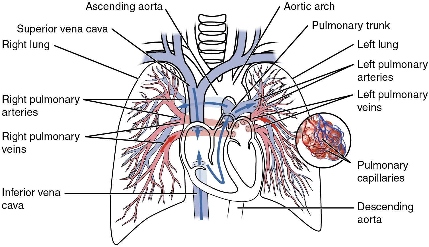
The heart pumps blood to two distinct but linked circulatory systems called the pulmonary and systemic circuits. The pulmonary circuit transports blood to and from the lungs, where it picks up oxygen and drops off carbon dioxide. The systemic circuit transports freshly oxygenated blood to virtually all of the tissues of the body and returns relatively deoxygenated blood and carbon dioxide to the heart to be sent back to the pulmonary circulation.
Did You Know?
- Blood that is carrying carbon dioxide and waste products from the body tissues is returned to the right atrium via the superior vena cava and the inferior vena cava.
- From the right atrium, the deoxygenated blood moves through the tricuspid valve into the right ventricle.
- The right ventricle pumps deoxygenated blood through the pulmonary valve into the pulmonary trunk, which splits into the right and left pulmonary arteries, leading toward the lungs. These arteries branch many times before reaching the pulmonary capillaries, where gas exchange occurs: carbon dioxide exits the blood and oxygen enters. The pulmonary arteries are the only arteries in the postnatal body that carry deoxygenated blood. Did you notice that they are often coloured blue on diagrams of the heart?
- Freshly oxygenated blood returns from the lungs to the left atrium via the pulmonary veins. These veins are the only postnatal veins in the body that carry highly oxygenated blood and are often coloured red on heart images.
- From the left atrium, the blood moves through the mitral valve into the left ventricle.
- The left ventricle pumps blood through the aortic valve, into the aorta, delivering blood to all parts of the body.
Concept Check 1
- On Figure 9.3 below, use your finger to trace the pathway of blood flowing through the right side of the heart, naming each each of the following structures as you encounter them: superior and inferior venae cavae, right atrium, tricuspid valve, right ventricle, pulmonary valve, right and left pulmonary arteries.
- Suggest what would happen if the aorta experienced a blockage or constriction.
Pulmonary Circuit
Blood exiting from the right ventricle flows into the pulmonary trunk, which bifurcates into the two pulmonary arteries. These vessels branch to supply blood to the pulmonary capillaries, where gas exchange occurs within the lung alveoli. Blood returns via the pulmonary veins to the left atrium.
Concept Check 2
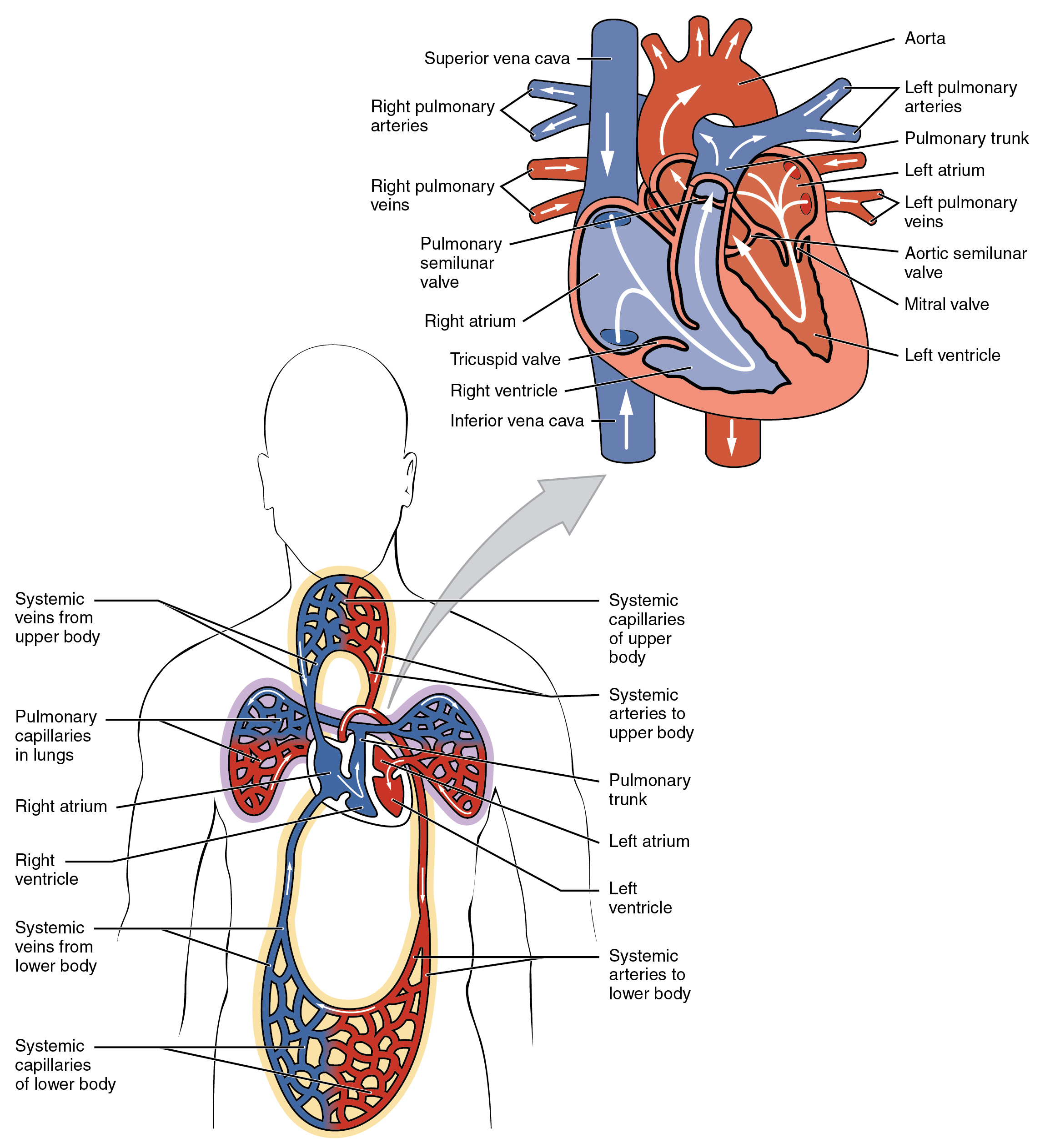
- On Figure 9.4 below, use your finger to trace the pathway of blood flowing through the left side of the heart, naming each of the following structures as you encounter them: right and left pulmonary veins, left atrium, mitral valve, left ventricle, aortic valve, aorta.
Cardiac Cycle
The process of pumping and circulating blood is active, coordinated, and rhythmic. Each heartbeat represents one cycle of the heart receiving blood and ejecting blood.
- Diastole is the portion of the cycle in which the heart is relaxed and the atria and ventricles are filling with blood. The AV valves are open so that blood can move from the atria to the ventricles.
- Systole is the portion of the cycle in which the heart contracts, AV valves slam shut, and the ventricles eject blood to the lungs and to the body through the open semilunar valves. Once this phase ends, the semilunar valves close in preparation for another filling phase.
2. The Heart as an Organ: The Coronary Blood Supply
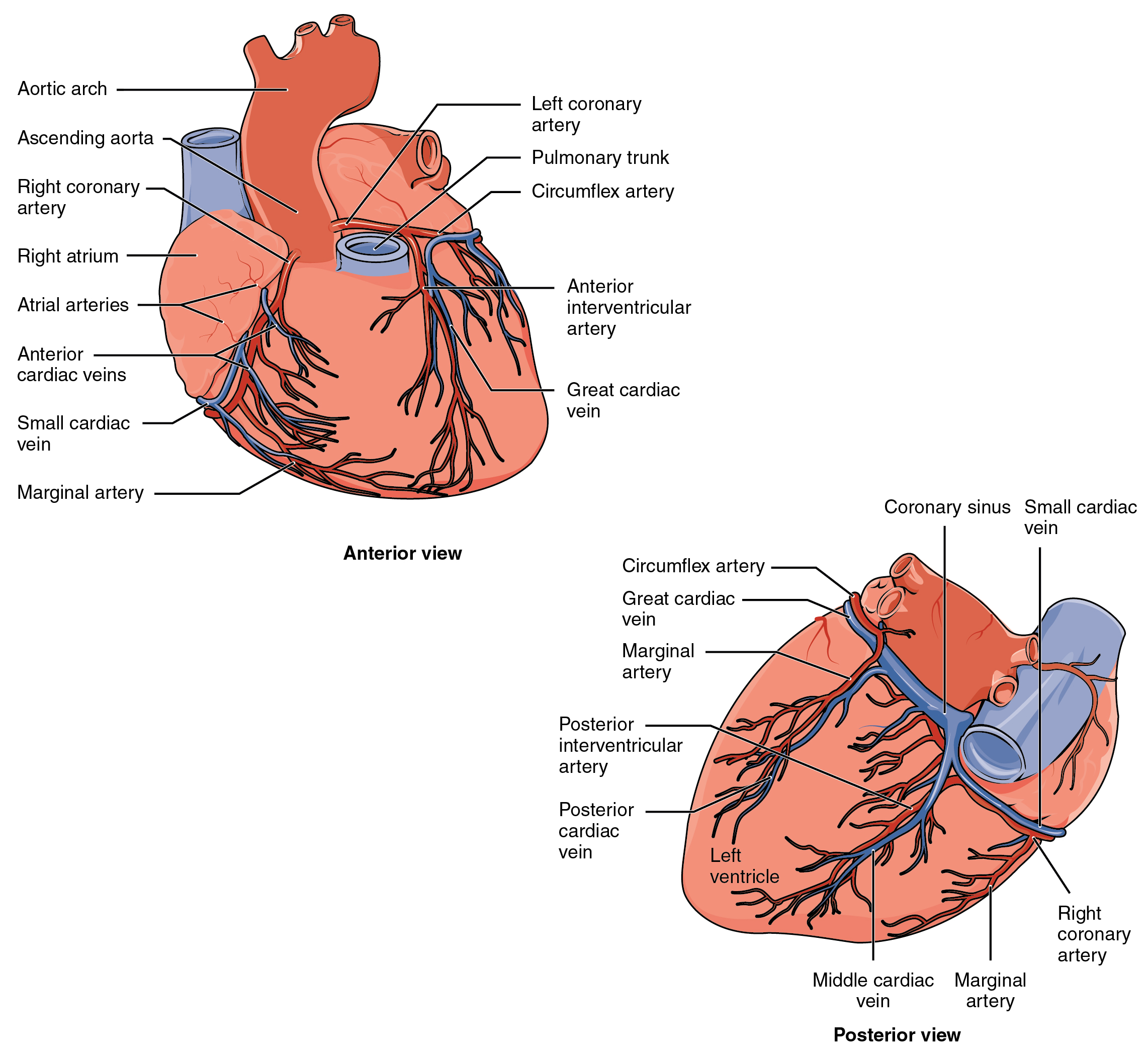
Myocardial cells require their own blood supply to carry out their function of contracting and relaxing the heart in order to pump blood. Their own blood supply provides nutrients and oxygen and carry away carbon dioxide and waste. These functions are provided by the coronary arteries and coronary veins.
Concept Check 3
On the image (Figure 9.5) below, locate the three main coronary arteries:
- Anterior interventricular artery (more commonly known as the left anterior descending artery, or LAD)
- Circumflex artery (Cx)
- Right coronary artery (RCA)
Follow the path of each of these three arteries to try to determine which parts of the myocardium each artery (along with its many smaller branches) supplies with blood.
3. The Heart’s Electrical Conduction System
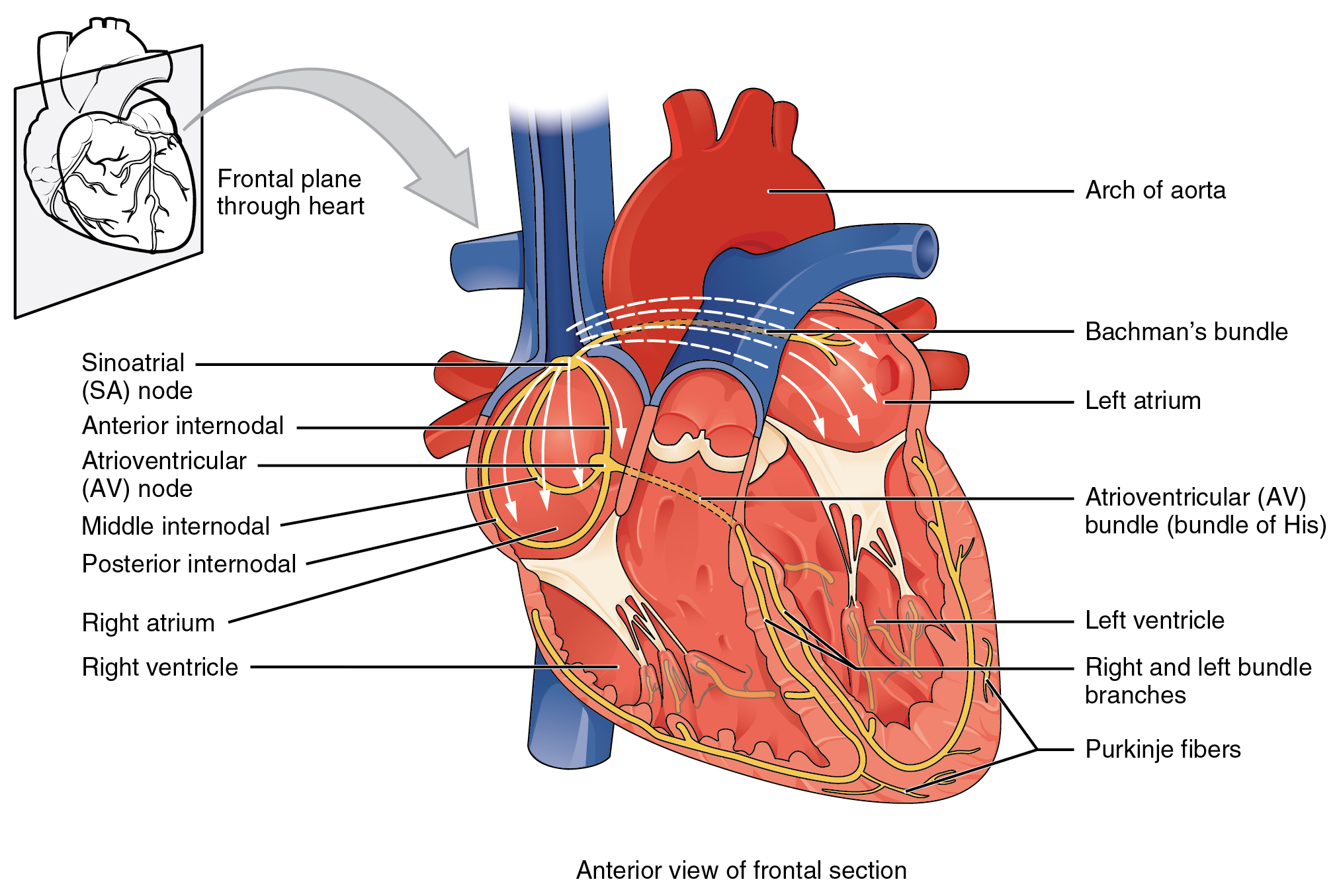
In order for all parts of the heart to work together to beat regularly and effectively, the heart has its own electrical system, which initiates and conducts each heartbeat through the entire myocardium. Specialized groups of heart cells perform this function all on their own, without requiring messages from the central nervous system.
Watch The Heart, Part 2 – Heart Throbs: Crash Course Anatomy & Physiology #26 (9:30 min) on YouTube
Media 9.2: CrashCourse. (2015, July 13). The heart, part 2 – Heart throbs: Crash Course anatomy & Pphysiology #26 [Video]. YouTube. https://youtu.be/FLBMwcvOaEo
Concept Check 4
- On the image (Figure 9.6) above, trace the electrical impulse generated by the heart’s pacemaker (the sinoatrial node, or SA node) through the rest of the conduction system, including the atrioventricular (AV) node, the atrioventricular bundle (bundle of His), the right and left bundle branches, and the Purkinje fibers.
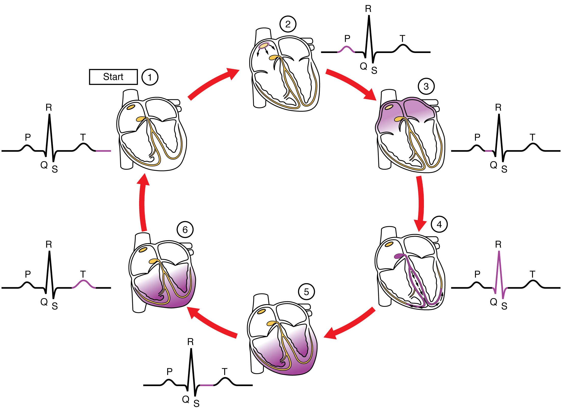
We can detect and record the electrical activity of the heart’s conduction system using an electrocardiogram (ECG or EKG). Figure 9.7 shows the electrical impulse originating in the SA node (step 2) and travelling through the heart’s conduction system, allowing the heart to complete one cardiac cycle. Each waveform on the ECG tracing represents electricity moving through and affecting a different part of the heart. Did you notice that the AV valves close when the electrical impulse reaches the ventricles, just before systole occurs?
Heart Medical Terms and Abbbreviations
Cardiovascular System – Heart Terms Not Easily Broken Down
Cardiovascular System – Heart Terms Not Easily Broken Down (Text Version)
Practice the following cardiovascular system words by breaking into word parts and pronouncing.
- arrhythmia
- deviation in the normal pattern (rhythmn) of a heartbeat
- congenital
- present at birth
- stethoscope
- An instrument used to hear heart and lung sounds
- aneurysm
- localized dilation of the wall of a blood vessel
- diastole
- Phase in the cardiac cycle where heart muscles relax allowing the chambers to fill with blood.
- bruit
- abnormal blowing, swishing heart sound heard on auscultation
- syncope
- brief lapse in consciousness (faint)
- auscultation
- listening to a patient’s heart sounds
- occlude
- block or close tightly
- sphygmomanometer
- instrument used to measure blood pressure
- diaphoresis
- profuse (excessive) sweating
- myocardial infarction (MI)
- heart attack, caused by lack of blood flow and oxygen to the heart
- systole
- Phase in cardiac cycle when ventricles contract and eject blood
Activity source: Cardiovascular System – Heart not easily broken down by Kimberlee Carter, from Building a Medical Terminology Foundation by Kimberlee Carter and Marie Rutherford, licensed under CC BY- 4.0. /Text version added.
Heart Abbreviations
Many terms and phrases related to the cardiovascular system- heart are abbreviated.
Learn these common abbreviations by expanding the list below.
Cardiovascular System – Heart Common Abbreviations
- ACS (acute coronary syndrome)
- AFib (atrial fibrillation)
- AV (atrioventricular)
- BP (blood pressure)
- CABG (coronary artery bypass graft)
- CAD (coronary artery disease)
- CCU (coronary care unit, cardiac care unit)
- CPR (cardiopulmonary resuscitation)
- DVT (deep vein thrombosis)
- ECG, EKG (electrocardiogram)
- ECHO (echocardiogram)
- HF (Heart Failure)
- HHD (hypertensive heart disease)
- HTN (hypertension)
- IV (intravenous)
- MI (Myocardial Infarction)
- PAD (peripheral artery disease)
- PTCA (percutaneous tranluminal coronary angioplasty)
- SPECT (single-photon emission computed tomography)
- TEE (transesophageal echocardiogram)
Activity source: Cardiovascular System – Heart Common Abbreviations by Kimberlee Carter, from Building a Medical Terminology Foundation, licensed under CC BY 4.0./ Text version added.
Image Descriptions
Figure 9.3 image description: This diagram shows the network of blood vessels in the lungs. Labels read (from top, clockwise (left-side of the body): aortic arch, pulmonary trunk, left lung, left pulmonary arteries, left pulmonary vein, pulmonary capillaries, descending aorta, (right side of body) inferior vena cava, right pulmonary veins, right pulmonary arteries, right lung, superior vena cava, ascending aorta. [Return to Figure 9.3].
Figure 9.4 image description: The top panel shows the human heart with the arteries and veins labeled (from top, clockwise): aorta, left pulmonary arteries, pulmonary trunk, left atrium, left pulmonary veins, aortic semilunar valve, mitral valve, left ventricle, inferior vena cava, right ventricle, tricuspid valve, right atrium, pulmonary semilunar valve, right pulmonary veins, right pulmonary arteries, superior vena cava. The bottom panel shows a rough map of the the human circulatory system. Labels read (from top, clockwise): systemic capillaries of upper body, systemic arteries to upper body, pulmonary trunk, left atrium, left ventricle, systemic arteries to lower body, systemic capillaries of lower body, systemic veins from lower body, right ventricle, right atrium, pulmonary capillaries in lungs, systemic veins from upper body. [Return to Figure 9.4].
Figure 9.5 image description: The top panel of this figure shows the anterior view of the heart while the bottom panel shows the posterior view of the heart. The different blood vessels are labeled. Anterior view labels (from top of diagram, clockwise): left coronary artery, pulmonary trunk, circumflex artery, anterior interventricular artery, great cardiac vein, small cardiac vein, anterior cardiac veins, atrial arteries, right atrium, right coronary artery, ascending aorta, aortic arch. Posterior view labels (from top of diagram, clockwise): coronary sinus, small cardiac vein, right coronary artery, marginal artery, middle cardiac vein, posterior cardiac vein, posterior interventricular artery, marginal artery, great cardiac vein, circumflex artery. [Return to Figure 9.5].
Figure 9.6 image description: This image shows the anterior view of the frontal section of the heart with the major parts labeled. Labels read (from top of diagram, clockwise) arch of aorta, Bachman’s bundle, atrioventricular bundle (bundle of His), left ventricle, right and left bundle branches, Purkinje fibers, right ventricle, right atrium, posterior intermodal, middle intermodal, atrioventricular node, anterior intermodal, Sinoatrial node. [Return to Figure 9.6].
Figure 9.7 image description: This diagram shows the six different stages of heart contraction and relaxation along with the stages in the QT cycle. [Return to Figure 9.7].
Attribution
Except where otherwise noted, this chapter is adapted from “Cardiovascular System – Heart” in Building a Medical Terminology Foundation by Kimberlee Carter and Marie Rutherford, licensed under CC BY 4.0. / A derivative of Betts et al., which can be accessed for free from Anatomy and Physiology (OpenStax). Adaptations: dividing Cardiovascular System – Heart chapter content into sub-chapters.
One of the two largest veins in the body. It carries deoxygenated blood from the head and upper extremities back to the heart.
One of the two largest veins in the body. It carries deoxygenated blood from the torso and legs back to the heart.
very large artery referred to as a trunk, a term indicating that the vessel gives rise to several smaller arteries
Atrioventricular valves: mitral (bicuspid) valve allows blood to flow from left atrium to left ventricle, tricuspid valve allows blood to flow from right atrium to right ventricle
