7.2 – Anatomy (Structures) of the Female Reproductive System
External Female Genitals
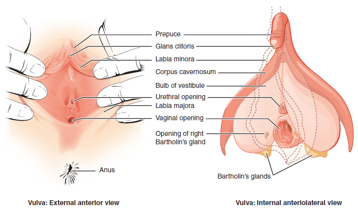
The external female reproductive structures are referred to collectively as the vulva and they include:
- The mons pubis is a pad of fat that is located at the anterior, over the pubic bone. After puberty, it becomes covered in pubic hair.
- The labia majora (labia = “lips”; majora = “larger”) are folds of hair-covered skin that begin just posterior to the mons pubis.
- The labia minora (labia = “lips”; minora = “smaller”) is thinner and more pigmented and extends medially to the labia majora.
- Although they naturally vary in shape and size from woman to woman, the labia minora serve to protect the female urethra and the entrance to the female reproductive tract.
- The superior, anterior portions of the labia minora come together to encircle the clitoris (or glans clitoris), an organ that originates from the same cells as the glans penis and has abundant nerves that make it important in sexual sensation and orgasm. The hymen is a thin membrane that sometimes partially covers the entrance to the vagina.
- The vaginal opening is located between the opening of the urethra and the anus. It is flanked by outlets to the Bartholin’s glands.
Internal Female Reproductive Organs
Vagina
The vagina is a muscular canal (approximately 10 cm long) that is the entrance to the reproductive tract. It also serves as the exit from the uterus during menses and childbirth. The outer walls of the anterior and posterior vagina are columns with ridges. The superior fornix meets the uterine cervix. The cervix is the opening to the uterus.
The walls of the vagina are lined with:
- An outer, fibrous adventitia
- A middle layer of smooth muscle
- An inner mucous membrane with transverse folds called rugae
Together, the middle and inner layers allow the expansion of the vagina to accommodate intercourse and childbirth. The thin, perforated hymen can partially surround the opening to the vaginal orifice. The Bartholin’s glands and the lesser vestibular glands (located near the clitoris) secrete mucus, which keeps the vestibular area moist.
The vagina has a normal population of microorganisms that help to protect against infection. There is both pathogenic bacteria and yeast in the vagina. In a healthy woman, the most predominant type of vaginal bacteria is from the genus Lactobacillus, which secretes lactic acid. The lactic acid protects the vagina by maintaining an acidic pH (below 4.5).
Lactic acid, in combination with other vaginal secretions, makes the vagina a self-cleansing organ. However, douching can disrupt the normal balance of healthy microorganisms, and increase a woman’s risk for infections and irritation. It is recommend that women do not douche and that they allow the vagina to maintain its normal healthy population of protective microbial flora.
Ovaries
The ovaries are the female gonads. There are two, one at each entrance to the fallopian tube. They are each about 2 to 3 cm in length, about the size of an almond. The ovaries are located within the pelvic cavity. The ovary itself is attached to the uterus via the ovarian ligament. The ovarian stroma forms the bulk of the adult ovary. Oocytes develop within the outer layer of this stroma, each surrounded by supporting cells. This grouping of an oocyte and its supporting cells is called a follicle.
The Fallopian Tubes
The fallopian tubes are the conduit of the oocyte from the ovary to the uterus. Each of the two fallopian tubes is close to, but not directly connected to, the ovary.
- The isthmus is the narrow medial end of each uterine tube that is connected to the uterus.
- The wide distal infundibulum flares out with slender, finger-like projections called fimbriae.
- The middle region of the tube, called the ampulla, is where fertilization often occurs.
The fallopian tubes have three layers:
- An outer serosa
- A middle smooth muscle layer
- An inner mucosal layer
- In addition to its mucus-secreting cells, the inner mucosa contains ciliated cells that beat in the direction of the uterus, producing a current that will be critical to moving the oocyte.
Did You Know?
The Uterus and Cervix
The uterus is the muscular organ that nourishes and supports the growing embryo. Its average size is approximately 5 cm wide by 7 cm long and it has three sections.
- The portion of the uterus superior to the opening of the uterine tubes is called the fundus.
- The middle section of the uterus is called the body of uterus (or corpus).
- The cervix is the narrow inferior portion of the uterus that projects into the vagina.
- The cervix produces mucus secretions that become thin and stringy under the influence of high systemic plasma estrogen concentrations, and these secretions can facilitate sperm movement through the reproductive tract.
The wall of the uterus is made up of three layers:
- Perimetrium: the most superficial layer and serous membrane.
- Myometrium: a thick layer of smooth muscle responsible for uterine contractions.
- Endometrium: the innermost layer containing a connective tissue lining covered by epithelial tissue that lines the lumen. It provides the site of implantation for a fertilized egg and sheds during menstruation if no egg is fertilized.
Check Your Knowledge of the Female Reproductive System
Female Reproductive System Medical Abbreviations
Learn the abbreviations using the list below.
Female Reproductive System Medical Abbreviations
- BC (birth control)
- BSO (bilateral salpingo-oophorectomy)
- Cx (cervix)
- D&C (dilation and curettage)
- FCC (fibrocystic changes to the breast)
- GYN (gynecology)
- HPV (human papillomavirus)
- HRT (hormone replacement therapy)
- HSG (hysterosalpingogram)
- IUD (intrauterine device)
- LAVH (laparoscopically assisted vaginal hysterectomy)
- PCOS (polycystic ovarian syndrome)
- PID (pelvic inflammatory disease)
- PMS (premenstrual syndrome)
- SGH (sonohysterography)
- TAH (total abdominal hysterectomy)
- TLH (total laparoscopic hysterectomy)
- TSS (toxic shock syndrome)
- TVH (total vaginal hysterectomy)
- TVS (transvaginal sonography)
- UAE (uterine artery embolization)
Activity source: Female Reproductive System Common Abbreviations by Kimberlee Carter, licensed under CC BY 4.0.
Concept Check
- Write or draw out the components of the pathway that an oocyte takes from beginning to end.
- Why do you think the fallopian tubes are not connected to the ovaries?
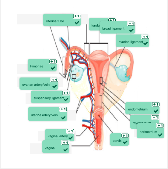
Anatomy Labeling Activity
Anatomy Labeling Activity (Text Version)
Label the diagram correctly with the following words:
- Broad Ligament
- Fundus
- Uterine Tube
- Uterine artery/vein
- Vaginal Artery
- Cervix
- Myometrium
- Ovarian Ligament
- Endometrium
- Perimetrium
- Ovarian artery/vein
- Fimbrae
- Suspensory Ligament
- Vagina
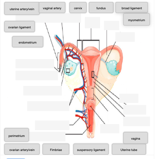
Anatomy Labeling Activity Diagram (Text Version)
Diagram of female reproductive system featuring anatomy organs and structures. The ______[Blank 1] also know as the fallopian tubes are positioned at the right top portion of the diagram. Extending from the uterine tube is finger-like projects known as ________[Blank 2]. The ______[Blank 3] is also known as the womb, is a hollow, muscular organ located in the pelvis between the bladder and rectum. The _______[Blank 4] supplies blood to the uterus. The _____[Blank 5] of uterus, also called the uterine fundus, refers to the dome-shaped, rounded superior part of the body of the uterus that lies above the opening. The _____[Blank 6] and the round ligaments of the uterus serve as secondary support for the uterus within the pelvis. The ________[Blank 7] is a fold of tissue arising from the peritoneum and extends out from the ovaries. The _______[Blank 8] connects the ovaries to the lateral surface of the uterus. The _______[Blank 9] supplies blood to the ovaries. The three layers of the uterus from outside to inside are the ______[Blank 10], ______[Blank 11], _______[Blank 12]. The neck of the uterus is called the cervix. The _______[Blank 13] is a muscular canal connecting the cervix of the uterus and serves as the birth canal during childbirth. The _______[Blank 14] supplies the vagina with blood.
Check your answers:[1]
Activity source: Female Reproductive System Anatomy by Kimberlee Carter, from Building a Medical Terminology Foundation, illustration from Anatomy and Physiology (OpenStax), licensed under CC BY 4.0./ Text version added.
Image Descriptions
Figure 7.2 image description: This figure shows the parts of the vulva. The right panel shows the external anterior view and the left panel shows the internal anteriolateral view. The major parts are labeled (from top): prepuce, glans clitoris, labia minora, corpus cavernosum, bulb of vestibule, urethral opening, labia majora, vaginal opening, opening of right Bartholin’s gland, Bartholin’s glands, anus. [Return to Figure 7.2].
Attribution
Except where otherwise noted, this chapter is adapted from “Female Reproductive System” in Building a Medical Terminology Foundation by Kimberlee Carter and Marie Rutherford, licensed under CC BY 4.0. / A derivative of Betts et al., which can be accessed for free from Anatomy and Physiology (OpenStax). Adaptations: dividing Female Reproductive System chapter content into sub-chapters.
-
↵
Anatomy Labeling Activity Diagram (Text Version)
Diagram of female reproductive system featuring anatomy organs and structures. The uterine tube also know as the fallopian tubes are positioned at the right top portion of the diagram. Extending from the uterine tube is finger-like projects known as fimbriae. The uterus is also known as the womb, is a hollow, muscular organ located in the pelvis between the bladder and rectum. The uterine artery/vein supplies blood to the uterus. The fundus of uterus, also called the uterine fundus, refers to the dome-shaped, rounded superior part of the body of the uterus that lies above the opening. The broad ligament and the round ligaments of the uterus serve as secondary support for the uterus within the pelvis. The suspensory ligament is a fold of tissue arising from the peritoneum and extends out from the ovaries. The ovarian ligament connects the ovaries to the lateral surface of the uterus. The ovarian artery/vein supplies blood to the ovaries. The three layers of the uterus from outside to inside are the endometrium, myometrium, perimetrium. The neck of the uterus is called the cervix. The vagina is a muscular canal connecting the cervix of the uterus and serves as the birth canal during childbirth. The vaginal artery supplies the vagina with blood.
superior portion of the vagina
Also known as greater vestibular glands they are responsible to secrete mucus to keep the vestibular area moist
washing the vagina with fluid
female gamete
pertaining to above
pertaining to below
