3.4 – Accessory Structures
Accessory structures of the skin include hair, nails, sweat glands, and sebaceous glands. These structures embryologically originate from the epidermis and can extend down through the dermis into the hypodermis.
Hair
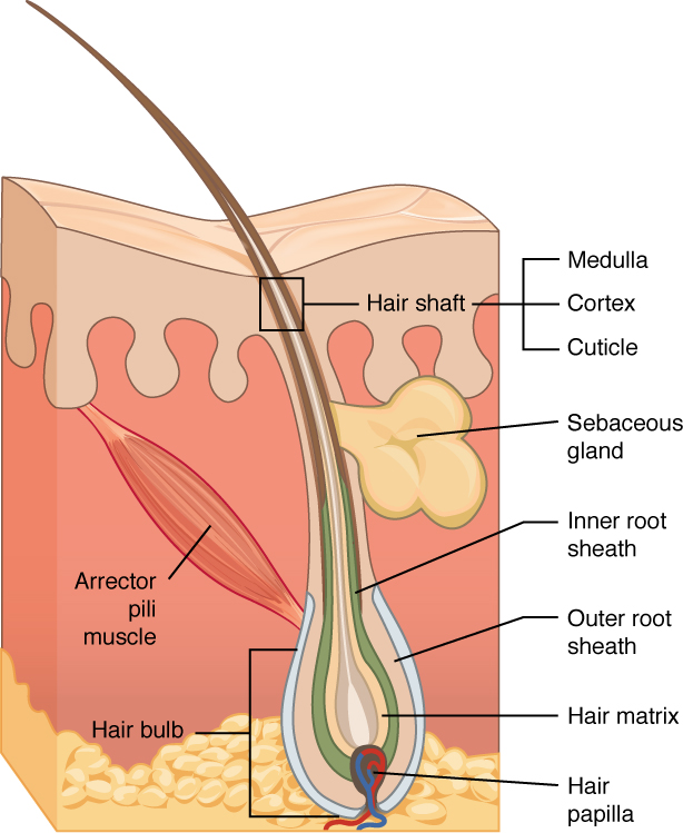
Hair is a keratinous filament growing out of the epidermis. It is primarily made of dead, keratinized cells. Strands of hair originate in an epidermal penetration of the dermis called the hair follicle. The hair shaft is the part of the hair not anchored to the follicle, and much of this is exposed at the skin’s surface. The rest of the hair, which is anchored in the follicle, lies below the surface of the skin and is referred to as the hair root. The hair root ends deep in the dermis at the hair bulb, and includes a layer of mitotically active basal cells called the hair matrix. The hair bulb surrounds the hair papilla, which is made of connective tissue and contains blood capillaries and nerve endings from the dermis (see Figure 3.6).
Hair Function
Hair serves a variety of functions, including protection, sensory input, thermoregulation, and communication. For example:
- Hair on the head protects the skull from the sun.
- Hair in the nose and ears, and around the eyes (eyelashes) defends the body by trapping and excluding dust particles that may contain allergens and microbes.
- Hair of the eyebrows prevents sweat and other particles from dripping into and bothering the eyes.
Hair also has a sensory function due to sensory innervation by a hair root plexus surrounding the base of each hair follicle. Hair is extremely sensitive to air movement or other disturbances in the environment, much more so than the skin surface. This feature is also useful for the detection of the presence of insects or other potentially damaging substances on the skin surface.
Each hair root is connected to a smooth muscle called the arrector pili that contracts in response to nerve signals from the sympathetic nervous system, making the external hair shaft “stand up.” The primary purpose for this is to trap a layer of air to add insulation. This is visible in humans as goose bumps and even more obvious in animals, such as when a frightened cat raises its fur. Of course, this is much more obvious in organisms with a heavier coat than most humans, such as dogs and cats.
Did You Know?
Hair Growth, Loss and Colour
Hair grows and is eventually shed and replaced by new hair. Hair typically grows at the rate of 0.3 mm per day. On average, 50 hairs are lost and replaced per day. Hair loss occurs if there is more hair shed than what is replaced and can happen due to hormonal or dietary changes. Hair loss can also result from the aging process, or the influence of hormones. Similar to the skin, hair gets its colour from the pigment melanin, produced by melanocytes in the hair papilla. Different hair color results from differences in the type of melanin. As a person ages, the melanin production decreases, and hair tends to lose its color and becomes gray and/or white.
Nails
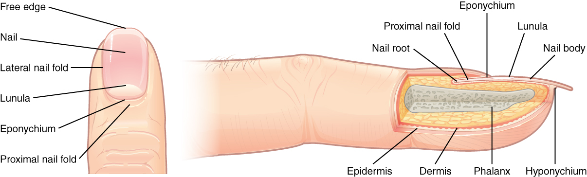
The nail bed is a specialized structure of the epidermis that is found at the tips of our fingers and toes. The nail body is formed on the nail bed, and protects the tips of our fingers and toes as they are the farthest extremities and the parts of the body that experience the maximum mechanical stress (see Figure 3.7). The nail body forms a back-support for picking up small objects with the fingers. The nail body is composed of densely packed dead keratinocytes.
The epidermis in this part of the body has evolved a specialized structure upon which nails can form. The nail body forms at the nail root, which has a matrix of proliferating cells from the stratum basale that enables the nail to grow continuously. The lateral nail fold overlaps the nail on the sides, helping to anchor the nail body. The nail fold that meets the proximal end of the nail body forms the nail cuticle, also called the eponychium.
The nail bed is rich in blood vessels, making it appear pink, except at the base, where a thick layer of epithelium over the nail matrix forms a crescent-shaped region called the lunula (the “little moon”). The area beneath the free edge of the nail, furthest from the cuticle, is called the hyponychium. It consists of a thickened layer of stratum corneum.
Sweat Glands
Sudoriferous Glands
When the body becomes warm, sudoriferous glands produce sweat to cool the body. Sweat glands develop from epidermal projections into the dermis and are classified as merocrine glands; that is, the secretions are excreted by exocytosis through a duct without affecting the cells of the gland. There are two types of sweat glands, each secreting slightly different products.
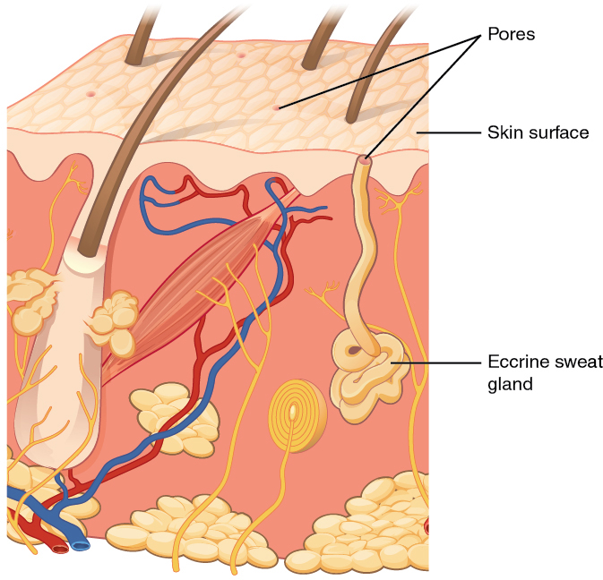
An eccrine sweat gland is a type of gland that produces a hypotonic sweat for thermoregulation as described previously. These glands are found all over the skin’s surface, but are especially abundant on the palms of the hand, the soles of the feet, and the forehead (Figure 3.8). They are coiled glands lying deep in the dermis, with the duct rising up to a pore on the skin surface, where the sweat is released. This type of sweat, released by exocytosis, is hypotonic and composed mostly of water, with some salt, antibodies, traces of metabolic waste, and dermicidin, an antimicrobial peptide. Eccrine glands are a primary component of thermoregulation in humans and thus help to maintain homeostasis.
An apocrine sweat gland is usually associated with hair follicles in densely hairy areas, such as armpits and genital regions. Apocrine sweat glands are larger than eccrine sweat glands and lie deeper in the dermis, sometimes even reaching the hypodermis, with the duct normally emptying into the hair follicle. In addition to water and salts, apocrine sweat includes organic compounds that make the sweat thicker and subject to bacterial decomposition and subsequent smell. The release of this sweat is under both nervous and hormonal control, and plays a role in the poorly understood human pheromone response. Most commercial antiperspirants use an aluminum-based compound as their primary active ingredient to stop sweat. When the antiperspirant enters the sweat gland duct, the aluminum-based compounds precipitate due to a change in pH and form a physical block in the duct, which prevents sweat from coming out of the pore.
Did You Know?
Aluminum-based compounds due to a change in pH form a physical block in the sweat gland duct. This prevents sweating.
Sebaceous Glands
A sebaceous gland is a type of oil gland that is found all over the body and helps to lubricate and waterproof the skin and hair. Most sebaceous glands are associated with hair follicles. They generate and excrete sebum, a mixture of lipids, onto the skin surface, thereby naturally lubricating the dry and dead layer of keratinized cells of the stratum corneum, keeping it pliable. The fatty acids of sebum also have antibacterial properties, and prevent water loss from the skin in low-humidity environments. The secretion of sebum is stimulated by hormones, many of which do not become active until puberty, thus sebaceous glands are relatively inactive during childhood.
The accessory structures also have lowered activity, generating thinner hair and nails, and reduced amounts of sebum and sweat. A reduced sweating ability can cause some elderly to be intolerant to extreme heat. Other cells in the skin, such as melanocytes and dendritic cells, also become less active, leading to a paler skin tone and lowered immunity. Wrinkling of the skin occurs due to breakdown of its structure, which results from decreased collagen and elastin production in the dermis, weakening of muscles lying under the skin, and the inability of the skin to retain adequate moisture.
Words Not Easily Broken into Word Parts
Integumentary System Terms Not Easily Broken into Word Parts
Integumentary Medical Terms (Text version)
- abscess
- AB-ses (Original Term)
- localized collection of pus
- abrasion
- ă-BRĀ-zhŏn (Original Term)
- scrape (by injury or mechanical process)
- acne
- AK-nē
- clogging of pores, which can lead to infection and inflammation
- adipocytes
- AD-ĭ-pō-sīts
- Fat cells
- adipose
- AD-ĭ-pōs
- Fat tissue
- albinism
- AL-bĭ-nizm
- genetic disorder that affects the coloring of skin, hair, and eyes.
- apocrine sweat gland
- AP-ŏ-krĕn swet gland
- A type of gland that is found in the skin, breast, eyelid, and ear
- autonomic
- ot-ŏ-NOM-ik
- unconsciously regulates
- bacteria, bacterium
- bak-TĒR-ē, bak-TĒR-ē-ŭm (Original Term)
- single-celled microorganisms that reproduce by cell division and may cause infection by invading body tissue
- basal cell carcinoma (BCC)
- BĀ-săl sel kar-sĭn-Ō-ma
- form of cancer that affects the mitotically active stem cells in the stratum basale of the epidermis
- benign
- bē-NĪN
- Noncancerous, harmless
- cancer
- KAN-sĕr
- A process where abnormal cells in the body divide uncontrollably
- cauterize, cauterization
- KAW-tĕr-īz (Original Term)
- to burn tissues by various means with the intent destroy damaged tissues, prevent infections or coagulate blood vessels
- cellulitis
- sel-yŭ-LĪT-ĭs (Original Term)
- bacterial infection of the skin and subcutaneous tissue, characterized by redness, pain, heat and swelling
- contusion
- kŏn-TOO-zhŏn (Original Term)
- bruise
- cyanosis
- sī-ă-NŌ-sĭs
- Abnormal condition of blue (bluish colour, lips and nail beds). Typically caused by low oxygenation
- cyst
- sist (Original Term)
- closed sac containing fluid or semisolid material
- debride, debridement
- di-BRĒD, di-BRĒD-mĕnt (Original Term)
- remove damaged tissues and cell debris from a wound or burn to prevent infection and promote healing.
- dehydration
- dē-hī-DRĀ-shŏn
- Loss of fluids/water is greater than what is taken in.
- dendritic cells
- den-DRIT-ik
- pertaining to dendrites
- dermabrasion
- DĔRM-ă-brā-zhŏn (Original Term)
- procedure to remove superficial scars using sandpaper or revolving wire brushes.
- diaphoresis
- dī-ă-fŏ-RĒ-sĭs (Original Term)
- condition of profuse, excessive sweating
- eccrine sweat gland
- ĔK-rĭn swet gland
- type of gland that produces a hypotonic sweat for thermoregulation
- eczema
- eg-ZĒ-mă (Original Term)
- noninfectious, inflammatory disease presents as redness, blisters, scabs and itching
- edema
- ĕ-DĒ-mă (Original Term)
- puffy swollen tissue due to accumulation of fluid
- excise, excision
- ĕk-SĪZ, ek-SIZH-ŏn (Original Term)
- surgical removal by cutting out
- fascia
- FASH-ē-ă
- Fibrous tissue
- frostbite
- FROST-bīt
- Conservation of core body heat results in the skin actually freezing
- gangrene
- GANG-grēn (Original Term)
- death of tissue due to blood supply loss
- incise, incision
- in-SĪZ, in-SIZH-ŏn (Original Term)
- surgical cut into or wound produced by a sharp instrument
- incision and drainage (I&D)
- in-SIZH-ŏn & DRĀN-ăj
- surgical cut made to allow the free flow of fluids from a lesion, wound, or cavity
- infection
- in-FEK-shŏn (Original Term)
- invasion of pathogens to body tissue
- jaundice, jaundiced
- JON-dĭs, JON-dĭsd (Original Term)
- yellow colouring of the mucous membranes and sclera
- keloid
- (KĒ-loyd)
- Formation of a raised or hypertrophic scar
- keratin
- (KER-ăt-ĭn)
- intracellular fibrous protein that gives hair, nails, and skin their hardness and water-resistant properties
- keratinocyte
- kĕ-RĂT-ĭ-nō-sīt
- Cell that manufactures and stores the protein keratin
- laceration
- las-ĕ-RĀ-shŏn (Original Term)
- torn, ragged-edged wound
- laser surgery
- LĀ-zĕr SŬRJ-ĕ-rē
- A surgical procedure using a powerful beam of light to cut or burn tissue.
- Lesion
- lĒ-zhŏn (Original Term)
- visible change in tissue resulting from injury or disease
- leukoplakia
- loo-kō-PLĀ-kē-ă
- white, thickened patches on mucus membrane tissue of the tongue or cheek
- macule
- MAK-ūl (Original Term)
- flat, coloured spot on the skin
- Meissner corpuscle
- MĪS-nĕr KOR-pŭs-ĕl
- Tactile corpuscle that responds to light and touch, touch receptor
- melanoma
- mel-ă-NŌ-mă
- cancer characterized by uncontrolled growth of melanocytes
- metastasize
- mĕ-TĂS-tă-sīz
- Production of cells that can mobilize and establish tumors in other organs of the body
- nevus
- NĒ-vŭs (Original Term)
- a pigmented skin blemish
- nodule
- NOJ-ool (Original Term)
- a small node-like structure
- Pacinian corpuscle
- pă-SIN-ē-ăn KOR-pŭs-ĕl
- Lamellated corpuscle that responds to vibration
- pallor
- PĂL-or (Original Term)
- paleness
- pathogens
- path-Ŏ-jĕns
- Disease-causing agents
- phagocytes
- făg-ō-SĬTS
- Cells that engulf and absorb bacteria and cell particles
- pruritus
- proo-RĪT-ŭs (Original Term)
- itching
- psoriasis
- sŏ-RĪ-ă-sĭs
- chronic autoimmune disorder that results in patches of thick red skin with the appearance of silvery scales
- pustule
- PŬS-tūl (Original Term)
- small elevation of the skin containing fluid
- reticulated
- rĕ-TIK-yŭ-lāt-ĕd
- constructed, arranged, or marked like a net or network.
- rickets
- RIK-ĕts
- A painful condition in children where bones are misshapen due to a lack of calcium, causing bow leggedness
- scar
- skăr
- Collagen-rich skin formed after the process of wound healing that differs from normal skin. Also known as a cicatrix.
- sebaceous gland
- sē-BĀ-shŭs gland
- type of oil gland that is found all over the body and helps to lubricate and waterproof the skin and hair.
- squamous cell carcinoma (SCC)
- SKWĀ-mŭs sel kar-sĭn-Ō-mă
- cancer that affects the keratinocytes of the stratum spinosum and presents as lesions commonly found on the scalp, ears, and hands
- stratum basale
- STRĀ-tŭm BĀS-al
- Deepest layer of the epidermal
- suture
- SOO-chŭr
- to stitch the edges of a wound
- sympathetic
- sĭm-pă-THĔT-ĭk
- Flight or fight response
- Sympathetic Nervous System
- sĭm-pă-THĔT-ĭk NĔR-vŭs SIS-tĕm
- Responsible for fight or flight responses
- tinea
- TIN-ē-ă (Original Term)
- A group of fungal skin diseases, charachterized by itching, scaling, and sometimes painful lesions.
- vascularized
- VAS-kyŭ-lă-rīzd
- Has numerous blood vessels
- verruca
- vĕr-ROO-kă
- Also known as a wart. An epidermal growth caused by a virus.
- virus
- VĪ-rŭs (Original Term)
- minute microorganism that may cuase infection by invading body tissue
Activity Source: Integumentary Medical Terms from Medical Terminology by Grimm et al., licensed under CC BY 4.0. /Re-recording of some H5P audio by Tania Deane and David McCuaig and text version added.
Common Integumentary System Abbreviations
Many terms and phrases related to the integumentary system are abbreviated. Learn these common abbreviations by expanding the list below.
Integumentary System Common Abbreviations
- BCC (basal cell carcinoma)
- SCC (squamous cell carcinoma)
- SLE (systemic lupus erythematosus)
- staph (staphylococcus)
- strep (streptococcus)
- subcut (subcutaneous)
- ID (intradermal)
- TD (transdermal)
- derm (dermatology)
- bx (biopsy)
- MRSA (methicillin-resistant Staphylococcus aureus)
Activity source: Integumentary System Common Abbreviations by Jesslyn Wilkinson, licensed under CC BY 4.0./ Converted to text.
Image Description
Figure 3.6 image description: A cross section of the skin containing a hair follicle. The follicle is teardrop shaped. Its enlarged base, labeled the hair bulb, is embedded in the hypodermis. The outermost layer of the follicle is the epidermis, which invaginates from the skin surface to envelope the follicle. Within the epidermis is the outer root sheath, which is only present on the hair bulb. It does not extend up the shaft of the hair. Within the outer root sheath is the inner root sheath. The inner root sheath extends about half of the way up the hair shaft, ending midway through the dermis. The hair matrix is the innermost layer. The hair matrix surrounds the bottom of the hair shaft where it is embedded within the hair bulb. The hair shaft, in itself, contains three layers: the outermost cuticle, a middle layer called the cortex, and an innermost layer called the medulla. [Return to Figure 3.6].
Figure 3.7 image description: The anatomy of the fingernail region. The top image shows a dorsal view of a finger. The proximal nail fold is the part underneath where the skin of the finger connects with the edge of the nail. The eponychium is a thin, pink layer between the white proximal edge of the nail (the lunula), and the edge of the finger skin. The lunula appears as a crescent-shaped white area at the proximal edge of the pink-shaded nail. The lateral nail folds are where the sides of the nail contact the finger skin. The distal edge of the nail is white and is called the free edge. An arrow indicates that the nail grows distally out from the proximal nail fold. The lower image shows a lateral view of the nail bed anatomy. In this view, one can see how the edge of the nail is located just proximal to the nail fold. This end of the nail, from which the nail grows, is called the nail root. [Return to Figure 3.7].
Figure 3.8 image description: An illustration of an eccrine sweat gland embedded in a cross section of skin tissue. The eccrine sweat gland is a bundle of white tubes embedded in the dermis. A single white tube travels up from the bundle and opens on to the surface of the epidermis. The opening is called a pore. There are several pores on the small block of skin portrayed in this diagram. [Return to Figure 3.8].
Attribution
Except where otherwise noted, this chapter is adapted from “Integumentary System” in Building a Medical Terminology Foundation by Kimberlee Carter and Marie Rutherford licensed under CC BY 4.0. / A derivative of Betts et al., which can be accessed for free from Anatomy and Physiology (OpenStax). Adaptations: dividing Integumentary System chapter content into sub-chapters.
Literally means below the dermis. The layer of the skin below the dermis that is composed mainly of loose connective and fatty tissues
outer layer of skin, made of closely packed epithelial cells
Specialized cells that produce melanin which is a dark pigment responsible for colouration of skin and hair.
cells that manufacture and store the protein keratin
Molecules are transported out of cells. A form of active transport
biological process that results in stable equilibrium
Pertaining to dendrites
