2.3 – Body Cavities and Serous Membranes
Body Planes
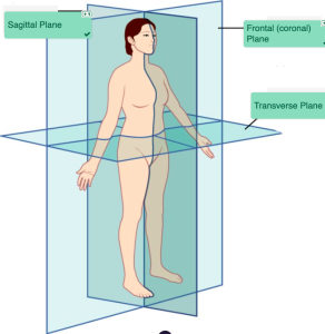
A section is a two-dimensional surface of a three-dimensional structure that has been cut. Modern medical imaging devices enable clinicians to obtain “virtual sections” of living bodies. We call these scans. Body sections and scans can be correctly interpreted, however, only if the viewer understands the plane along which the section was made. A plane is an imaginary two-dimensional surface that passes through the body. There are three planes commonly referred to in anatomy and medicine:
- The sagittal plane is the plane that divides the body or an organ vertically into right and left sides. If this vertical plane runs directly down the middle of the body, it is called the midsagittal or median plane. If it divides the body into unequal right and left sides, it is called a parasagittal plane or, less commonly, a longitudinal section.
- The frontal plane is the plane that divides the body or an organ into an anterior (front) portion and a posterior (rear) portion. The frontal plane is often referred to as a coronal plane (“corona” is Latin for “crown”).
- The transverse plane is the plane that divides the body or organ horizontally into upper and lower portions. Transverse planes produce images referred to as cross sections.
Can You Locate the Planes?
Body Planes (Text Version)
Label the diagram with correct words listed below:
- Sagittal Plane
- Frontal (coronal) Plane
- Transverse Plane
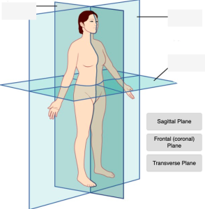
Body Planes Diagram (Text Version)
This illustration activity shows the human body standing upright in the anatomical position. Three anatomical planes are illustrated with transparent lines identifying the location of the three planes: the _______[Blank 1] which divides the left and right side of the body, the _______[Blank 2] dividing front and back portions of the body, and the _______[Blank 3] dividing top and bottom portion of the body.
Check your answers: [1]
Activity source: Medical language with the Context of Anatomy and Physiology Body by Tiffany Hunt, illustration from Anatomy and Physiology (OpenStax) licensed under CC BY 4.0./ Text version added.
Body Cavities and Serous Membranes
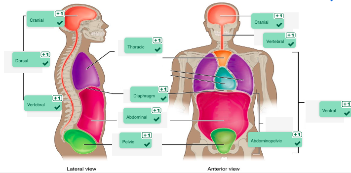
The body maintains its internal organization by means of membranes, sheaths, and other structures that separate compartments. The dorsal (posterior) cavity and the ventral (anterior) cavity are the largest body compartments (Figure 2.6). These cavities contain and protect delicate internal organs, and the ventral cavity allows for significant changes in the size and shape of the organs as they perform their functions. The lungs, heart, stomach, and intestines, for example, can expand and contract without distorting other tissues or disrupting the activity of nearby organs.
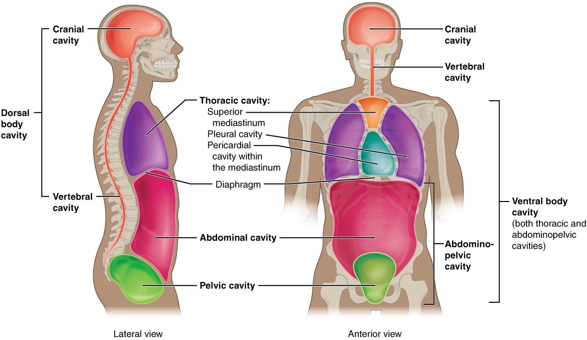
Subdivisions of the Posterior (Dorsal) and Anterior (Ventral) Cavities
The posterior (dorsal) and anterior (ventral) cavities are each subdivided into smaller cavities.
The posterior (dorsal) cavity has two main subdivisions:
- In the posterior (dorsal) cavity, the cranial cavity houses the brain.
- Protected by the bones of the skulls and cerebrospinal fluid.
- The spinal cavity (or vertebral cavity) encloses the spinal cord.
- Protected by the vertebral column and cerebrospinal fluid.
The anterior (ventral) cavity has two main subdivisions:
- The thoracic cavity is the more superior subdivision of the anterior cavity, and it is enclosed by the rib cage.
- The thoracic cavity contains the lungs and the heart, which is located in the mediastinum.
- The diaphragm forms the floor of the thoracic cavity and separates it from the more inferior abdominopelvic cavity.
- The abdominopelvic cavity is the largest cavity in the body.
- No membrane physically divides the abdominopelvic cavity.
- The abdominal cavity houses the digestive organs, the pelvic cavity, and the reproductive organs.
Practice Locating Cavities
Body Cavities (Text Version)
Label the diagram with correct words listed below:
- Abdominopelvic
- Abdominal
- Thoracic
- Pelvic
- Cranial
- Ventral
- Vertebral
- Diaphragm
- Dorsal
- Cranial
- Vertebral
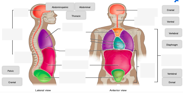
Body Cavities Diagram (Text Version)
This diagram activity shows two views of the human head and torso. The right image shows the lateral view, and the left image shows an anterior view of the human head and torso. The image is highlighting the several cavities or hollowed out spaces where organs and structures are positioned. The lateral view show four cavities: the head cavity known as the _______[Blank 1], the upper back cavity known as the _______[Blank 2], the lower back area known as the _______[Blank 3], and the chest cavity known as the ______[Blank 4] cavity. Adjoining lines for both views identify the ________[Blank 5] which is a muscular partition dividing the lungs from the location below it known as the _________[Blank 6] and followed by the lower _______[Blank 7]. The anterior view identifies the _______[Blank 8], _____[Blank 9], ________[Blank 10],________[Blank 11] cavities, and sections.
Check your answers: [2]
Activity source: Medical language with the Context of Anatomy and Physiology Dorsal by Tiffany Hunt, illustration from Anatomy and Physiology (OpenStax) licensed under CC BY 4.0./Text version added.
Abdominal Regions and Quadrants
To promote clear communication, for instance about the location of a patient’s abdominal pain or a suspicious mass, health care providers typically divide up the cavity into either nine regions or four quadrants.
Practice Locating the Quadrants
Locating the quadrants (Text Version)
Label the diagram with correct words listed below:
- Umbilical
- Epigastric
- Left Hypochondriac
- Right Lumbar
- Right Lower Quadrant
- Right Hypochondriac
- Right Iliac
- Left Lower Quadrant
- Left Lumbar
- Right Upper Quadrant
- Left Upper Quadrant
- Hypogastric
- Left Iliac
- Diaphragm
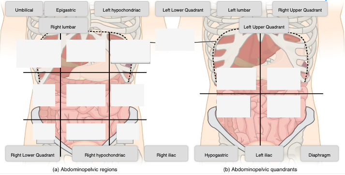
Locating the quadrants Diagram (Text version)
This diagram activity shows two views of the abdominopelvic region. Image A shows the abdominopelvic region divided in nine sections with nine boxes. Image B shows the abdominopelvic region divided in four sections known as quadrants with a line dividing the four sections for each quadrant. For image A at the top are three white boxes identified working from right to left labeled as _______[Blank 1], followed by ______[Blank 2], and ________[Blank 3] region. The middle three boxes working from right to left identified as _______[Blank 4], followed by the _______[Blank 5], and ________[Blank 6] region. The three lower boxes working from right to left is the_______[Blank 7], followed by ________[Blank 8], and _________[Blank 9] region. Image B shows two upper white boxes and are from right to left is the _________[Blank 10] followed by the _________[Blank 11]. The two lower boxes from right to left is the _________[Blank 12] followed by _______[Blank 13].
Check your answers: [3]
Activity source: Medical language with the Context of Anatomy and Physiology Dorsal by Tiffany Hunt, illustration from Anatomy and Physiology (OpenStax) licensed under CC BY 4.0./Text version added.
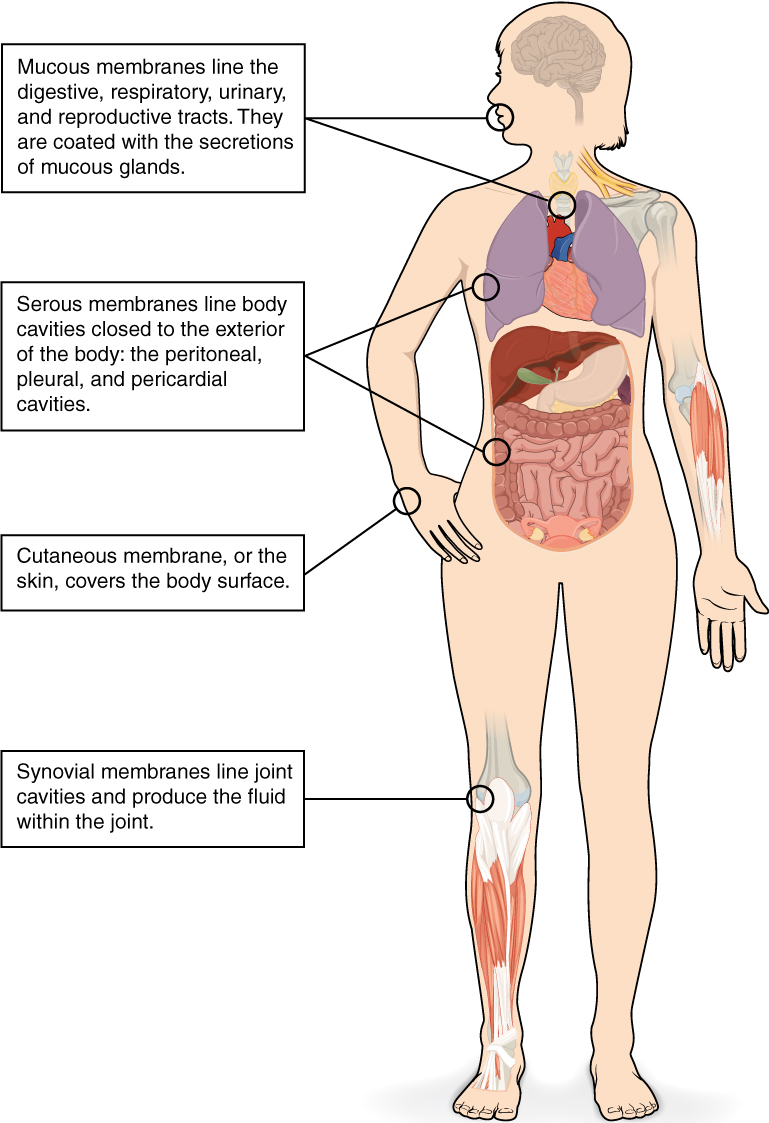
Tissue Membranes
A tissue membrane is a thin layer or sheet of cells that covers the outside of the body (for example, skin), the organs (for example, pericardium), internal passageways that lead to the exterior of the body (for example, abdominal mesenteries), and the lining of the movable joint cavities. There are two basic types of tissue membranes: connective tissue and epithelial membranes (Figure 2.7).
Did You Know?
Synovial membranes line cavities which hold synovial fluid.
Synovial fluid lubricates the joints for movement.
Connective Tissue Membranes
- The connective tissue membrane is formed solely from connective tissue.
- These membranes encapsulate organs, such as the kidneys, and line our movable joints.
- A synovial membrane is a type of connective tissue membrane that lines the cavity of a freely movable joint.
- For example, synovial membranes surround the joints of the shoulder, elbow, and knee.
Epithelial Membranes
- The epithelial membrane is composed of epithelium attached to a layer of connective tissue.
- For example, your skin.
- The mucous membrane is also a composite of connective and epithelial tissues.
- Sometimes called mucosae, these epithelial membranes line the body cavities and hollow passageways that open to the external environment, and include the digestive, respiratory, excretory, and reproductive tracts.
- Mucus, produced by the epithelial exocrine glands, covers the epithelial layer.
- The underlying connective tissue, called the lamina propria (literally “own layer”), help support the fragile epithelial layer.
- The skin is an epithelial membrane also called the cutaneous membrane.
- It is a stratified squamous epithelial membrane resting on top of connective tissue. The apical surface of this membrane is exposed to the external environment and is covered with dead, keratinized cells that help protect the body from desiccation and pathogens.
Membranes of the Anterior (Ventral) Body Cavity
- A serous membrane (also referred to as serosa) is an epithelial membrane composed of mesodermally derived epithelium called the mesothelium that is supported by connective tissue. These membranes line the coelomic cavities of the body and they cover the organs located within those cavities. They are essentially membranous bags, with mesothelium lining the inside and connective tissue on the outside.
- Parietal layers: line the walls of the body cavity.
- Visceral layer: covers the organs (the viscera).
- Between the parietal and visceral layers is a very thin, fluid-filled serous space.
There are three serous cavities and their associated membranes. Serous membranes provide additional protection to the viscera they enclose by reducing friction that could lead to inflammation of the organs.
- Pleura: surrounds the lungs in the pleural cavity and reduces friction between the lungs and the body wall.
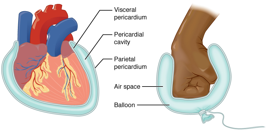
- Pericardium: surrounds the heart in the pericardial cavity and reduces friction between the heart and the wall of the pericardium.
- Peritoneum: surrounds several organs in the abdominopelvic cavity. The peritoneal cavity reduces friction between the abdominal and pelvic organs and the body wall.
Image Descriptions
Figure 2.6 image description: This illustration shows a lateral and anterior view of the body and highlights the body cavities with different colors. The cranial cavity is a large, bean-shaped cavity filling most of the upper skull where the brain is located. The vertebral cavity is a very narrow, thread-like cavity running from the cranial cavity down the entire length of the spinal cord. Together the cranial cavity and vertebral cavity can be referred to as the dorsal body cavity. The thoracic cavity consists of three cavities that fill the interior area of the chest. The two pleural cavities are situated on both sides of the body, anterior to the spine and lateral to the breastbone. The superior mediastinum is a wedge-shaped cavity located between the superior regions of the two thoracic cavities. The pericardial cavity within the mediastinum is located at the center of the chest below the superior mediastinum. The pericardial cavity roughly outlines the shape of the heart. The diaphragm divides the thoracic and the abdominal cavities. The abdominal cavity occupies the entire lower half of the trunk, anterior to the spine. Just under the abdominal cavity, anterior to the buttocks, is the pelvic cavity. The pelvic cavity is funnel shaped and is located inferior and anterior to the abdominal cavity. Together the abdominal and pelvic cavity can be referred to as the abdominopelvic cavity while the thoracic, abdominal, and pelvic cavities together can be referred to as the ventral body cavity. [Return to Figure 2.6].
Figure 2.7 image description: This illustrations shows the silhouette of a human female from an anterior view. Several organs are showing in her neck, thorax, abdomen left arm and right leg. Text boxes point out and describe the mucous membranes in several different organs. The topmost box points to the mouth and trachea. It states that mucous membranes line the digestive, respiratory, urinary and reproductive tracts. They are coated with the secretions of mucous glands. The second box points to the outside edge of the lungs as well as the large intestine and states that serous membranes line body cavities that are closed to the exterior of the body, including the peritoneal, pleural and pericardial cavities. The third box points to the skin of the hand. It states that cutaneous membrane, also known as the skin, covers the body surface. The fourth box points to the right knee. It states that synovial membranes line joint cavities and produce the fluid within the joint.[Return to Figure 2.7]
Figure 2.8 image description: This diagram shows the pericardium on the left next to an analogy of a hand punching a balloon on the right. The pericardium is a two-layered sac that surrounds the entire heart except where the blood vessels emerge on the heart’s superior side. The pericardium has two layers because it folds over itself in the shape of the letter U. The inner layer that borders the heart is the visceral pericardium while the outer layer is the parietal pericardium. The space between the two layers is called the pericardial cavity. The heart sits in the cavity much like a fist punching into a balloon. The balloon surrounds the lower part of the fist with a two-layered sac, with the top of the balloon, where it contacts the fist, being analogous to the visceral pericardium. The bottom of the balloon, where it is tied off, is analogous to the parietal pericardium. The air within the balloon is analogous to the pericardial cavity. [Return to Figure 2.8].
Attribution
Except where otherwise noted, this chapter is adapted from “Medical Language Within the Context of Anatomy and Physiology” in Building a Medical Terminology Foundation by Kimberlee Carter and Marie Rutherford, licensed under CC BY 4.0. / A derivative of Betts et al., which can be accessed for free from Anatomy and Physiology (OpenStax). Adaptations: dividing Medical Language Within the Context of Anatomy and Physiology chapter content into sub-chapters.
-
↵
Check your answers: Body Planes Diagram
This illustration activity shows the human body standing upright in the anatomical position. Three anatomical planes are illustrated with transparent lines identifying the location of the three planes: the sagittal plane which divides the left and right side of the body, the frontal plane dividing front and back portions of the body, and the transverse plane dividing top and bottom portion of the body. -
↵
Check your answers: Body Cavities Diagram (Text Version)
This diagram activity shows two views of the human head and torso. The right image shows the lateral view, and the left image shows an anterior view of the human head and torso. The image is highlighting the several cavities or hollowed out spaces where organs and structures are positioned. The lateral view show four cavities: the head cavity known as the cranial, the upper back cavity known as the dorsal, the lower back area known as the vertebral, and the chest cavity known as the thoracic cavity. Adjoining lines for both views identify the diaphragm which is a muscular partition dividing the lungs from the location below it known as the abdominal cavity and followed by the lower pelvic cavity. The anterior view identifies the cranial, vertebral, ventral, abdominopelvic cavities, and sections. -
↵
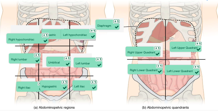
Check your answers: Locating the quadrants Diagrams (Text Version)
This diagram activity shows two views of the abdominopelvic region. Image A shows the abdominopelvic region divided in nine sections with nine boxes. Image B shows the abdominopelvic region divided in four sections known as quadrants with a line dividing the four sections for each quadrant. For image A at the top are three white boxes identified working from right to left labeled as right hypochondriac, followed by epigastric, and left hypochondriac region. The middle three boxes working from right to left identified as right lumbar, followed by the umbilical, and left lumbar region. The three lower boxes working from right to left is the right iliac, followed by hypogastric, and left iliac region. Image B show two upper white boxes and are from right to left is the right upper quadrant followed by the left upper quadrant. The two lower boxes from right to left is the right lower quadrant followed by left lower quadrant.
Produced by the brain is a colourless fluid that cushions the brain and spinal cord.
cavities that do not open to the outside
