17.2 – Anatomy (Structures) of the Endocrine System
The Endocrine Gland
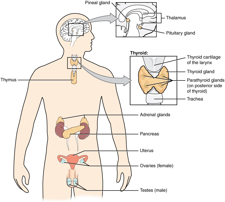
The endocrine system consists of cells, tissues, and organs that secrete hormones as a primary or secondary function. The endocrine gland is the major player in this system. The primary function of the endocrine gland is to secrete hormones directly into the surrounding fluid. The surrounding fluid (interstitial fluid) and the blood vessels then transport the hormones throughout the body. The endocrine system includes the pituitary, thyroid, parathyroid, adrenal, and pineal glands (see Figure 17.2). Some of these glands have both endocrine and non-endocrine functions. For example, the pancreas contains cells that function in digestion as well as cells that secrete the endocrine hormones like insulin and glucagon, which regulate blood glucose levels. The hypothalamus, thymus, heart, kidneys, stomach, small intestine, liver, skin, female ovaries, and male testes are other organs that contain cells with endocrine function. Moreover, fat (adipose) tissue has long been known to produce hormones, and recent research has revealed that even bone tissue has endocrine functions.
The ductless endocrine glands are not to be confused with the body’s exocrine system, whose glands release their secretions through ducts. Examples of exocrine glands include the sebaceous and sweat glands of the skin. As just noted, the pancreas also has an exocrine function: most of its cells secrete pancreatic juice through the pancreatic and accessory ducts to the lumen of the small intestine.
Did You Know?
The pancreas acts as a endocrine and exocrine gland.
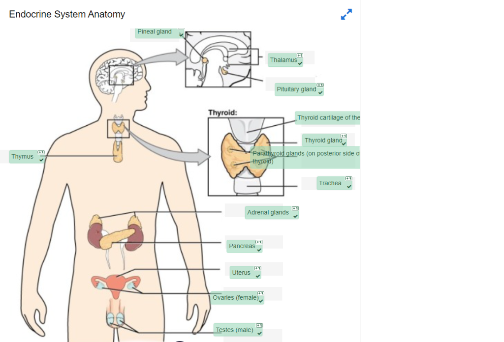
Endocrine System Anatomy labeling activity
Endocrine System Anatomy labeling activity (Text Version)
Label the diagram with words listed below:
- Trachea
- Testes (male)
- Pancreas
- Thyroid cartilage of the larynx
- Pineal gland
- Thyroid gland
- Thalamus
- Adrenal glands
- Uterus
- Ovaries (female)
- Parathyroid glands (on posterior side of thyroid)
- Thymus
- Pituitary gland
Endocrine System Diagram (Text Version)
This diagram shows the endocrine glands and cells that are located throughout the body. The endocrine system organs shown from top to bottom include the pea size structure known as the ______[Blank 1] as well as the primary glandular structure of the endocrine system found enclosed within the ______[Blank 2] known as the _______[Blank 3]. The pituitary is located on the anterior side of the thalamus while the pineal gland is located on the posterior side of the thalamus. The ______[Blank 4] is a shield shaped cartilage that forms part of the laryngeal skeleton. This is a butterfly-shaped gland that wraps around the _______[Blank 5] within the neck. Four small, disc-shaped ________[Blank 6] are embedded into the posterior side of the thyroid. The _______[Blank 7] are located on top of the kidneys. The ________[Blank 8] is located at the center of the abdomen. In females, the ______[Blank 9] connects the two ______[Blank 10] a by two long, curved, tubes in the pelvic region. In males, the two _______[Blank 11] are in the scrotum below the penis. One the left side of the diagram and located in the center of the chest is the _______[Blank 12], a glandular structure responsible for the secretion of a hormone called thymosin.
Check your answers [1]
Activity source: Endocrine System Anatomy by Gisele Tuzon, from Building a Medical Terminology Foundation by Kimberlee Carter and Marie Rutherford, licensed under CC BY- 4.0. /Text version added.
Image Descriptions
Figure 17.2 image description: This diagram shows the endocrine glands and cells that are located throughout the body. The endocrine system organs include the pineal gland and pituitary gland in the brain. The pituitary is located on the anterior side of the thalamus while the pineal gland is located on the posterior side of the thalamus. The thyroid gland is a butterfly-shaped gland that wraps around the trachea within the neck. Four small, disc-shaped parathyroid glands are embedded into the posterior side of the thyroid. The adrenal glands are located on top of the kidneys. The pancreas is located at the center of the abdomen. In females, the two ovaries are connected to the uterus by two long, curved, tubes in the pelvic region. In males, the two testes are located in the scrotum below the penis. [Return to Figure 17.2].
Attribution
Except where otherwise noted, this chapter is adapted from “Endocrine System” in Building a Medical Terminology Foundation by Kimberlee Carter and Marie Rutherford, licensed under CC BY 4.0. / A derivative of Betts et al., which can be accessed for free from Anatomy and Physiology (OpenStax). Adaptations: dividing Endocrine System chapter content into sub-chapters.
-
Check your answers: Endocrine System Diagram (Text Version)
This diagram shows the endocrine glands and cells that are located throughout the body. The endocrine system organs shown from top to bottom include the pea size structure known as the pineal gland as well as the primary glandular structure of the endocrine system found enclosed within the thalamus known as the pituitary gland. The pituitary is located on the anterior side of the thalamus while the pineal gland is located on the posterior side of the thalamus. The thyroid cartilage of the larynx is a shield shaped cartilage that forms part of the laryngeal skeleton. The thyroid gland is a butterfly-shaped gland that wraps around the trachea within the neck. Four small, disc-shaped parathyroid glands are embedded into the posterior side of the thyroid. The adrenal glands are located on top of the kidneys. The pancreas is located at the center of the abdomen. In females, the uterus connects the two ovaries a by two long, curved, tubes in the pelvic region. In males, the two testes are in the scrotum below the penis. One the left side of the diagram and located in the center of the chest is the thymus, a glandular structure responsible for the secretion of a hormone called thymosin. ↵
