5 Medical Language Within the Context of Anatomy and Physiology
Learning Objectives
- Connect medical language learning to the context of anatomy and physiology
- Introduce the basic architecture and levels of organization of the human body
- Evaluate the anatomical position, regional terms, directional terms, body planes, and body quadrants for anatomical positioning
- Describe body cavities and the functions of associated membranes
As you memorize the language components of medical terminology it is important to support that learning within the context of anatomy and physiology. Proceeding through the body system chapters you will learn word parts, whole medical terms, and common abbreviations. It is important to put into context where in the body the medical term is referencing, and then consider how it works within the body.
Anatomy focuses on structure and physiology focuses on function. Much of the study of physiology centers on the body’s tendency toward homeostasis .
Consider the structures of the body in terms of fundamental levels of organization that increase in complexity: subatomic particles, atoms, molecules, organelles, cells, tissues, organs, organ systems, organisms, and biosphere (Figure 5.1).
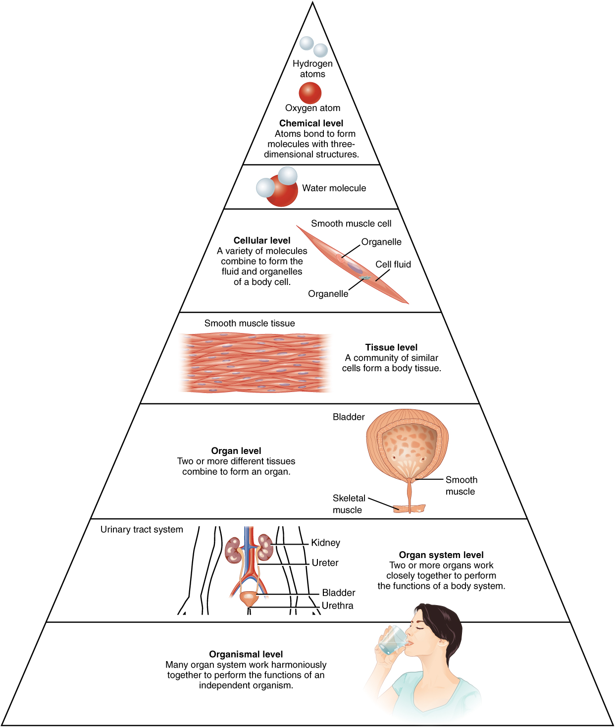
The Levels of Organization
All matter in the universe is composed of one or more unique pure substances called elements, familiar examples are hydrogen, oxygen, carbon, nitrogen, calcium, and iron.
- The smallest unit of any of these pure substances (elements) is an atom.
- Atoms are made up of subatomic particles such as the proton, electron, and neutron.
- Two or more atoms combine to form a molecule, such as the water molecules, proteins, and sugars found in living things.
- Molecules are the chemical building blocks of all body structures.
- A cell is the smallest independently functioning unit of a living organism.
- Even bacteria, which are extremely small, independently-living organisms, have a cellular structure. Each bacterium is a single cell. All living structures of human anatomy contain cells, and almost all functions of human physiology are performed in cells or are initiated by cells
- A human cell typically consists of flexible membranes that enclose cytoplasm, a water-based cellular fluid, together with a variety of tiny functioning units called organelles. In humans, as in all organisms, cells perform all functions of life.
- A tissue is a group of many similar cells (though sometimes composed of a few related types) that work together to perform a specific function.
- An organ is an anatomically distinct structure of the body composed of two or more tissue types. Each organ performs one or more specific physiological functions.
An organ system is a group of organs that work together to perform major functions or meet the physiological needs of the body.
Did you know?
- Organs are very collaborative and work with multiple body systems.
- For example, the heart (cardiovascular system) and lungs (respiratory system) work together to deliver oxygen throughout the body and remove carbon dioxide from the body.
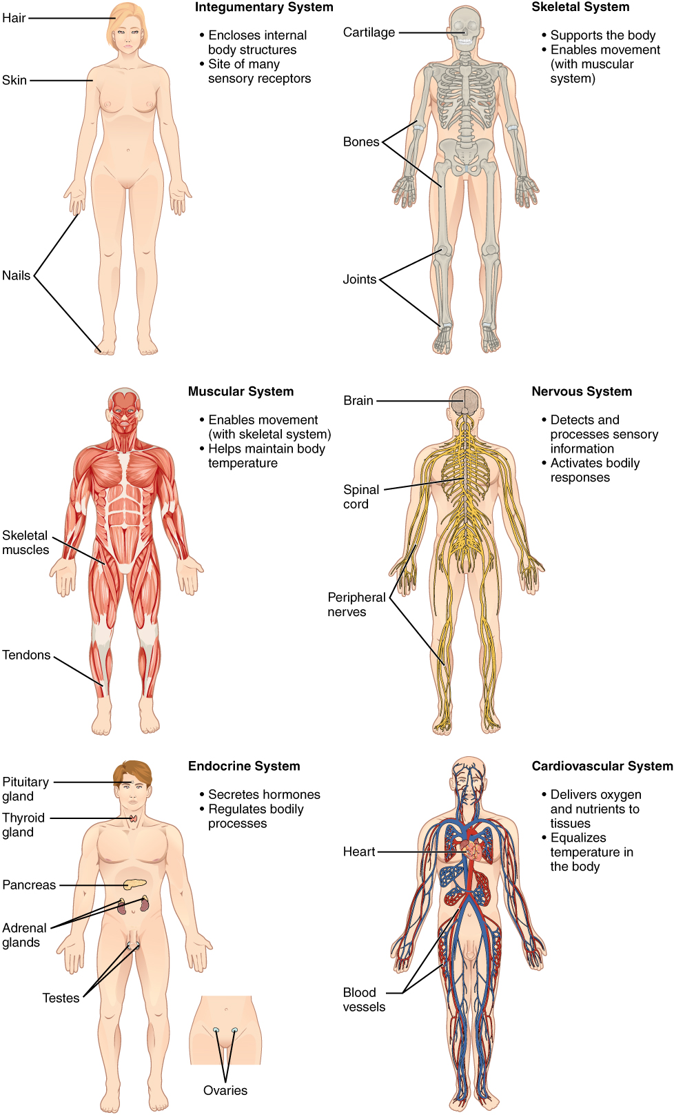
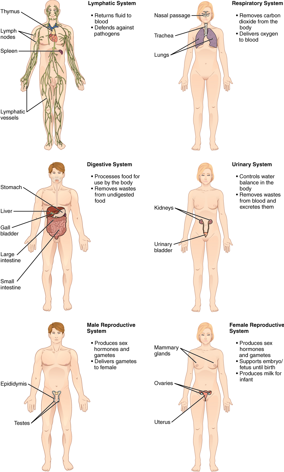
Consider the breakdown into eleven distinct organ systems in the human body (Figure 5.2 and Figure 5.3). Assigning organs to organ systems can be imprecise since organs that “belong” to one system can also have functions integral to another system. In fact, most organs contribute to more than one system.
The organism level is the highest level of organization. An organism is a living being that has a cellular structure and that can independently perform all physiologic functions necessary for life. In multicellular organisms, including humans, all cells, tissues, organs, and organ systems of the body work together to maintain the life and health of the organism.
Watch this video:
Media 5.1. Introduction to Anatomy & Physiology: Crash Course A&P #1 [Online video]. Copyright 2015 by CrashCourse.
Anatomical Position
Anatomists and health care providers use terminology for the purpose of precision and to reduce medical errors. For example, is a scar “above the wrist” located on the forearm two or three inches away from the hand? Or is it at the base of the hand? Is it on the palm-side or back-side? By using precise anatomical terminology, we eliminate ambiguity. Anatomical terms derive from ancient Greek and Latin words.
To further increase precision, anatomists standardize the way in which they view the body. Just as maps are normally oriented with north at the top, the standard body “map,” or anatomical position, is that of the body standing upright, with the feet at shoulder width and parallel, toes forward. The upper limbs are held out to each side, and the palms of the hands face forward as illustrated.
Using this standard position reduces confusion. It does not matter how the body being described is oriented, the terms are used as if it is in anatomical position. For example, a scar in the “anterior (front) carpal (wrist) region” would be present on the palm side of the wrist. The term “anterior” would be used even if the hand were palm down on a table.
A body that is lying down is described as either prone or supine. These terms are sometimes used in describing the position of the body during specific physical examinations or surgical procedures.
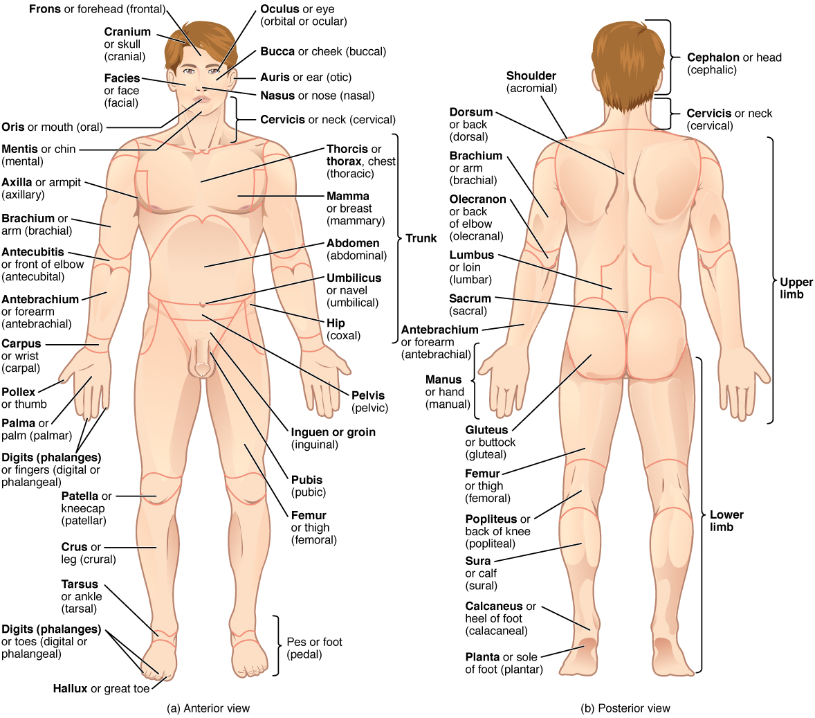
Regional Terms
The human body’s numerous regions have specific terms to help increase precision. Notice that the term “brachium” or “arm” is reserved for the “upper arm” and “antebrachium” or “forearm” is used rather than “lower arm.” Similarly, “femur” or “thigh” is correct, and “leg” or “crus” is reserved for the portion of the lower limb between the knee and the ankle. You will be able to describe the body’s regions using the terms from the anatomical position.
Directional Terms
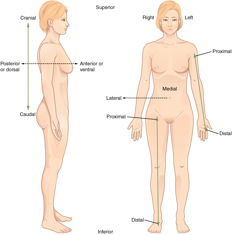
Directional terms are essential for describing the relative locations of different body structures. For instance, an anatomist might describe one band of tissue as “inferior to” another or a physician might describe a tumor as “superficial to” a deeper body structure. Commit these terms to memory to avoid confusion when you are studying or describing the locations of particular body parts.
- Anterior (or ventral) describes pertaining to the front or direction toward the front of the body. The toes are anterior to the foot.
- Posterior (or dorsal) describes pertaining to the back or direction toward the back of the body. The popliteus is posterior to the patella.
- Superior (or cranial) describes pertaining to a position above or higher than another part of the body proper. The orbits are superior to the oris.
- Inferior (or caudal) describes a position pertaining to below or lower than another part of the body proper; near or toward the tail (in humans, the coccyx, or lowest part of the spinal column). The pelvis is inferior to the abdomen.
- Lateral describes the side or direction pertaining to toward the side of the body. The thumb (pollex) is lateral to the digits.
- Medial describes the middle or direction pertaining to toward the middle of the body. The hallux is the medial toe.
- Proximal describes a position pertaining to a limb that is nearer to the point of attachment or the trunk of the body. The brachium is proximal to the antebrachium.
- Distal describes a position pertaining to a limb that is farther from the point of attachment or the trunk of the body. The crus is distal to the femur.
- Superficial describes a position pertaining to closer to the surface of the body. The skin is superficial to the bones.
- Deep describes a position farther from the surface of the body. The brain is deep to the skull.
Practice these directional terms.
Concept Check
- Find a partner and take turns choosing two body parts on your or your partner’s body.
- Using directional terms, describe the location of those body parts relative to one another.
Body Planes
A section is a two-dimensional surface of a three-dimensional structure that has been cut. Modern medical imaging devices enable clinicians to obtain “virtual sections” of living bodies. We call these scans. Body sections and scans can be correctly interpreted, however, only if the viewer understands the plane along which the section was made. A plane is an imaginary two-dimensional surface that passes through the body. There are three planes commonly referred to in anatomy and medicine:
- The sagittal plane is the plane that divides the body or an organ vertically into right and left sides. If this vertical plane runs directly down the middle of the body, it is called the midsagittal or median plane. If it divides the body into unequal right and left sides, it is called a parasagittal plane or less commonly a longitudinal section.
- The frontal plane is the plane that divides the body or an organ into an anterior (front) portion and a posterior (rear) portion. The frontal plane is often referred to as a coronal plane. (“Corona” is Latin for “crown.”)
- The transverse plane is the plane that divides the body or organ horizontally into upper and lower portions. Transverse planes produce images referred to as cross sections.
Can you locate the planes?
Body Cavities and Serous Membranes
The body maintains its internal organization by means of membranes, sheaths, and other structures that separate compartments. The dorsal (posterior) cavity and the ventral (anterior) cavity are the largest body compartments (Figure 5.6). These cavities contain and protect delicate internal organs, and the ventral cavity allows for significant changes in the size and shape of the organs as they perform their functions. The lungs, heart, stomach, and intestines, for example, can expand and contract without distorting other tissues or disrupting the activity of nearby organs.
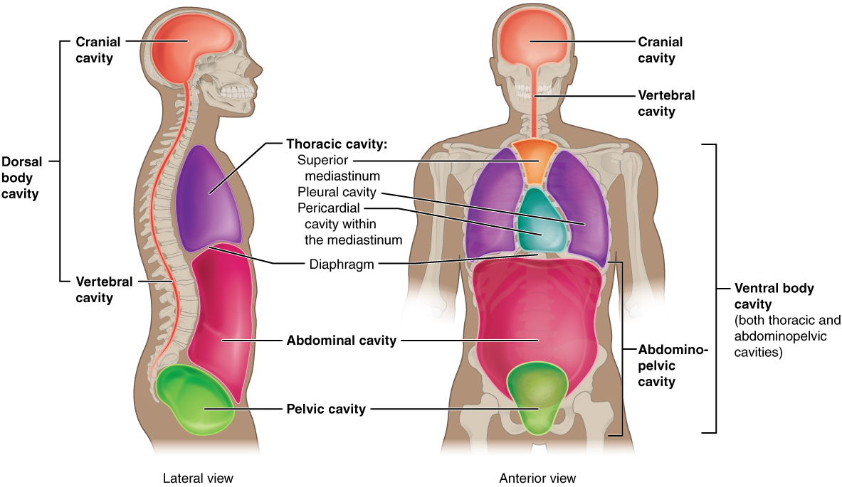
Subdivisions of the Posterior (Dorsal) and Anterior (Ventral) Cavities
The posterior (dorsal) and anterior (ventral) cavities are each subdivided into smaller cavities:
The posterior (dorsal) cavity has two main subdivisions:
- In the posterior (dorsal) cavity, the cranial cavity houses the brain
- Protected by the bones of the skulls and cerebrospinal fluid
- The spinal cavity (or vertebral cavity) encloses the spinal cord.
- Protected by the vertebral column and cerebrospinal fluid
The anterior (ventral) cavity has two main subdivisions:
- The thoracic cavity is the more superior subdivision of the anterior cavity, and it is enclosed by the rib cage.
- The thoracic cavity contains the lungs and the heart, which is located in the mediastinum.
- The diaphragm forms the floor of the thoracic cavity and separates it from the more inferior abdominopelvic cavity.
- The abdominopelvic cavity is the largest cavity in the body.
- No membrane physically divides the abdominopelvic cavity.
- The abdominal cavity houses the digestive organs, the pelvic cavity, and the reproductive organs.
Practice locating cavities.
Abdominal Regions and Quadrants
To promote clear communication, for instance about the location of a patient’s abdominal pain or a suspicious mass, health care providers typically divide up the cavity into either nine regions or four quadrants.
Practice locating the quadrants.
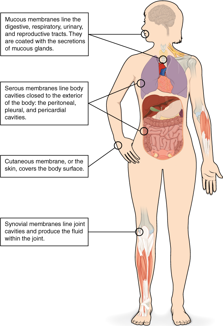
Tissue Membranes
A tissue membrane is a thin layer or sheet of cells that covers the outside of the body (for example, skin), the organs (for example, pericardium), internal passageways that lead to the exterior of the body (for example, abdominal mesenteries), and the lining of the movable joint cavities. There are two basic types of tissue membranes: connective tissue and epithelial membranes (Figure 5.7).
Connective Tissue Membranes
- The connective tissue membrane is formed solely from connective tissue.
- These membranes encapsulate organs, such as the kidneys, and line our movable joints.
- A synovial membrane is a type of connective tissue membrane that lines the cavity of a freely movable joint.
- For example, synovial membranes surround the joints of the shoulder, elbow, and knee.
Epithelial Membranes
- The epithelial membrane is composed of epithelium attached to a layer of connective tissue.
- For example, your skin.
- The mucous membrane is also a composite of connective and epithelial tissues.
- Sometimes called mucosae, these epithelial membranes line the body cavities and hollow passageways that open to the external environment, and include the digestive, respiratory, excretory, and reproductive tracts.
- Mucus, produced by the epithelial exocrine glands, covers the epithelial layer.
- The underlying connective tissue, called the lamina propria (literally “own layer”), help support the fragile epithelial layer.
- The skin is an epithelial membrane also called the cutaneous membrane.
- It is a stratified squamous epithelial membrane resting on top of connective tissue. The apical surface of this membrane is exposed to the external environment and is covered with dead, keratinized cells that help protect the body from desiccation and pathogens.
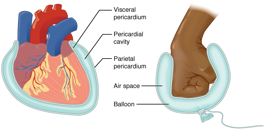
Membranes of the Anterior (Ventral) Body Cavity
- A serous membrane (also referred to as serosa) is an epithelial membrane composed of mesodermally derived epithelium called the mesothelium that is supported by connective tissue. These membranes line the coelomic cavities of the body and they cover the organs located within those cavities. They are essentially membranous bags, with mesothelium lining the inside and connective tissue on the outside.
- Parietal layers: line the walls of the body cavity.
- Visceral layer: covers the organs (the viscera).
- Between the parietal and visceral layers is a very thin, fluid-filled serous space.
There are three serous cavities and their associated membranes. Serous membranes provide additional protection to the viscera they enclose by reducing friction that could lead to inflammation of the organs.
- Pleura: surrounds the lungs in the pleural cavity and reduces friction between the lungs and the body wall.
- Pericardium: surrounds the heart in the pericardial cavity and reduces friction between the heart and the wall of the pericardium.
- Peritoneum: surrounds several organs in the abdominopelvic cavity. The peritoneal cavity reduces friction between the abdominal and pelvic organs and the body wall.
Test Yourself
References
[CrashCourse]. (2015, January 6). Introduction to anatomy & physiology: Crash course A&P #1 [Video]. YouTube. https://youtu.be/uBGl2BujkPQ
Image Descriptions
Figure 5.1 image description: This illustration shows biological organization as a pyramid. The chemical level is at the apex of the pyramid where atoms bond to form molecules with three dimensional structures. An example is shown with two white hydrogen atoms bonding to a red oxygen atom to create water. The next level down on the pyramid is the cellular level, as illustrated with a long, tapered, smooth muscle cell. At this level, a variety of molecules combine to form the interior fluid and organelles of a body cell. The next level down is the tissue level. A community of similar cells forms body tissue. The example given here is a section of smooth muscle tissue, which contains many smooth muscle cells closely bound side by side. The next level down is the organ level, as illustrated with the bladder and urethra. The bladder contains smooth muscle while the urethra contains skeletal muscle. These are both examples of muscle tissues. The next level down is the organ system level, as illustrated by the entire urinary system containing the kidney, ureters, bladder and urethra. At this level, two or more organs work closely together to perform the functions of a body system. At the base of the pyramid is the organismal level illustrated with a woman drinking water. At this level, many organ systems work harmoniously together to perform the functions of an independent organism. [Return to Figure 5.1].
Figure 5.2 image description: This illustration shows eight silhouettes of a human female, each showing the components of a different organ system. The integumentary system encloses internal body structures and is the site of many sensory receptors. The integumentary system includes the hair, skin, and nails. The skeletal system supports the body and, along with the muscular system, enables movement. The skeletal system includes cartilage, such as that at the tip of the nose, as well as the bones and joints. The muscular system enables movement, along with the skeletal system, but also helps to maintain body temperature. The muscular system includes skeletal muscles, as well as tendons that connect skeletal muscles to bones. The nervous system detects and processes sensory information and activates bodily responses. The nervous system includes the brain, spinal cord, and peripheral nerves, such as those located in the limbs. The endocrine system secretes hormones and regulates bodily processes. The endocrine system includes the pituitary gland in the brain, the thyroid gland in the throat, the pancreas in the abdomen, the adrenal glands on top of the kidneys, and the testes in the scrotum of males as well as the ovaries in the pelvic region of females. The cardiovascular system delivers oxygen and nutrients to the tissues as well as equalizes temperature in the body. The cardiovascular system includes the heart and blood vessels.[Return to Figure 5.2].
Figure 5.3 image description: The lymphatic system returns fluid to the blood and defends against pathogens. The lymphatic system includes the thymus in the chest, the spleen in the abdomen, the lymphatic vessels that spread throughout the body, and the lymph nodes distributed along the lymphatic vessels. The respiratory system removes carbon dioxide from the body and delivers oxygen to the blood. The respiratory system includes the nasal passages, the trachea, and the lungs. The digestive system processes food for use by the body and removes wastes from undigested food. The digestive system includes the stomach, the liver, the gall bladder (connected to the liver), the large intestine, and the small intestine. The urinary system controls water balance in the body and removes and excretes waste from the blood. The urinary system includes the kidneys and the urinary bladder. The reproductive system of males and females produce sex hormones and gametes. The male reproductive system is specialized to deliver gametes to the female while the female reproductive system is specialized to support the embryo and fetus until birth and produce milk for the infant after birth. The male reproductive system includes the two testes within the scrotum as well as the epididymis which wraps around each testis. The female reproductive system includes the mammary glands within the breasts and the ovaries and uterus within the pelvic cavity. [Return to Figure 5.3]
Figure 5.4 image description: This illustration shows an anterior and posterior view of the human body. The cranial region encompasses the upper part of the head while the facial region encompasses the lower half of the head beginning below the ears. The eyes are referred to as the ocular region. The cheeks are referred to as the buccal region. The ears are referred to as the auricle or otic region. The nose is referred to as the nasal region. The chin is referred to as the mental region. The neck is referred to as the cervical region. The trunk of the body contains, from superior to inferior, the thoracic region encompassing the chest, the mammary region encompassing each breast, the abdominal region encompassing the stomach area, the coxal region encompassing the belt line, and the pubic region encompassing the area above the genitals. The umbilicus, or naval, is located at the center of the abdomen. The pelvis and legs contain, from superior to inferior, the inguinal or groin region between the legs and the genitals, the pubic region surrounding the genitals, the femoral region encompassing the thighs, the patellar region encompassing the knee, the crural region encompassing the lower leg, the tarsal region encompassing the ankle, the pedal region encompassing the foot and the digital/phalangeal region encompassing the toes. The great toe is referred to as the hallux. The regions of the upper limbs, from superior to inferior, are the axillary region encompassing the armpit, the brachial region encompassing the upper arm, the antecubital region encompassing the front of the elbow, the antebrachial region encompassing the forearm, the carpal region encompassing the wrist, the palmar region encompassing the palm, and the digital/phalangeal region encompassing the fingers. The thumb is referred to as the pollux. The posterior view contains, from superior to inferior, the cervical region encompassing the neck, the dorsal region encompassing the upper back and the lumbar region encompassing the lower back. The regions of the back of the arms, from superior to inferior, include the cervical region encompassing the neck, acromial region encompassing the shoulder, the brachial region encompassing the upper arm, the olecranal region encompassing the back of the elbow, the antebrachial region encompasses the back of the arm, and the manual region encompassing the palm of the hand. The posterior regions of the legs, from superior to inferior, include the gluteal region encompassing the buttocks, the femoral region encompassing the thigh, the popliteus region encompassing the back of the knee, the sural region encompassing the back of the lower leg, and the plantar region encompassing the sole of the foot. Some regions are combined into larger regions. These include the trunk, which is a combination of the thoracic, mammary, abdominal, naval, and coxal regions. The cephalic region is a combination of all of the head regions. The upper limb region is a combination of all of the arm regions. The lower limb region is a combination of all of the leg regions. [Return to Figure 5.4].
Figure 5.5 image description: This illustration shows two diagrams: one of a side view of a female and the other of an anterior view of a female. Each diagram shows directional terms using double-sided arrows. The cranial-distal arrow runs vertically behind the torso and lower abdomen. The cranial arrow is pointing toward the head while the caudal arrow is pointing toward the tail bone. The posterior/anterior arrow is running horizontally through the back and chest. The posterior or dorsal arrow is pointing toward the back while the anterior, or ventral arrow, is pointing toward the abdomen. On the anterior view, the proximal/distal arrow is on the right arm. The proximal arrow is pointing up toward the shoulder while the distal arrow is pointing down toward the hand. The lateral-medial arrow is a horizontal arrow on the abdomen. The medial arrow is pointing toward the navel while the lateral arrow is pointing away from the body to the right. Right refers to the right side of the woman’s body from her perspective while left refers to the left side of the woman’s body from her perspective. [Return to Figure 5.5].
Figure 5.6 image description: This illustration shows a lateral and anterior view of the body and highlights the body cavities with different colors. The cranial cavity is a large, bean-shaped cavity filling most of the upper skull where the brain is located. The vertebral cavity is a very narrow, thread-like cavity running from the cranial cavity down the entire length of the spinal cord. Together the cranial cavity and vertebral cavity can be referred to as the dorsal body cavity. The thoracic cavity consists of three cavities that fill the interior area of the chest. The two pleural cavities are situated on both sides of the body, anterior to the spine and lateral to the breastbone. The superior mediastinum is a wedge-shaped cavity located between the superior regions of the two thoracic cavities. The pericardial cavity within the mediastinum is located at the center of the chest below the superior mediastinum. The pericardial cavity roughly outlines the shape of the heart. The diaphragm divides the thoracic and the abdominal cavities. The abdominal cavity occupies the entire lower half of the trunk, anterior to the spine. Just under the abdominal cavity, anterior to the buttocks, is the pelvic cavity. The pelvic cavity is funnel shaped and is located inferior and anterior to the abdominal cavity. Together the abdominal and pelvic cavity can be referred to as the abdominopelvic cavity while the thoracic, abdominal, and pelvic cavities together can be referred to as the ventral body cavity. [Return to Figure 5.6].
Figure 5.7 image description: This illustrations shows the silhouette of a human female from an anterior view. Several organs are showing in her neck, thorax, abdomen left arm and right leg. Text boxes point out and describe the mucous membranes in several different organs. The topmost box points to the mouth and trachea. It states that mucous membranes line the digestive, respiratory, urinary and reproductive tracts. They are coated with the secretions of mucous glands. The second box points to the outside edge of the lungs as well as the large intestine and states that serous membranes line body cavities that are closed to the exterior of the body, including the peritoneal, pleural and pericardial cavities. The third box points to the skin of the hand. It states that cutaneous membrane, also known as the skin, covers the body surface. The fourth box points to the right knee. It states that synovial membranes line joint cavities and produce the fluid within the joint.[Return to Figure 5.7]
Figure 5.8 image description: This diagram shows the pericardium on the left next to an analogy of a hand punching a balloon on the right. The pericardium is a two-layered sac that surrounds the entire heart except where the blood vessels emerge on the heart’s superior side. The pericardium has two layers because it folds over itself in the shape of the letter U. The inner layer that borders the heart is the visceral pericardium while the outer layer is the parietal pericardium. The space between the two layers is called the pericardial cavity. The heart sits in the cavity much like a fist punching into a balloon. The balloon surrounds the lower part of the fist with a two-layered sac, with the top of the balloon, where it contacts the fist, being analogous to the visceral pericardium. The bottom of the balloon, where it is tied off, is analogous to the parietal pericardium. The air within the balloon is analogous to the pericardial cavity. [Return to Figure 5.8].
Unless otherwise indicated, this chapter contains material adapted from Anatomy and Physiology (on OpenStax), by Betts, et al. and is used under a a CC BY 4.0 international license. Download and access this book for free at https://openstax.org/books/anatomy-and-physiology/pages/1-introduction.
biological process that results in stable equilibrium
is that of the body standing upright, with the feet at shoulder width and parallel, toes forward. The upper limbs are held out to each side, and the palms of the hands face forward (Betts, et al 2013)
face-down
face up
Produced by the brain is a colourless fluid that cushions the brain and spinal cord.
cavities that do not open to the outside
