Choosing Initial Settings for a Control Mode
When setting the ventilator, a “one-size-fits-all” mentality cannot be used. We have talked a lot about mechanical ventilation as a trauma. Every patient is different and must be approached based on their individual needs and the pathophysiology that they are dealing with. A clinician must consider their patient and the reason for intubation when they are setting up their initial settings. Let’s walk through a systematic approach to choosing your control mode settings based on your patient and their presenting illness.
PEEP and FiO2
We have discussed in depth the importance of maintaining an SPO2 of more than 92% but less than 100%, so we do not over-oxygenate and cause the release of oxygen free radicals (see Chapter 2). We have also discussed the relationship between PEEP and FiO2 and how they both contribute to oxygenation (see Chapter 2). FiO2 can be increased or decreased to deliver a higher concentration into the lungs with every breath. The higher the oxygen level being delivered, the more oxygen will be present to diffuse across the alveolar-capillary membrane into the blood. PEEP contributes by recruiting (opening) collapsed alveoli—allowing more lung surface to exchange oxygen with every breath and increasing the oxygen getting into the blood with every breath, as well as increasing the driving pressure to push the oxygen across the alveolar-capillary membrane. Both FiO2 and PEEP can directly increase the amount of oxygen that gets into the blood to circulate to the vital organs. If any of this is unclear, review Chapter 2, and then come back and read this section again.
So how do you approach setting FiO2 and PEEP? The easiest way to approach FiO2 is to start at 0.5 (50%) or 1.00 (100%). If the patient was requiring high oxygen before intubation, start at 1.00. If the patient was not requiring a lot or any oxygen prior to intubation, then start at 0.50. But you don’t stop here! Within minutes of starting ventilation, titrate (increase or decrease) the FiO2 by 10% based on the SpO2 as you learned in Chapter 2. Within 5 minutes, you should be able to settle on an FiO2 that correlates to an SpO2 >92% (and <100%). Leave the FiO2 at this level until you get arterial blood gas (ABG) results and can make further changes from there.
Key Concept
Initial PEEP settings should be anywhere from 5-10 cmH20. Remember, the minimum PEEP is 5 cmH20. It is always better to start low and go up after you take an ABG and the patient has had some time on the ventilator. People with healthy lungs should be started at a PEEP of 5 cmH20. For clinicians without advanced ventilator training, PEEPs of 8 or 10 should only be considered for initial settings when you have patients with known atelectasis (collapse of some alveoli), pulmonary edema or evidence of thickening of the alveolar-capillary membrane in their diagnosis.
Remember, increasing PEEP is not without its dangers (see Chapter 2). If PEEP is increased too much, it can decrease blood return to the heart and also decrease lung compliance. Ensure the patient’s pathophysiology would benefit from PEEP prior to initiating at 8 or 10. You can always increase the PEEP after you take a blood gas. It is better to allow the patient to settle on the ventilator with lower PEEPs and gradually increase later, if you are not sure whether they would benefit from higher PEEPs.
Refer to this summary table for FIO2 and PEEP initial settings:
| Setting | Patient Status | Initial Settings |
|---|---|---|
| FiO2 | Hypoxic prior to intubation | 1.0 (100%) |
| No/little need for supplemental oxygen | 0.5 (50%) | |
| PEEP | Most patients | 5 cmH20 |
| Known atelectasis or thickening of their alveolar-capillary membrane | 8-10 cmH20 |
Respiratory Rate
One of the cornerstones of both control modes—both volume and pressure—is that the clinician sets the minimum respiratory rate the patient must breathe every minute. Remember, the patient can trigger additional breaths above that set rate, but all breaths will be delivered the exact same based on what the clinician sets in the other settings.
When choosing a respiratory rate for adult patients, you always want to be within the normal physiologic respiratory rates. An adult person breathing normally at rest usually breathes 12-20 bpm when there are no issues with their lungs. When setting the respiratory rate on the ventilator, initial respiratory rates should be chosen within that range.
So how do you choose what number to actually set? The best way to do this is to look at how your patient was breathing prior to intubation and think about what physiologic process was going on. Do they have an issue with their lungs? Why did we intubate them?
If the patient was breathing normally and was only intubated for airway protection, but their respiratory rate was in the lower end of normal, this patient can safely be initiated with a lower RR (still within those normal limits).
If the patient has compromised lungs or was breathing rapidly prior to intubation and is being intubated due to oxygenation or ventilation failure, we know the chemoreceptors in the brain were stimulating them to breathe rapidly, most likely because of elevated CO2 levels or low oxygen. They will be tachypneic, breathing at a higher RR than normal (>25 bpm usually). The patient may also be showing signs of increased work of breathing with accessory muscle use. When you note signs of tachypnea in a patient prior to intubation, they are most likely requiring the higher RR to fix an abnormality in their CO2 or O2 levels. Even without an ABG to confirm this diagnosis, initial settings can still be set based on this observation. We, as clinicians, need to mimic a patient’s physiologic breathing.
Now, the aim of mimicking physiologic breathing does not mean that you should always copy a patient’s RR. Some of these patients are breathing faster than 30 bpm. With positive pressure ventilation, that is difficult to do without causing extra damage to the lungs because, remember, we are pushing the air into the lungs, and the patient is not spontaneously pulling the air in which is less traumatic to their alveoli (see Chapter 1). Therefore, do not copy a too-high rate of breathing, which would cause trauma. Instead, choose a RR on the high side of normal. So, if normal is 12-20 bpm, a clinician should start at 18-20 bpm for this patient’s RR.
With positive pressure ventilation, an RR of higher than 24 bpm can start causing patient asynchrony and potentially contribute to gas trapping and damage to the lungs. It takes a trained eye to look at ventilator waveforms and patient respiratory efforts to ensure this outcome is not happening. Clinicians who are not as experienced with ventilation should try to stay below 24 bpm. A physician and/or RRT should be consulted to ensure a higher RR is appropriate.
Remember, these are your initial settings only. You would start with this RR and then do an ABG to assess how well the CO2 and O2 levels are after 30-60 minutes on the ventilator and make changes accordingly (we will discuss this further in Chapters 8 and 9).
| Setting | Patient Status | Initial Settings |
|---|---|---|
| RR | Normal lungs/intubated for airway protection only or slow RR prior to intubation. |
14 bpm |
| Compromised lungs/intubated due to oxygenation or ventilation issues or tachypnea prior to intubation. |
18-20 bpm |
Key Concept
Mode specific settings: Tidal Volume or Pressure Control
We have reviewed the settings for PEEP, FiO2 and also the frequency of breath delivery (RR). The final setting the clinician needs to decide on in control ventilation is how big of a breath the patient will need. You need to control the tidal volume (VT) that will be delivered with every breath.
We’ll talk about appropriate volumes first, on the next page, because this will dictate what you will set—either tidal volume or pressure control and Itime.

Media Attributions
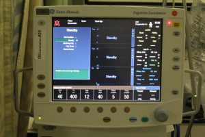
- Ventilator control panel © quinn.anya is licensed under a CC BY-SA (Attribution ShareAlike) license

