Week 8 – Histology Module
This module was originally designed by Dr. Sunita Nadella (School of Interdisciplinary Science). It has been revised by Ms. Devon Jones, Mr. Ryan Belowitz and Dr. Ana Tomljenovic-Berube in the School of Interdisciplinary Science.
Module Overview
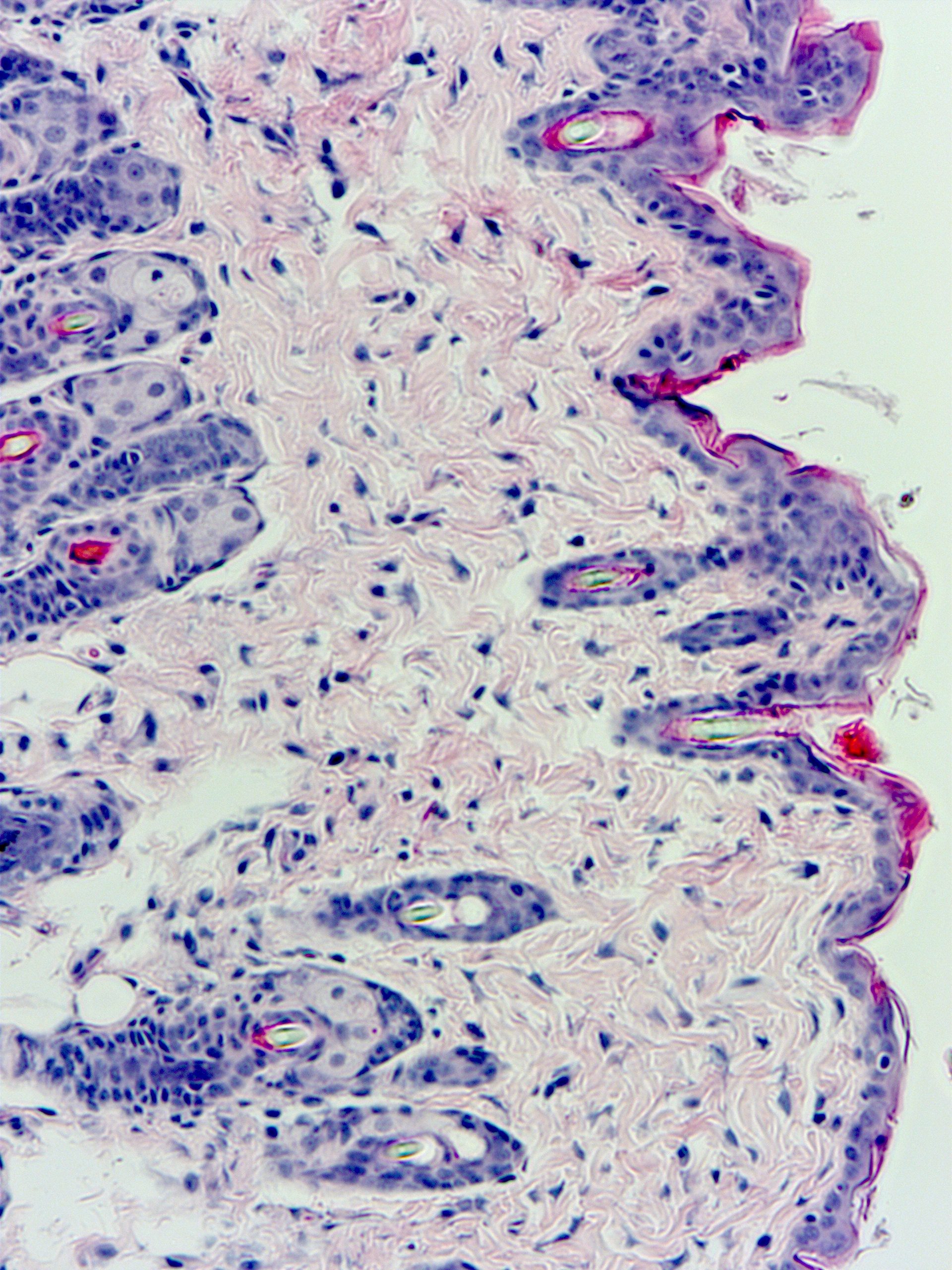
In this Histology Module, we will use the integumentary system (skin) to introduce you to the field of histology and the techniques used in the preparation of tissues for observation and image acquisition using light microscopy, and subsequent analysis of these images using software. The lecture will provide general background information on histological techniques, the components of different tissues, the structure of skin, and age related effects on skin structure. In lab, you will conduct a hematoxylin and eosin (H&E) histological staining protocol and try coverslipping prepared microscope slides. You will also use the compound light microscopes to obtain images of prepared mouse skin samples. In tutorial, you will complete a case study showing how microscopy can be used in diagnostic tests in the health care field.
Learning Objectives
By the end of this module, students should be able to:
- Describe the different sectional planes and the various internal views achieved with each plane.
- Explain why accurate representations of tissues and their structures is important for histology, and how various factors can change normal tissue appearance.
- Follow an H&E staining protocol and coverslip slides they have stained using permount.
- Use the Zeiss Primostar compound microscope to capture images of prepared mouse skin tissue.
- Use ImageJ to analyze images captured in the lab.
- Create a graph that compares measurements of the epidermis and dermis of mouse skin at different ages.
Missed work
If you miss the lab, tutorial or any work associated with this module, here are the details on accommodations.
