7.5 Muscles of the Pectoral Girdle and Upper Limbs
Learning Objectives
By the end of this section, you will be able to:
- Identify the muscles of the pectoral girdle and upper limbs
- Identify the movement and function of the pectoral girdle and upper limbs
Muscles of the shoulder and upper limb can be divided into four groups: muscles that stabilize and position the pectoral girdle, muscles that move the arm, muscles that move the forearm, and muscles that move the wrists, hands, and fingers. The pectoral girdle, or shoulder girdle, consists of the lateral ends of the clavicle and scapula, along with the proximal end of the humerus, and the muscles covering these three bones to stabilize the shoulder joint. The girdle creates a base from which the head of the humerus, in its ball-and-socket joint with the glenoid fossa of the scapula, can move the arm in multiple directions.
Muscles That Position the Pectoral Girdle
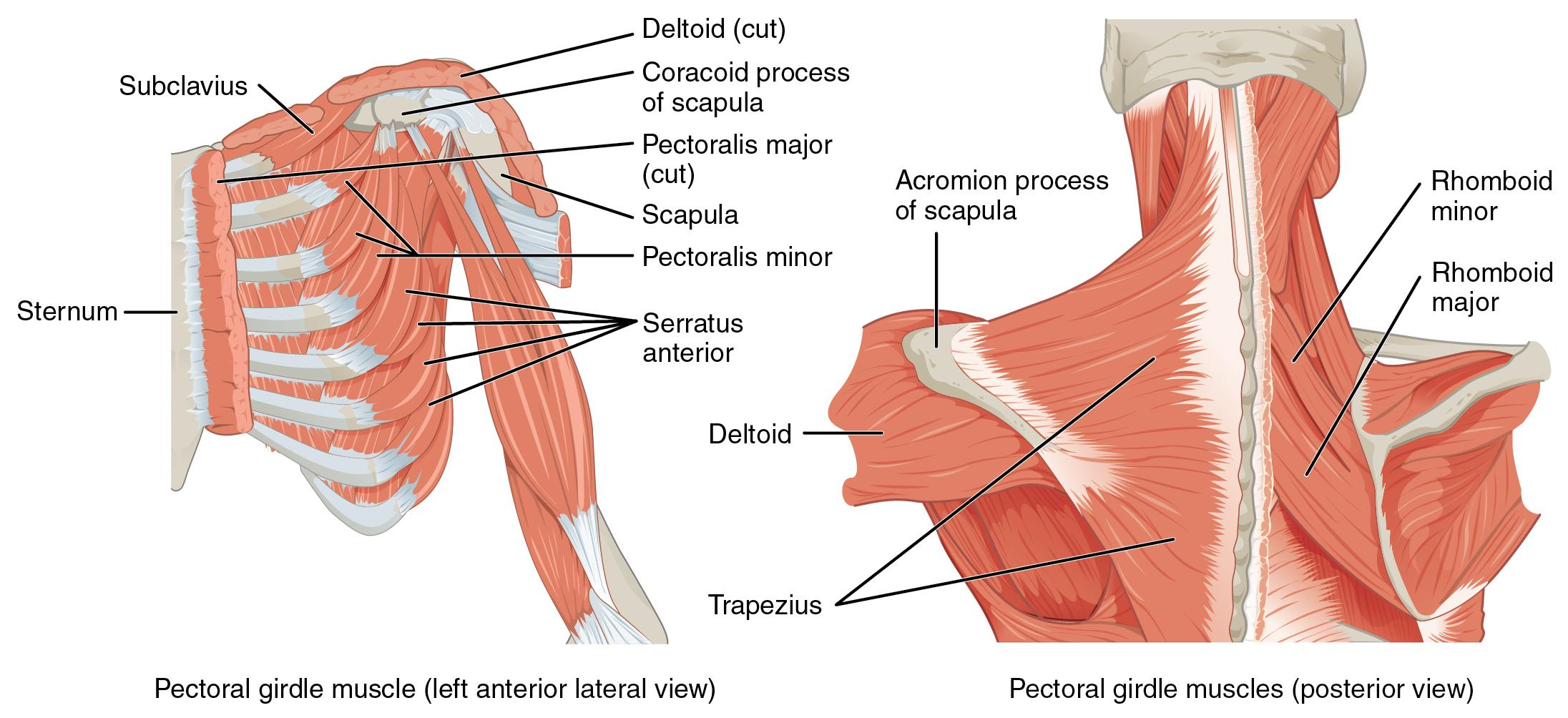
Muscles that position the pectoral girdle are located either on the anterior thorax or on the posterior thorax (Figure 7.12 and Table 7.6). The anterior muscles include the subclavius, pectoralis minor, and serratus anterior. The posterior muscles include the trapezius, rhomboid major, and rhomboid minor. When the rhomboids are contracted, your scapula moves medially, which can pull the shoulder and upper limb posteriorly.
|
Prime mover |
Origin |
Insertion |
Movement |
|
Subclavius |
First rib |
Inferior surface of clavicle |
Depression of the clavicle; stabilizes the clavicle during shoulder movement |
|
Levator scapula |
Transverse processes C1-4 |
Medial border of scapula |
Elevation of the scapula; cervical side flexion |
|
Pectoralis minor |
Ribs 3-5 |
Coracoid process of the scapula |
Stabilizes the scapula against the rib cage by pulling anteriorly; assists with inhalation |
|
Serratus anterior |
Lateral aspects of ribs 1-9 |
Anterior surface of lateral border of scapula |
Scapular protraction |
|
Trapezius |
Occipital bone; nuchal ligament; spinous process of C7-T12 |
Lateral ⅓ of clavicle; spine of scapula; acromion process |
Upper fibers – scapular elevation; cervical side flexion Middle fibers – scapular retraction Lower fibers – scapular depression |
|
Rhomboid major |
Thoracic vertebrae T2-5 |
Medial border of scapula |
Scapular retraction; scapular downward rotation |
|
Rhomboid minor |
C7 and T1 |
Medial border of scapula |
Scapular retraction; scapular downward rotation |
Muscles That Move the Humerus
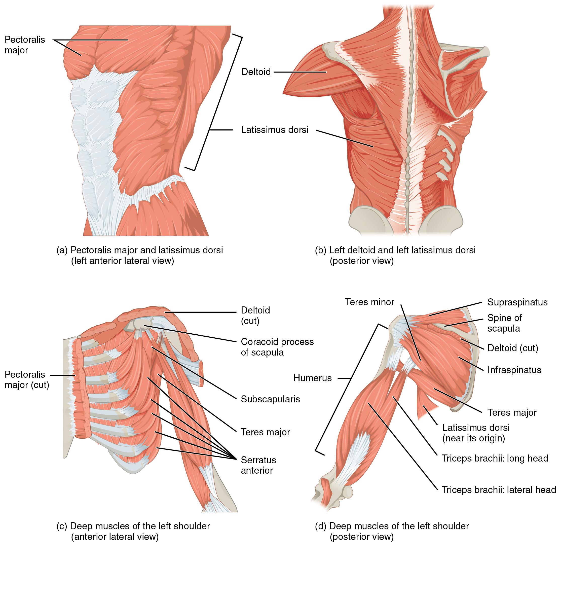
Similar to the muscles that position the pectoral girdle, muscles that cross the shoulder joint and move the humerus bone of the arm include both axial and scapular muscles (Figure 7.13 and Table 7.7). The two axial muscles are the pectoralis major and the latissimus dorsi. The pectoralis major is thick and fan-shaped, covering much of the superior portion of the anterior thorax. The broad, triangular latissimus dorsi is located on the inferior part of the back, where it inserts into a thick connective tissue sheath called an aponeurosis.
|
Prime mover |
Origin |
Insertion |
Movement |
|
Axial muscles |
|||
|
Pectoralis major |
Clavicle; sternum; cartilage of certain ribs (1-6/7); aponeurosis of external oblique muscle |
Greater tubercle of humerus |
Shoulder flexion, adduction, and medial rotation |
|
Latissimus dorsi |
T7-T12; lumbar vertebrae; ribs 9-12; iliac crest |
Bicipital groove of humerus |
Shoulder extension, adduction, and medial rotation Scapular depression |
|
Appendicular muscles |
|||
|
Deltoid |
Lateral ⅓ of clavicle; acromion of scapula; spine of scapula |
Deltoid tuberosity of humerus |
Anterior fibers – shoulder flexion and medial rotation Middle fibers – shoulder abduction Lateral fibers – shoulder extension and lateral rotation |
|
Subscapularis |
Subscapular fossa of scapula |
Lesser tubercle of humerus |
Centers head of humerus in glenoid fossa Shoulder medial rotation |
|
Supraspinatus |
Supraspinous fossa of scapula |
Greater tubercle of humerus |
Centers head of humerus in glenoid fossa Shoulder lateral rotation |
|
Infraspinatus |
Infraspinous fossa of scapula |
Greater tubercle of humerus |
Centers head of humerus in glenoid fossa Shoulder lateral rotation |
|
Teres major |
Inferior angle and lateral border of scapula |
Bicipital groove of humerus |
Shoulder extension and adduction |
|
Teres minor |
Lateral border of scapula |
Greater tubercle of humerus |
Centers head of humerus in glenoid fossa Shoulder lateral rotation |
|
Coracobrachialis |
Coracoid process of scapula |
Medial surface of humeral shaft |
Shoulder flexion and adduction |
The rest of the shoulder muscles originate on the scapula. The anatomical and ligamental structure of the shoulder joint and the arrangements of the muscles covering it, allows the arm to carry out different types of movements. The deltoid, the thick muscle that creates the rounded lines of the shoulder is the major abductor of the arm, but it also facilitates flexing and medial rotation, as well as extension and lateral rotation. The subscapularis originates on the anterior scapula and medially rotates the arm. Named for their locations, the supraspinatus (superior to the spine of the scapula) and the infraspinatus (inferior to the spine of the scapula) abduct the arm, and laterally rotate the arm, respectively. The thick and flat teres major is inferior to the teres minor and extends the arm, and assists in adduction and medial rotation of it. The long teres minor laterally rotates and extends the arm. Finally, the coracobrachialis flexes and adducts the arm.
The tendons of the deep subscapularis, supraspinatus, infraspinatus, and teres minor connect the scapula to the humerus, forming the rotator cuff (musculotendinous cuff), the circle of tendons around the shoulder joint. When baseball pitchers undergo shoulder surgery it is usually on the rotator cuff, which becomes pinched and inflamed, and may tear away from the bone due to the repetitive motion of bring the arm overhead to throw a fast pitch.
Muscles That Move the Elbow and Forearm
The forearm, made of the radius and ulna bones, has four main types of action at the hinge of the elbow joint: flexion, extension, pronation, and supination. The elbow flexors include the biceps brachii, brachialis, and brachioradialis. The elbow extensor is the triceps brachii. The forearm pronators are the pronator teres and the pronator quadratus, and the supinator is the only one that supinates the forearm (turns the palm of the hand anteriorly).
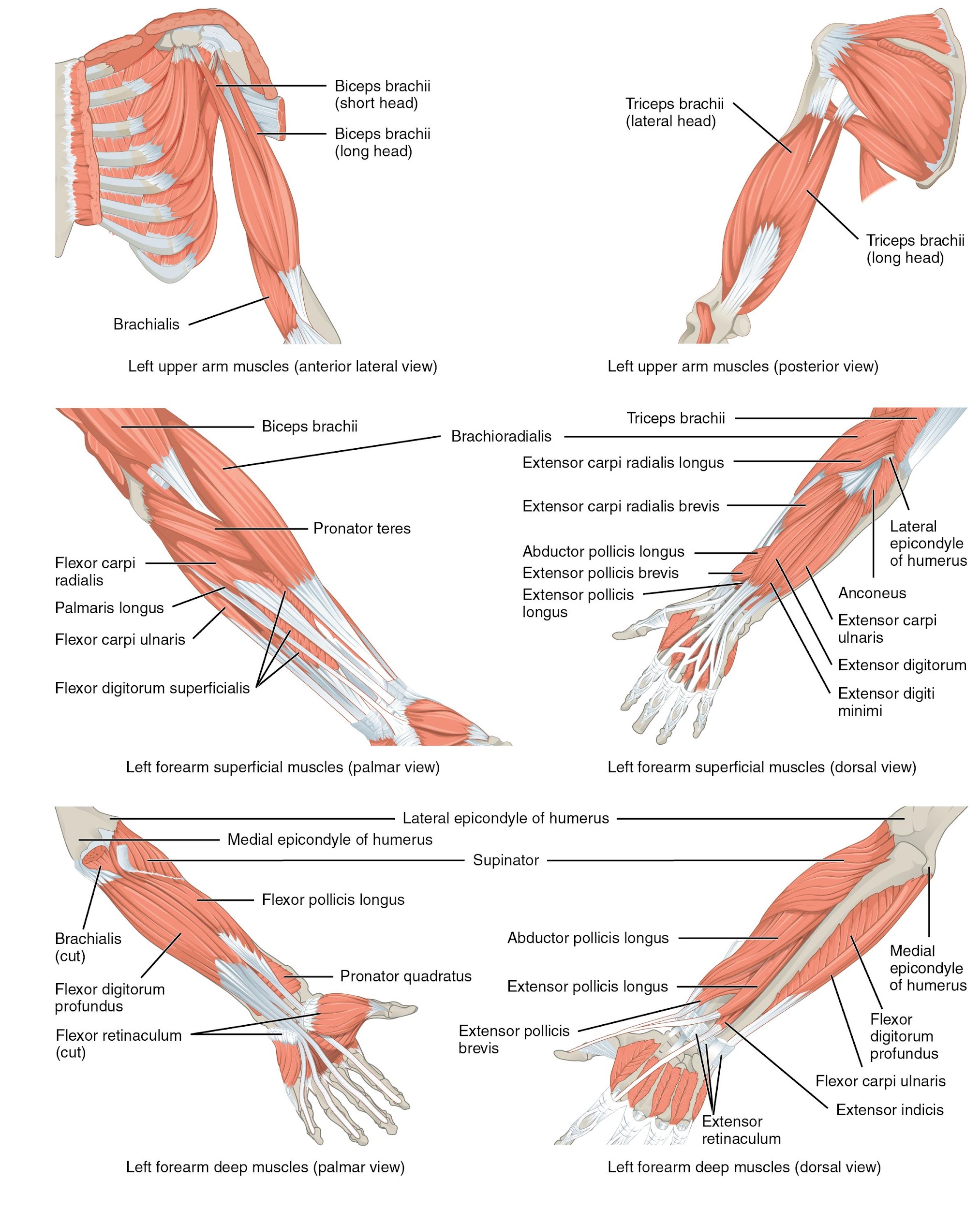
The biceps brachii, brachialis, and brachioradialis flex the elbow. The two-headed biceps brachii crosses the shoulder and elbow joints to flex the elbow, also taking part in supinating the forearm at the radioulnar joints and flexing the arm at the shoulder joint. Deep to the biceps brachii, the brachialis provides additional power in flexing the elbow. Finally, the brachioradialis can flex the elbow quickly or help lift a load slowly. These muscles and their associated blood vessels and nerves form the anterior compartment of the arm (anterior flexor compartment of the arm) (Figure 7.14 and Table 7.8).
|
Prime mover |
Origin |
Insertion |
Movement |
|
Anterior muscles (flexion) |
|||
|
Biceps brachii |
Long head – supraglenoid tubercle of scapula Short head – coracoid process of scapula |
Radial tuberosity |
Elbow flexion and supination Shoulder flexion (long head only) |
|
Brachialis |
Anterior distal shaft of humerus |
Coronoid process of ulna |
Elbow flexion |
|
Brachioradialis |
Lateral distal shaft of humerus |
Radial styloid process |
Elbow flexion Assists forearm supination when pronated Assists forearm pronation when supinated |
|
Posterior muscles (extension) |
|||
|
Triceps brachii |
Long head – infraglenoid tubercle of scapula Medial head – medial shaft of humerus Lateral head – lateral shaft of humerus |
Olecranon process of ulna |
Elbow extension Shoulder extension (long head only) |
|
Anconeus |
Lateral epicondyle of humerus |
Lateral aspect of olecranon process of ulna |
Elbow extension |
|
Anterior muscles (pronation) |
|||
|
Pronator teres |
Medial epicondyle of humerus; coronoid process of ulna |
Lateral mid-shaft of radius |
Forearm pronation |
|
Pronator quadratus |
Anterior distal shaft of ulna |
Anterior distal shaft of radius |
Forearm pronation |
|
Posterior muscles (supination) |
|||
|
Supinator |
Lateral epicondyle of humerus; proximal ulna |
Lateral proximal shaft of radius |
Forearm supination |
Muscles That Move the Wrist, Hand, and Fingers
Wrist, hand, and finger movements are facilitated by two groups of muscles. The forearm is the origin of the extrinsic muscles of the hand. The palm is the origin of the intrinsic muscles of the hand.
Muscles of the Arm That Move the Wrists, Hands, and Fingers
The muscles in the anterior compartment of the forearm (anterior flexor compartment of the forearm) originate on the humerus and insert into different parts of the hand. These make up the bulk of the forearm. From lateral to medial, the superficial anterior compartment of the forearm includes the flexor carpi radialis, palmaris longus, flexor carpi ulnaris, and flexor digitorum superficialis. The flexor digitorum superficialis flexes the hand as well as the digits at the knuckles, which allows for rapid finger movements, as in typing or playing a musical instrument (see Table 7.9 and Table 7.10). However, poor ergonomics can irritate the tendons of these muscles as they slide back and forth with the carpal tunnel of the anterior wrist and pinch the median nerve, which also travels through the tunnel, causing Carpal Tunnel Syndrome. The deep anterior compartment produces wrist and digit flexion, bending the fingers to make a fist. These are the flexor pollicis longus and the flexor digitorum profundus.
The muscles in the superficial posterior compartment of the forearm (superficial posterior extensor compartment of the forearm) originate on the humerus. These are the extensor radialis longus, extensor carpi radialis brevis, extensor digitorum, extensor digiti minimi, and the extensor carpi ulnaris.
The muscles of the deep posterior compartment of the forearm (deep posterior extensor compartment of the forearm) originate on the radius and ulna. These include the abductor pollicis longus, extensor pollicis brevis, extensor pollicis longus, and extensor indicis (see Table 7.9).
The tendons of the forearm muscles attach to the wrist and extend into the hand. Fibrous bands called retinacula sheath the tendons at the wrist. The flexor retinaculum extends over the palmar surface of the hand while the extensor retinaculum extends over the dorsal surface of the hand.
|
Prime mover |
Origin |
Insertion |
Movement |
|
Superficial anterior compartment of forearm |
|||
|
Flexor carpi radialis |
Medial epicondyle of humerus |
Base of 2nd and 3rd metacarpals |
Wrist flexion and abduction |
|
Flexor carpi ulnaris |
Medial epicondyle of humerus |
Pisiform, hamate, base of 5th metacarpal |
Wrist flexion and adduction |
|
Flexor digitorum superficialis |
Medial epicondyle of humerus; anterior shaft of radius |
Middle phalanges of digits 2-5 |
Wrist flexion; finger flexion digits 2-5 |
|
Palmaris longus |
Medial epicondyle of humerus |
Palmar aponeurosis |
Wrist flexion |
|
Deep anterior compartment of forearm |
|||
|
Flexor digitorum profundus |
Coronoid process; anteromedial surface of ulna |
Distal phalanges of digits 2-5 |
Wrist flexion; finger flexion digits 2-5 |
|
Flexor pollicis longus |
Anterior surface of radius |
Distal phalanx of thumb |
Thumb flexion |
|
Superficial posterior compartment of forearm |
|||
|
Extensor carpi radialis longus |
Lateral supracondylar ridge of humerus |
Base of 2nd metacarpal |
Wrist extension and abduction |
|
Extensor carpi radialis brevis |
Lateral epicondyle of humerus |
Base of 3rd metacarpal |
Wrist extension and abduction |
|
Extensor carpi ulnaris |
Lateral epicondyle of humerus |
Base of 5th metacarpal |
Wrist extension and adduction |
|
Extensor digitorum |
Lateral epicondyle of humerus |
Extensor expansions; distal phalanges of digits 2-5 |
Wrist extension; finger extension |
|
Extensor digiti minimi |
Lateral epicondyle of humerus |
Extensor expansions; distal phalanx of digit 5 |
Finger extension of digit 5 |
|
Deep posterior compartment of forearm |
|||
|
Abductor pollicis longus |
Posterior surface of radius and ulna |
Base of first metacarpal; trapezium |
Thumb abduction and extension Wrist abduction |
|
Extensor pollicis longus |
Posterior shaft of radius and ulna |
Base of distal phalanx of thumb |
Thumb extension |
|
Extensor pollicis brevis |
Posterior shaft of radius and ulna |
Base of proximal phalanx of thumb |
Thumb extension |
|
Extensor indicis |
Posterior surface of radius and ulna |
Tendon of extensor digitorum of index finger |
Finger extension of digit 2 |
Intrinsic Muscles of the Hand
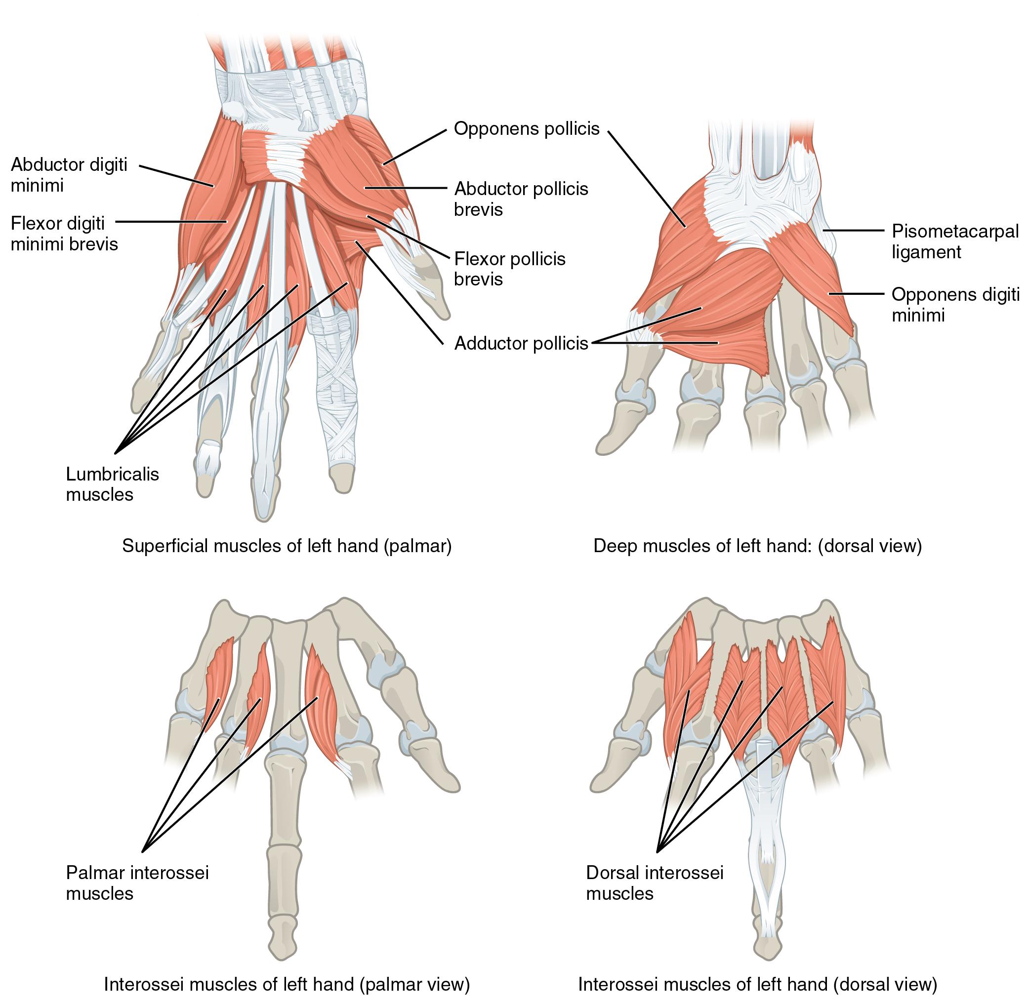
The intrinsic muscles of the hand both originate and insert within it (Figure 7.15). These muscles allow your fingers to also make precise movements for actions, such as typing or writing. These muscles are divided into three groups. The thenar muscles are on the radial aspect of the palm. The hypothenar muscles are on the medial aspect of the palm, and the intermediate muscles are midpalmar.
The thenar muscles include the abductor pollicis brevis, opponens pollicis, flexor pollicis brevis, and the adductor pollicis. These muscles form the thenar eminence, the rounded contour of the base of the thumb, and all act on the thumb. The movements of the thumb play an integral role in most precise movements of the hand.
The hypothenar muscles include the abductor digiti minimi, flexor digiti minimi brevis, and the opponens digiti minimi. These muscles form the hypothenar eminence, the rounded contour of the little finger, and as such, they all act on the little finger. Finally, the intermediate muscles act on all the fingers and include the lumbrical, the palmar interossei, and the dorsal interossei.
|
Prime mover |
Origin |
Insertion |
Movement |
|
Abductor pollicis brevis |
Flexor retinaculum; nearby carpals |
Lateral base of proximal phalanx of thumb |
Thumb abduction |
|
Opponens pollicis |
Flexor retinaculum; trapezium |
Anterior shaft of first metacarpal |
Thumb opposition |
|
Flexor pollicis brevis |
Flexor retinaculum; trapezium |
Base of proximal phalanx of thumb |
Thumb flexion |
|
Adductor pollicis |
Capitate; bases of metacarpals 2-4 |
Medial base of proximal phalanx of thumb |
Thumb adduction |
|
Abductor digiti minimi |
Pisiform |
Medial side of proximal phalanx of digit 5 |
Abduction of digit 5 |
|
Flexor digiti minimi brevis |
Hamate; flexor retinaculum |
Medial side of proximal phalanx of digit 5 |
Flexion of digit 5 |
|
Opponens digiti minimi |
Hamate; flexor retinaculum |
Medial side of 5th metacarpal |
Opposition of digit 5 |
|
Lumbricals |
Lateral sides of tendons in flexor digitorum profundus |
Lateral edges of extensor expansion on first phalanges |
Flexion at metacarpo-phalangeal joint and extension of interphalangeal joint of digits 2-5 |
|
Palmar interossei |
Sides of each metacarpal that faces metacarpal 3 |
Extensor expansion on first phalanx of each expect (expect digit 3) on side facing digit 3 |
Adducts the fingers (except digit 3) |
|
Dorsal interossei |
Sides of metacarpals |
Both sides of digit 3; for each other finger extensor expansion over first phalanx on side opposite digit 3 |
Abducts the three middle fingers (digits 2-4) |
