2.7 Cartilaginous Joints
Learning Objectives
By the end of this section, you will be able to:
- Describe the structural features of cartilaginous joints
- Distinguish between a synchondrosis and symphysis
- Give an example of each type of cartilaginous joint
As the name indicates, at a cartilaginous joint, the adjacent bones are united by cartilage, a tough but flexible type of connective tissue. These types of joints lack a joint cavity and involve bones that are joined together by either hyaline cartilage or fibrocartilage (Figure 2.19). There are two types of cartilaginous joints. A synchondrosis is a cartilaginous joint where the bones are joined by hyaline cartilage. Also classified as a synchondrosis are places where bone is united to a cartilage structure, such as between the anterior end of a rib and the costal cartilage of the thoracic cage. The second type of cartilaginous joint is a symphysis, where the bones are joined by fibrocartilage.
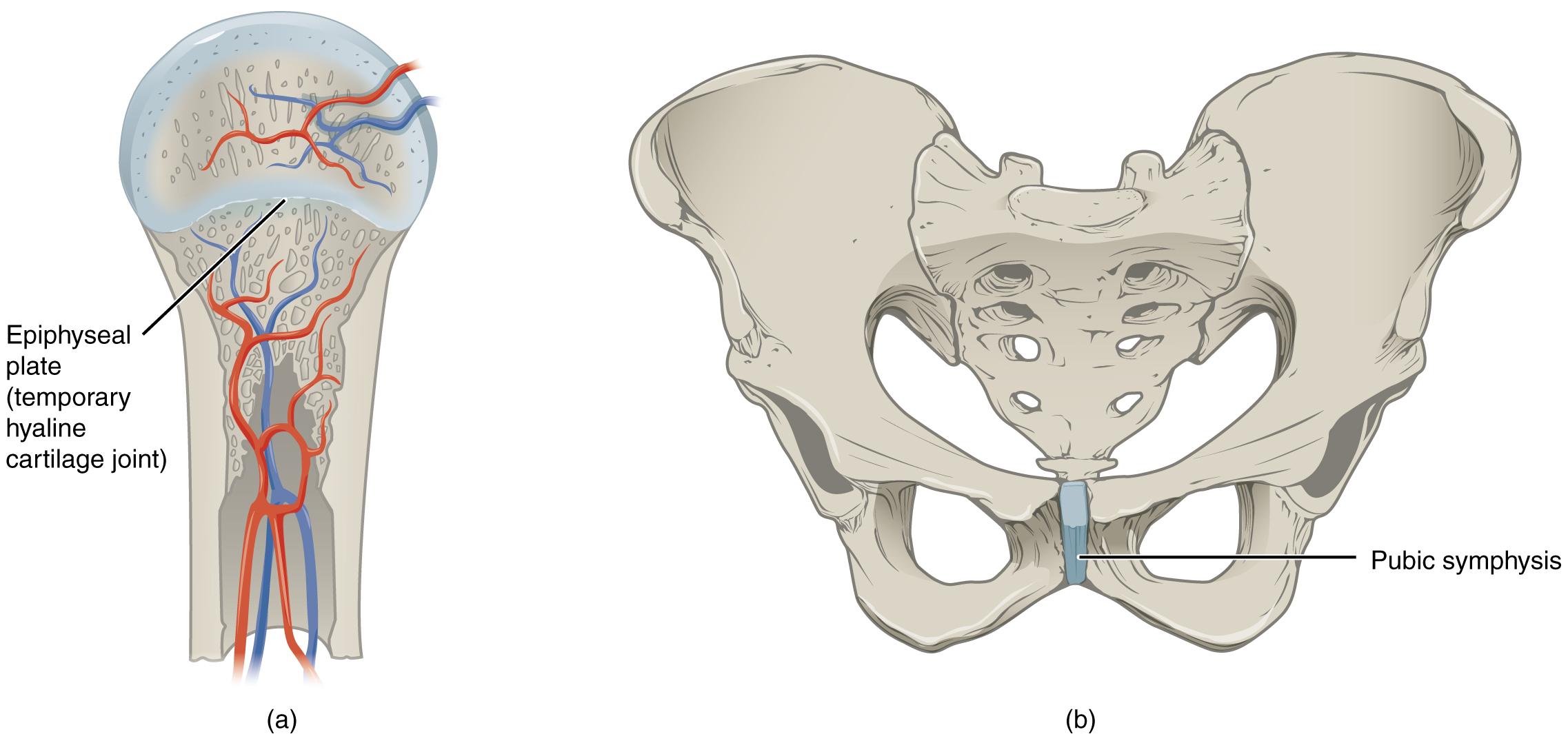
Synchondrosis
A synchondrosis (“joined by cartilage”) is a cartilaginous joint where bones are joined together by hyaline cartilage, or where bone is united to hyaline cartilage. A synchondrosis may be temporary or permanent. A temporary synchondrosis is the epiphyseal plate (growth plate) of a growing long bone. The epiphyseal plate is the region of growing hyaline cartilage that unites the diaphysis (shaft) of the bone to the epiphysis (end of the bone). The epiphyseal plate is considered to be a temporary synchondrosis because in the late teens growth of the cartilage eventually stops and the epiphyseal plate is completely replaced by bone, fusing the epiphysis and diaphysis into one single adult bone.
Examples of permanent synchondroses are found in the thoracic cage. One example is the first sternocostal joint, where the first rib is anchored to the manubrium by its costal cartilage. (The articulations of the remaining costal cartilages to the sternum are all synovial joints.) Additional synchondroses are formed where the anterior end of the other 11 ribs is joined to its costal cartilage. Unlike the temporary synchondroses of the epiphyseal plate, these permanent synchondroses retain their hyaline cartilage and thus do not ossify with age. Due to the lack of movement between the bone and cartilage, both temporary and permanent synchondroses are functionally classified as a synarthrosis.
Interactive Link
Visit this website to view a radiograph (X-ray image) of a child’s hand and wrist. The growing bones of child have an epiphyseal plate that forms a synchondrosis between the shaft and end of a long bone. Being less dense than bone, the area of epiphyseal cartilage is seen on this radiograph as the dark epiphyseal gaps located near the ends of the long bones, including the radius, ulna, metacarpal, and phalanx bones. Which of the bones in this image do not show an epiphyseal plate (epiphyseal gap)?
Symphysis
A cartilaginous joint where the bones are joined by fibrocartilage is called a symphysis (“growing together”). Fibrocartilage is very strong because it contains numerous bundles of thick collagen fibers, thus giving it a much greater ability to resist pulling and bending forces when compared with hyaline cartilage. This gives symphyses the ability to strongly unite the adjacent bones, but can still allow for limited movement to occur. Thus, a symphysis is functionally classified as an amphiarthrosis.
The gap separating the bones at a symphysis may be narrow or wide. Examples in which the gap between the bones is narrow include the pubic symphysis and the manubriosternal joint. At the pubic symphysis, the pubic portions of the right and left hip bones of the pelvis are joined together by fibrocartilage across a narrow gap. Similarly, at the manubriosternal joint, fibrocartilage unites the manubrium and body portions of the sternum.
The intervertebral symphysis is a wide symphysis located between the bodies of adjacent vertebrae of the vertebral column. Here a thick pad of fibrocartilage called an intervertebral disc strongly unites the adjacent vertebrae by filling the gap between them. The width of the intervertebral symphysis is important because it allows for small movements between the adjacent vertebrae. In addition, the thick intervertebral disc provides cushioning between the vertebrae, which is important when carrying heavy objects or during high-impact activities such as running or jumping.
