7.6 Appendicular Muscles of the Pelvic Girdle and Lower Limbs
Learning Objectives
By the end of this section, you will be able to:
- Identify the appendicular muscles of the pelvic girdle and lower limb
- Identify the movement and function of the pelvic girdle and lower limb
The appendicular muscles of the lower body position and stabilize the pelvic girdle, which serves as a foundation for the lower limbs. Comparatively, there is much more movement at the pectoral girdle than at the pelvic girdle. There is very little movement of the pelvic girdle because of its connection with the sacrum at the base of the axial skeleton. The pelvic girdle has less range of motion because it was designed to stabilize and support the body.
Muscles of the Thigh
What would happen if the pelvic girdle, which attaches the lower limbs to the torso, were capable of the same range of motion as the pectoral girdle? For one thing, walking would expend more energy if the heads of the femurs were not secured in the acetabula of the pelvis. The body’s center of gravity is in the area of the pelvis. If the center of gravity were not to remain fixed, standing up would be difficult as well. Therefore, what the leg muscles lack in range of motion and versatility, they make up for in size and power, facilitating the body’s stabilization, posture, and movement.
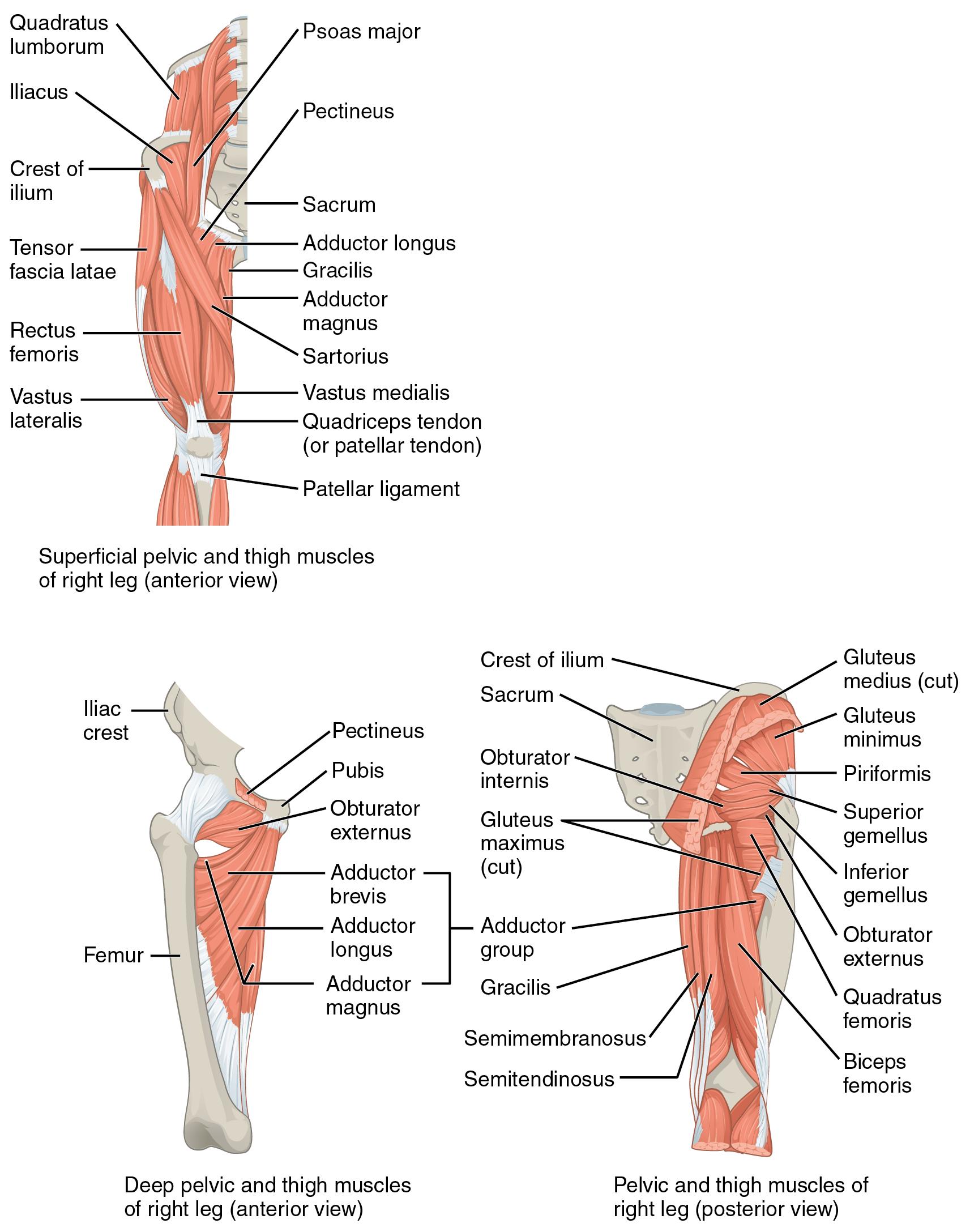
Most muscles that insert on the femur (the thigh bone) and move it, originate on the pelvic girdle. The psoas major and iliacus make up the iliopsoas muscle. Some of the largest and most powerful muscles in the body are the gluteal muscles or gluteal group. The gluteus maximus is the largest; deep to the gluteus maximus is the gluteus medius, and deep to the gluteus medius is the gluteus minimus, the smallest of the trio (Figure 7.16 and Table 7.11).
The tensor fascia latae is a thick, squarish muscle in the superior aspect of the lateral thigh. It acts as a synergist of the gluteus medius in abducting the thigh, and as a synergist of the iliopsoas in flexing the thigh. It also helps stabilize the lateral aspect of the knee by pulling on the iliotibial tract (band), making it taut. Deep to the gluteus maximus, the piriformis, obturator internus, obturator externus, superior gemellus, inferior gemellus, and quadratus femoris laterally rotate the femur at the hip. These muscles are often grouped together and referred to as the deep lateral rotator muscle group.
The pectineus, adductor longus, adductor brevis, adductor magnus, and gracilis are muscles in the medial compartment of the thigh with the gracilis originating most medially and the pectineus originating most laterally. The muscles in the medial compartment of the thigh are responsible for adducting the femur at the hip. The pectineus flexes the femur at the hip as well. The pectineus is located in the femoral triangle, which is formed at the junction between the hip and the leg and also includes the femoral nerve, the femoral artery, the femoral vein, and the deep inguinal lymph nodes. The adductor longus, adductor brevis, and adductor magnus can both medially and laterally rotate the thigh depending on the placement of the foot. The adductor longus flexes the thigh, whereas the adductor magnus extends it. The strap-like gracilis additionally flexes the leg at the knee (Table 7.12).
|
Prime mover |
Origin |
Insertion |
Movement |
|
Iliopsoas group |
|||
|
Psoas major |
T12, L1-5 |
Lesser trochanter of femur |
Hip flexion, external rotation |
|
Iliacus |
Iliac fossa; iliac crest |
Lesser trochanter of femur |
Hip flexion, external rotation |
|
Gluteal group |
|||
|
Gluteus maximus |
Dorsal ilium; sacrum; coccyx |
Gluteal tuberosity of femur; iliotibial band |
Hip extension, external rotation, abduction |
|
Gluteus medius |
Iliac crest |
Greater trochanter of femur |
Hip abduction Pelvic stability in weight bearing |
|
Gluteus minimus |
Ilium |
Greater trochanter of femur |
Hip abduction |
|
Tensor fascia latae |
Anterior superior iliac spine |
Iliotibial band |
Assists with hip flexion and hip abduction |
|
Lateral rotators |
|||
|
Piriformis |
Anterolateral surface of sacrum |
Greater trochanter of femur |
Hip external rotation |
|
Obturator internus |
Inner surface of obturator membrane; greater sciatic notch; margins of obturator foramen |
Greater trochanter of femur |
Hip external rotation |
|
Obturator externus |
Outer surface of obturator membrane; pubic and ischium; margins of obturator foramen |
Trochanteric fossa of femur |
Hip external rotation |
|
Superior gemellus |
Ischial spine |
Greater trochanter of femur |
Hip external rotation |
|
Inferior gemellus |
Ischial tuberosity |
Greater trochanter of femur |
Hip external rotation |
|
Quadratus femoris |
Ischial tuberosity |
Trochanteric crest of femur |
Hip external rotation |
|
Adductors |
|||
|
Gracilis |
Pubis near pubic symphysis |
Tibial shaft at pes anserine |
Hip adduction Knee flexion |
|
Adductor longus |
Pubis near pubic symphysis |
Linea aspera |
Hip adduction |
|
Adductor brevis |
Body of pubis; inferior ramus of pubis |
Linea aspera, more proximal than adductor longus |
Hip adduction |
|
Adductor magnus |
Pubic ramus; ischial tuberosity |
Linea aspera of femur gluteal tuberosity; adductor tubercle of femur |
Hip adduction and extension |
|
Pectineus |
Pectineal line of pubis |
Proximal medial shaft of femur, just inferior to lesser trochanter |
Hip adduction and flexion |
|
Prime mover |
Origin |
Insertion |
Movement |
|
Anterior compartment of thigh |
|||
|
Rectus femoris |
Anterior inferior iliac spine |
Patella; tibial tuberosity |
Knee extension Hip flexion |
|
Vastus lateralis |
Greater trochanter; intertrochanteric line; linea aspera |
Patella; tibial tuberosity |
Knee extension |
|
Vastus medialis |
Linea aspera; intertrochanteric line |
Patella; tibial tuberosity |
Knee extension |
|
Vastus intermedius |
Proximal shaft of femur |
Patella; tibial tuberosity |
Knee extension |
|
Sartorius |
Anterior superior iliac spine |
Medial aspect of proximal tibia |
Hip flexion, abduction and external rotation Knee flexion |
|
Posterior compartment of thigh |
|||
|
Biceps femoris |
Ischial tuberosity; linea aspera; distal femur |
Head of fibula; lateral condyle of tibia |
Knee flexion Hip extension and external rotation |
|
Semitendinosus |
Ischial tuberosity |
Upper medial tibial shaft |
Knee flexion Hip extension and internal rotation |
|
Semimembranosus |
Ischial tuberosity |
Medial condyle of tibia |
Knee flexion Hip extension and internal rotation |
The muscles of the anterior compartment of the thigh flex the hip and extend the knee. This compartment contains the quadriceps femoris group, which actually comprises four muscles that extend and stabilize the knee. The rectus femoris is on the anterior aspect of the thigh, the vastus lateralis is on the lateral aspect of the anterior thigh, the vastus medialis is on the medial aspect of the anterior thigh, and the vastus intermedius is between the vastus lateralis and vastus medialis and deep to the rectus femoris. The tendon common to all four is the quadriceps tendon, which inserts into the patella and continues below it as the patellar ligament. The patellar ligament attaches to the tibial tuberosity. In addition to the quadriceps femoris, the sartorius is a band-like muscle that extends from the anterior superior iliac spine to the medial side of the proximal tibia. This versatile muscle flexes the leg at the knee and flexes, abducts, and laterally rotates the leg at the hip. This muscle allows us to sit cross-legged. It is the longest muscle in the human body.
The posterior compartment of the thigh includes muscles that flex the leg and extend the thigh. The three long muscles on the back of the knee are the hamstring group, which flexes the knee. These are the biceps femoris, semitendinosus, and semimembranosus. The tendons of these muscles form the popliteal fossa, the diamond-shaped space at the back of the knee.
Muscles That Move the Feet and Toes
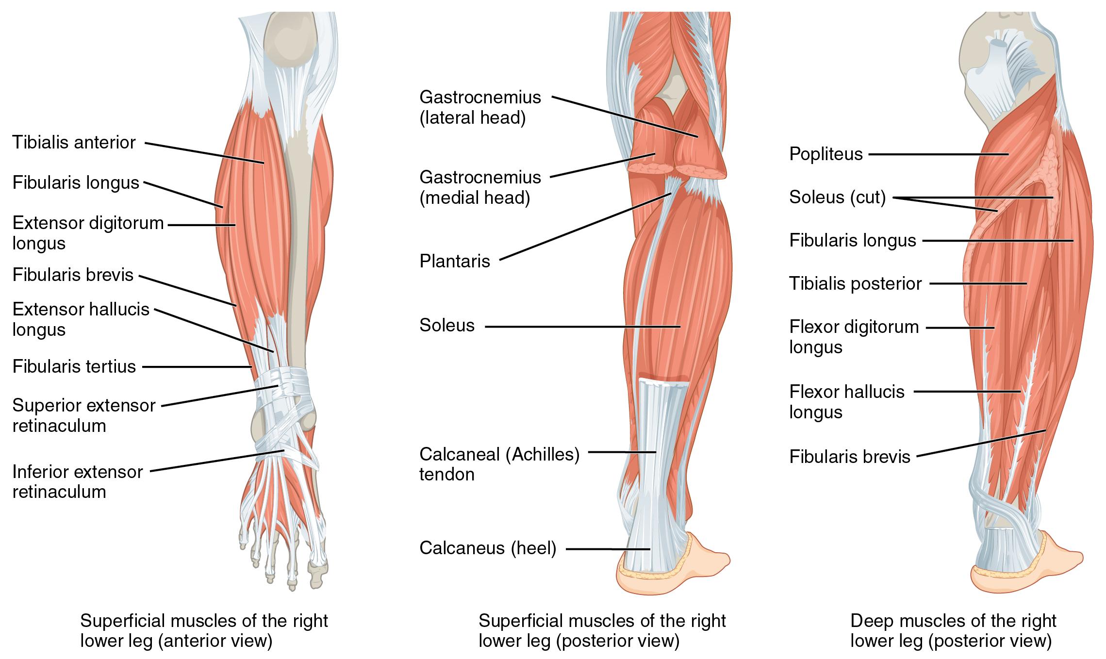
Similar to the thigh muscles, the muscles of the leg are divided by deep fascia into compartments, although the leg has three: anterior, lateral, and posterior (Figure 7.17 and Table 7.13).
|
Prime mover |
Origin |
Insertion |
Movement |
|
Anterior compartment of the leg |
|||
|
Tibialis anterior |
Anterior shaft of tibia; interosseous membrane |
First metatarsal; medial cuneiform |
Ankle dorsiflexion Foot inversion |
|
Extensor hallucis longus |
Anteromedial fibular shaft; interosseous membrane |
Distal phalanx of big toe |
Big toe extension Ankle dorsiflexion |
|
Extensor digitorum longus |
Lateral condyle of tibia; interosseous membrane |
Middle and distal phalanges of toes 2-5 |
Toe extension toes 2-5 Ankle dorsiflexion |
|
Lateral compartment of the leg |
|||
|
Fibularis longus |
Upper portion of lateral fibula |
First metatarsal; medial cuneiform |
Ankle plantarflexion Foot eversion |
|
Fibularis brevis |
Lateral shaft of distal fibula |
Fifth metatarsal |
Ankle plantarflexion Foot eversion |
|
Posterior compartment of the leg: superficial muscles |
|||
|
Gastrocnemius |
Medial and lateral condyles of femur |
Posterior calcaneus |
Ankle plantarflexion Knee flexion |
|
Soleus |
Posterior shaft of tibia and fibula; interosseous membrane |
Posterior calcaneus |
Ankle plantarflexion |
|
Plantaris |
Posterior femur above lateral condyle |
Calcaneus or Achilles tendon |
Ankle plantarflexion Knee flexion |
|
Tibialis posterior |
Posterior shaft of tibia and fibula; interosseous membrane |
Plantar surfaces of navicular; metatarsals 2-4 |
Ankle plantarflexion Foot inversion Supports medial longitudinal arch |
|
Posterior compartment of the leg: deep muscles |
|||
|
Popliteus |
Lateral condyle of femur; lateral meniscus |
Proximal medial tibia |
Knee flexion “Unlocks” the knee from full extension |
|
Flexor digitorum longus |
Posterior tibia |
Distal phalanges of toes 2-5 |
Toe flexion toes 2-5 Ankle plantarflexion |
|
Flexor hallucis longus |
Midshaft of fibula; interosseous membrane |
Distal phalanx of big toe |
Big toe flexion Ankle plantarflexion |
The muscles in the anterior compartment of the leg: the tibialis anterior, a long and thick muscle on the lateral surface of the tibia, the extensor hallucis longus, deep to the tibialis anterior, and the extensor digitorum longus, lateral to the tibialis anterior. All three muscles contribute to ankle dorsiflexion – raising the front of the foot when they contract. Thick bands of connective tissue called the superior extensor retinaculum (transverse ligament of the ankle) and the inferior extensor retinaculum, hold the tendons of these muscles in place during dorsiflexion.
The lateral compartment of the leg includes two muscles: the fibularis longus (peroneus longus) and the fibularis brevis (peroneus brevis). Both of these muscles contribute to foot eversion.
The superficial muscles in the posterior compartment of the leg all insert onto the calcaneal tendon (Achilles tendon), a strong tendon that inserts into the calcaneal bone of the ankle. The muscles in this compartment are large and strong and keep humans upright. The most superficial and visible muscle of the calf is the gastrocnemius. Deep to the gastrocnemius is the wide, flat soleus. The plantaris runs obliquely between the two; some people may have two of these muscles, whereas no plantaris is observed in about seven percent of other cadaver dissections. There are four deep muscles in the posterior compartment of the leg as well: the popliteus, flexor digitorum longus, flexor hallucis longus, and tibialis posterior.
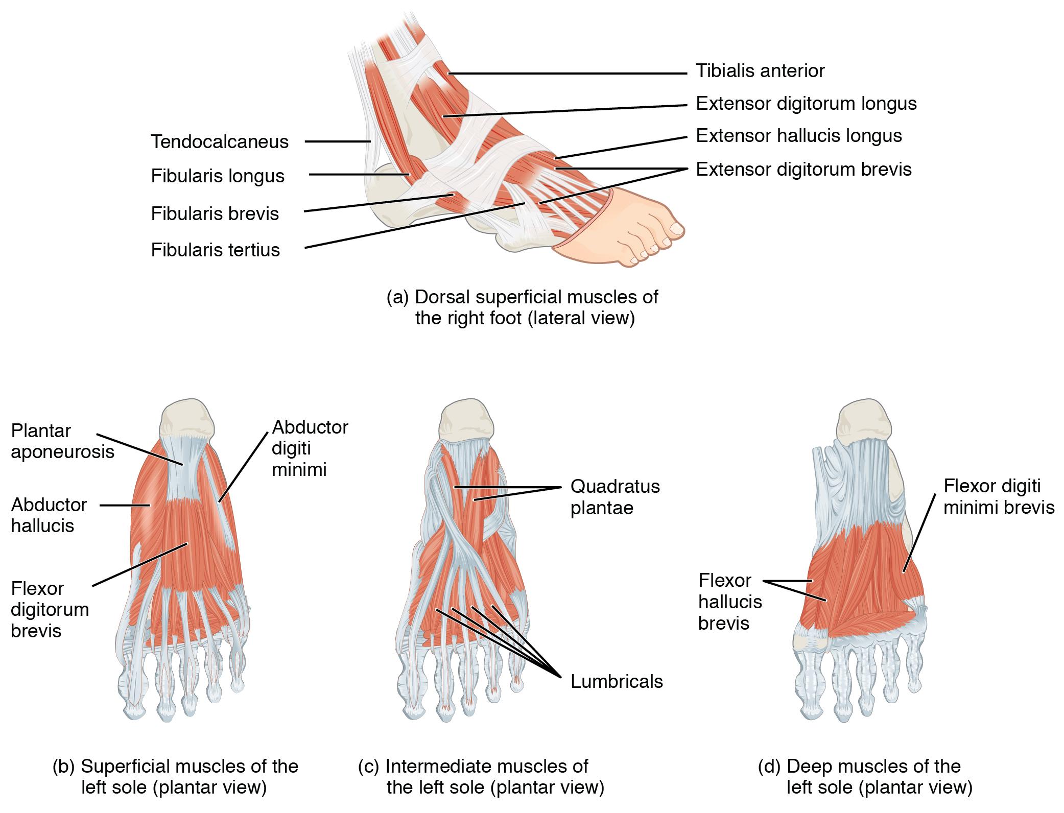
The foot also has intrinsic muscles, which originate and insert within it (similar to the intrinsic muscles of the hand). These muscles primarily provide support for the foot and its arch, and contribute to movements of the toes (Figure 7.18 and Table 7.14). The principal support for the longitudinal arch of the foot is a deep fascia called plantar aponeurosis, which runs from the calcaneus bone to the toes (inflammation of this tissue is the cause of “plantar fasciitis,” which can affect runners). The intrinsic muscles of the foot consist of two groups. The dorsal group includes only one muscle, the extensor digitorum brevis. The second group is the plantar group, which consists of four layers, starting with the most superficial.
|
Prime mover |
Origin |
Insertion |
Movement |
|
Dorsal group |
|||
|
Extensor digitorum brevis |
Calcaneus; extensor retinaculum |
Extensor expansion toes 2-5 |
Toe extension toes 2-5 |
|
Plantar group (layer 1) |
|||
|
Abductor hallucis |
Calcaneal tuberosity; flexor retinaculum |
Proximal phalanx of big toe |
Big toe adduction and flexion |
|
Flexor digitorum brevis |
Calcaneal tuberosity |
Middle phalanx of toes 2-4 |
Toe flexion toes 2-4 |
|
Abductor digiti minimi |
Calcaneal tuberosity |
Proximal phalanx of middle toe |
Toe abduction and flexion toe 5 |
|
Plantar group (layer 2) |
|||
|
Quadratus plantae |
Medial and lateral sides of calcaneus |
Tendon of flexor digitorum longus |
Toe flexion toes 2-5 |
|
Lumbricals |
Tendons of flexor digitorum longus |
Medial side of proximal phalanx toes 2-5 |
Extension at interphalangeal joint and flexion at metatarsophalangeal joint toes 2-5 |
|
Plantar group (layer 3) |
|||
|
Flexor hallucis brevis |
Lateral cuneiform; cuboid |
Base of proximal phalanx of big toe |
Big toe flexion |
|
Adductor hallucis |
Bases of metatarsals 2-4 |
Base of proximal phalanx of big toe |
Big toe adduction and flexion |
|
Flexor digiti minimi brevis |
Base of metatarsal 5 |
Base of proximal phalanx of toe 5 |
Toe flexion toe 5 |
|
Plantar group (layer 4) |
|||
|
Dorsal interossei |
Sides of metatarsals |
Both sides of toe 2; for other toes the extensor expansion on side opposite of toe 2 |
Toe abduction and flexion |
|
Plantar interossei |
Side of each metatarsal that faces toe 2 |
Extensor expansion on side facing toe 2 |
Toe abduction and flexion toes 3-5 |
