9.2 – C-arm X-Ray Machine
Introduction to X-rays
X-rays belong to the group of electromagnetic rays, hence, they follow the rules of electromagnetic radiation. Electromagnetic radiation transports energy, also called radiant energy, through space by waves and photons, just as radio waves, the visible light or microwaves. X-rays are electromagnetic radiation produced in an evacuated glass tube (see Fig. 9.2.1). A spinning tungsten anode is the target for high velocity electrons that have been accelerated across the vacuum in the tube by a large voltage differential between the cathode and the anode. When the electrons strike the anode, x-rays are liberated along with heat. X-rays are only produced when there is a voltage differential applied across the cathode and anode, therefore, the x-ray tube is dormant until activated by the Medical Radiation Technologist (MRT). This physical, on/off, arrangement is used for all modalities that employ x-rays for imaging (X-ray, CT, Fluoroscopy, Angiography).
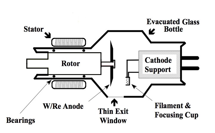
Figure 9.2.1 Schematic of X-ray tube
The x-rays that result from the electron bombardment of the anode are constrained within a heavy lead housing that contains circulating oil to cool the tube. The dispersal of the x-ray beam is limited to a small port in the lead housing of the x-ray tube and by mechanical metal filters that collimate the x-ray beam and prevent the uncontrolled, dissemination of x-rays (see Fig. 9.2.2). Therefore, the emitted x-rays expose only the desired region of the patient’s anatomy in a controlled manner. The limitation of the x-rays by the lead housing and the collimation filters is how small part x-rays, i.e. wrist, scaphoid bone, can be done, as well as, large field x-rays such as a full field chest x-ray.
![]()
Figure 9.2.2 X-ray Tube, Lead Housing with Portal for x-ray emission, bench top image
The x-rays pass through the patient to reach an x-ray detector on the other side of the patient (see Fig. 9.2.3). The image detectors used today rely on complex electronic systems that converts x-rays into electrical signals that then results in pixels of different intensity on the monitor of a digital display system.
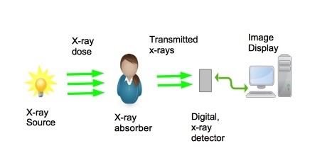
Figure 9.2.3 X-ray Image Creation and Display
Radiography describes the process of creating two dimensional projection images by exposing an anatomy of interest to X-rays and measuring the attenuation they undergo when passing through the object. It is a very common form of X-ray imaging and is used in clinics around the globe.
The main application area is the examination of fractures and changes of the skeletal system. Here, the high attenuation coefficient of bones compared to the surrounding tissue delivers a good contrast and allows for distinct detection and classification of fractures. Moreover, radiography can be used to detect changes of a bone’s consistency or density. There are several different types of x-ray machines but the BTEC 315 labs will focus on the portable C-arm x-ray imaging device (Fig. 9.2.4).
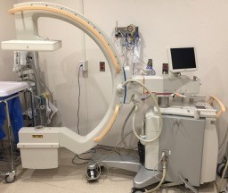
Figure 9.2.4 Portable C-arm x-ray device
Radiation Exposure: X-rays
X-rays are ionizing radiation. Thus, x-rays can cause tissue ionization and this can result in genetic alterations. The majority of the genetic alterations that occur do not lead to cellular damage and lead to no adverse genetic events or genetic mutation. However, it is practical, and appropriate, to minimize radiation exposure whenever possible. The, As Low As Reasonably Achievable approach (the ALARA principle) is an excellent strategy to employ in order to reduce the exposure of patients to x-rays. In addition, Appropriateness Guidelines (CAR, ACR, ESR), and strategies such as, “Choosing Wisely” and , “Imaging Gently” are also geared towards minimizing x-ray utilization if not clinically safe, appropriate. or helpful in managing the patient’s medical condition.
Attributes
This chapter is adapted from the following:
- Chapter 19 in Undergraduate Diagnostic Imaging Fundamentals by Brent Burbridge is licensed under CC BY-NC-SA 4.0
- Chapter 7 in Medical Imaging Systems: An Introductory Guide by M. Berger, Q. Yang and A Maier is licensed under CC BY 4.0 courtesy of National Center for Biotechnology Information
Figures
- 9.2.1 RotatingAnodeXRT by Kieranmaher is in the Public Domain courtesy of Wikimedia Commons
- 9.2.2 Omhulling-ro-buis by Rschiedon is available under a CC-BY-SA 3.0 Unported License courtesy of Wikimedia Commons
- 9.2.3 and 9.2.4 are by Dr. Brent Burbridge MD, FRCPC, University Medical Imaging Consultants, College of Medicine, University of Saskatchewan is used under a CC-BY-NC-SA 4.0 license
