1.2 – Common Biomedical Equipment
Common Biomedical Equipment Found in a Hospital
Although there is a plethora of different equipment that can be found in a hospital, some machines are more commonly used than others. Below is a list of common biomedical equipment found in hospitals. Keep in mind this is only an overview and not an exhaustive list.
Biomedical Equipment students will work with in BTEC 315
- Defibrillators (Fig. 1.2.1.1), Infusion pumps (Fig. 1.2.1.2 and 1.2.1.3), Electrosurgical Units (Fig. 1.2.1.4)
Biomedical Equipment students may work with during other coursework
- Patient/physiological monitors (Fig. 1.2.1.5), dialysis machines (Fig. 1.2.1.6), ventilators (Fig. 1.2.1.7), syringe pumps (Fig. 1.2.1.8), vital signs monitor (heart rate, SpO2, temperature, blood pressure), anesthesia machine, infant incubator, infant warmer, portable water treatment system for dialysis, x-ray machine and portable ultrasound unit
Examples of biomedical equipment you may interact with in a medical setting
- Heart-lung machines, Diagnostic imaging equipment i.e. x-rays, Computerized axial tomography or functional Magnetic Resonance Imaging machines, laboratory equipment, sterilizers, physiotherapy equipment, laser equipment, laparoscopic equipment, Infant incubators, anesthesia equipment.
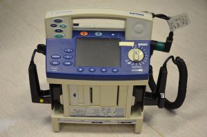
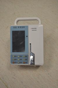
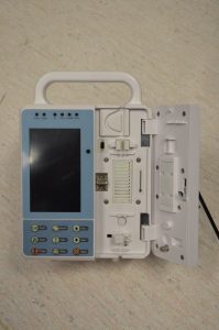
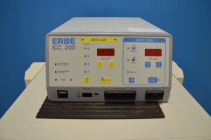
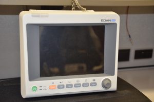
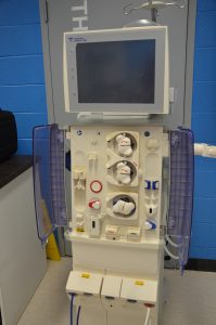
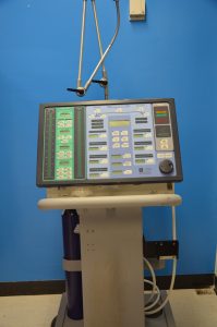
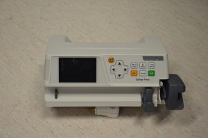
Figure 1.2.1.1 – 1.2.1.8 – A series of photographs that depict biomedical equipment Centennial College will provide you with hands-on experience. Equipment includes: infusion pumps, ventilators, defibrillators, electrosurgical units, patient monitors, dialysis machines and syringe pumps.
Although you are going to be learning about most of this equipment, the machines listed in bold (Defibrillators, Infusion pumps and Electrosurgical units) are the machines that you are going to have hands on experience with in this course (see above photos). The equipment listed in italics (Dialysis machines, physiological monitors, ventilators and syringe pumps) are equipment that you will likely get hands-on experience with in other BTEC courses. There are obviously dozens of different equipment that can be found in a hospital. The list found above only provide a general overview of what you may experience on the job-site.
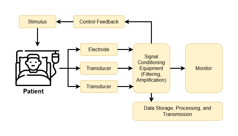
Figure 1.2.2 demonstrates the general principles of any piece of biomedical equipment using a block diagram method. Lets discuss each of these terms so we can have a general understanding of how biomedical equipment looks. Although this may appear simple, understanding the different components is integral to troubleshooting as the different errors a user receives will help you narrow your search to one of these general functions. For example, if there is no readings shown on one part of the screen, then you should start your troubleshooting at the mechanisms involved in output display.
Generalized Medical Instrumentation System
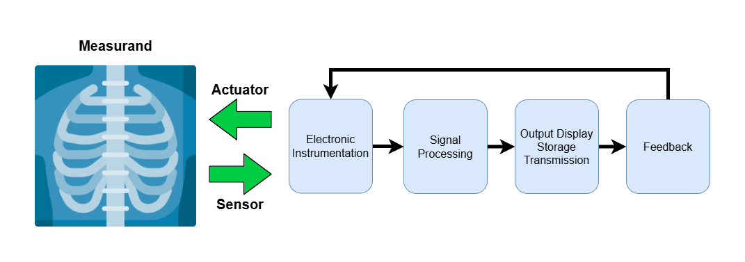
Measurand: Physical quantity, property or condition that the system measures. Below are a few examples of biomedical measurands:
- Internal – Blood pressure
- Body surface – ECG or EEG potentials
- Peripheral – Infrared radiation
- Offline – Extract tissue sample, blood analysis, or biopsy
Typical biomedical measurand quantities: Biopotential, pressure, flow, dimensions (imaging), displacement (velocity, acceleration and force), impedance, temperature and chemical concentration.
A sensor converts a physical measurand to an electrical output. For example, a heart rate monitor will take the measurand of your heart beat and will send it to the electronic components that will convert it to an electronic signal that can be viewed on a monitor. The most common types of sensors in biomedical systems are displacement sensors and pressure sensors. General sensor requirements:
- Selective: It should respond to a specific form of energy in the measurand. I.e. a heart rate monitor should not be affected by breathing rate
- Minimally invasive: invasive is requiring entry into a part of the body. A sensor should not enter the body any more than is necessary
- Sensors should not affect the response of the living tissue. The sensor itself should not distort or modify the signal but simply provide an accurate representation of the measurand
Signal Processing/Conditioning is the amplification and filtering of the signal (we will discuss these issues in a future lecture). This component acquires a signal from the sensor to make it suitable for display. It does so by making the signal large enough to see (amplification) and removes any signal that may be interfering with the measurement (filtering. Ex. If you are taking an electrical signal from the biceps muscles, a signal from the triceps muscle is not being shown).
Figure 1.2.3 is a block diagram of the above schematic. As you can see, a sensor (electrode or transducer) measures a measurand. This signal is then sent to the signal processing unit to be processed/conditioned (Ex. amplified and filtered). The signal can then be sent to the output display (monitor) or for data storage/transmission. In some machines (not all of them), devices can provide feedback based on the signal and increase/decrease the stimulus to the patient. For example: combined EMS/EMG machines. If the electrostimulation is so low that no signal is being observed, then the machine can slowly increase the amount of stimulation being applied to the patient.
