Module 4: Histology
Learning Objectives
By the end of this lab, students should be able to:
- Describe the major steps of the paraffin method
- Explain the importance of histological work in contributing to our understanding of development
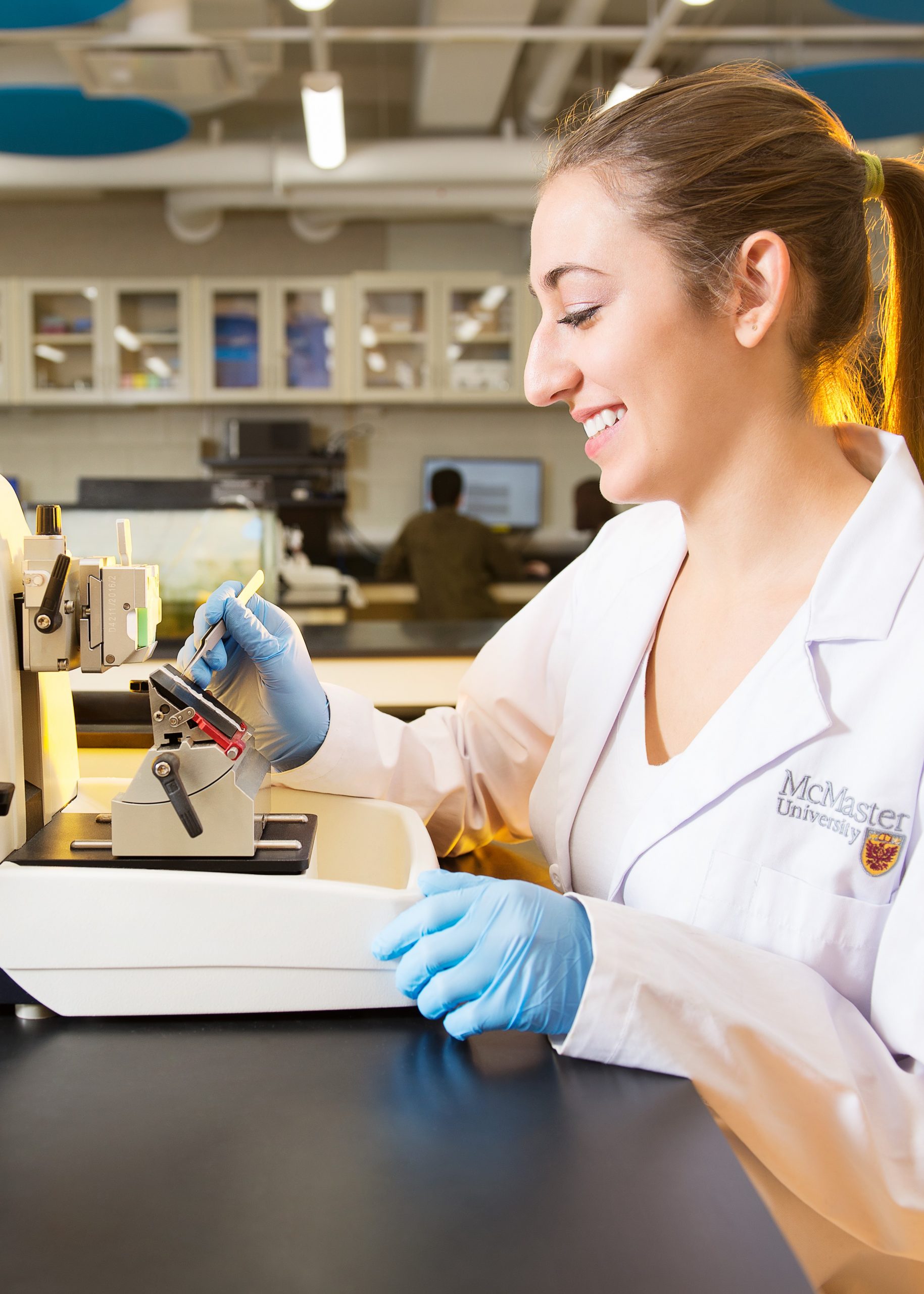
- Prepare a paraffin-wax embedded piece of mouse tissue in the proper orientation for preparing longitudinal or cross-sectional sections
- Practice sectioning embedded tissues using a microtome, and mounting the sections onto a microscope slide
- Prepare a permanently mounted slide of mouse tissue which has been stained with haematoxylin and eosin.
- Use the ‘Cell and Tissue Biology Atlas’ to assist you in identifying structures from student-prepared slides of mouse tissue
- Compose high quality images of stained mouse tissue using the compound microscope, image capturing software (Zen) and image analysis software (ImageJ)
- As a class, contribute to science communication by creating an open-access virtual slide library of mouse tissues
