Learning Objectives
By the end of this section, you will be able to:
Describe the bones of the lower limb, including the bones of the thigh, leg, ankle, and foot
- Appropriately name the regions of the lower limb and list the bones in each region
Like the upper limb, the lower limb is divided into three regions. The thigh is that portion of the lower limb located between the hip joint and knee joint. The leg is specifically the region between the knee joint and the ankle joint. Distal to the ankle is the foot. The lower limb contains 30 bones. These are the femur, patella, tibia, fibula, tarsal bones, metatarsal bones, and phalanges (see Chapter 8.1 Figure 8.2). The femur is the single bone of the thigh. The patella is the kneecap and articulates with the distal femur. The tibia is the larger, weight-bearing bone located on the medial side of the leg, and the fibula is the thin bone of the lateral leg. The bones of the foot are divided into three groups. The posterior portion of the foot is formed by a group of seven tarsal bones, whereas the mid-foot contains five elongated metatarsal bones. The toes contain 14 small phalanges.
Femur
The femur, or thigh bone, is the single bone of the thigh region (Figure 8.4.1). It is the longest and strongest bone of the body, and accounts for approximately one-quarter of a person’s total height. The rounded, proximal end is the head of the femur, which articulates with the acetabulum of the hip bone to form the hip joint.
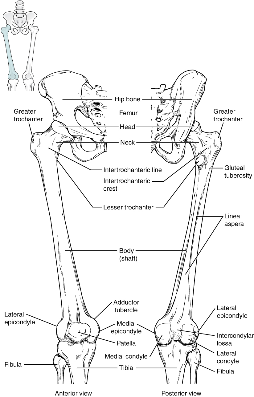
The narrowed region below the head is the neck of the femur. This is a common area for fractures of the femur. The greater trochanter is the large, upward, bony projection located above the base of the neck. Multiple muscles that act across the hip joint attach to the greater trochanter, which, because of its projection from the femur, gives additional leverage to these muscles. The greater trochanter can be felt just under the skin on the lateral side of your upper thigh.
The elongated shaft of the femur has a slight anterior bowing or curvature. Multiple muscles of the hip and thigh regions make long, thin attachments to the femur.
External Website

Watch this video to view how a fracture of the mid-femur is surgically repaired. How are the two portions of the broken femur stabilized during surgical repair of a fractured femur?
Patella
The patella (kneecap) is largest sesamoid bone of the body (see Figure 8.4.1). A sesamoid bone is a bone that is incorporated into the tendon of a muscle where that tendon crosses a joint. The sesamoid bone articulates with the underlying bones to prevent damage to the muscle tendon due to rubbing against the bones during movements of the joint. The patella is found in the tendon of the quadriceps femoris muscle, the large muscle of the anterior thigh that passes across the anterior knee to attach to the tibia. The patella articulates with the patellar surface of the femur and thus prevents rubbing of the muscle tendon against the distal femur. The patella also lifts the tendon away from the knee joint, which increases the leverage power of the quadriceps femoris muscle as it acts across the knee. The patella does not articulate with the tibia.
Homeostatic Imbalances – Runner’s Knee
Runner’s knee, also known as patellofemoral pain syndrome, is the most common overuse injury among runners. It is most frequent in adolescents and young adults, and is more common in females. It often results from excessive running, particularly downhill, but may also occur in athletes who do a lot of knee bending, such as jumpers, skiers, cyclists, weight lifters, and soccer players. It is felt as a dull, aching pain around the front of the knee and deep to the patella. The pain may be felt when walking or running, going up or down stairs, kneeling or squatting, or after sitting with the knee bent for an extended period.
Patellofemoral pain syndrome may be initiated by a variety of causes, including individual variations in the shape and movement of the patella, a direct blow to the patella, or flat feet or improper shoes that cause excessive turning in or out of the feet or leg. These factors may cause in an imbalance in the muscle pull that acts on the patella, resulting in an abnormal tracking of the patella that allows it to deviate too far toward the lateral side of the patellar surface on the distal femur.
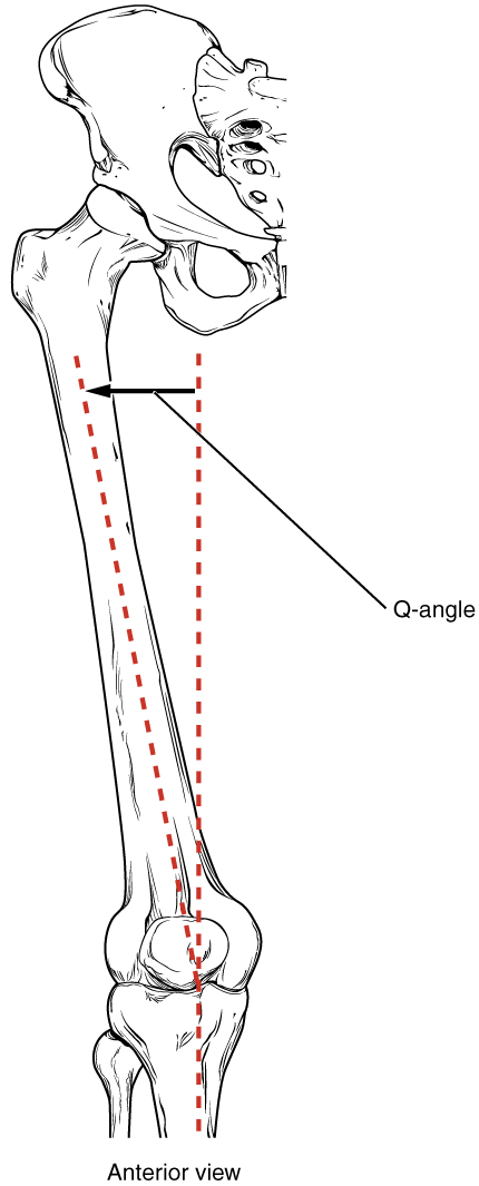
Because the hips are wider than the knee region, the femur has a diagonal orientation within the thigh, in contrast to the vertically oriented tibia of the leg (Figure 8.4.2). The Q-angle is a measure of how far the femur is angled laterally away from vertical. The Q-angle is normally 10–15 degrees, with females typically having a larger Q-angle due to their wider pelvis. During extension of the knee, the quadriceps femoris muscle pulls the patella both superiorly and laterally, with the lateral pull greater in women due to their large Q-angle. This makes women more vulnerable to developing patellofemoral pain syndrome than men. Normally, the large lip on the lateral side of the patellar surface of the femur compensates for the lateral pull on the patella, and thus helps to maintain its proper tracking. However, if the pull produced by the medial and lateral sides of the quadriceps femoris muscle is not properly balanced, abnormal tracking of the patella toward the lateral side may occur. With continued use, this produces pain and could result in damage to the articulating surfaces of the patella and femur, and the possible future development of arthritis. Treatment generally involves stopping the activity that produces knee pain for a period of time, followed by a gradual resumption of activity. Proper strengthening of the medial oblique portion of the quadriceps femoris muscle to correct for imbalances is also important to help prevent reoccurrence.
Tibia
The tibia (shin bone) is the medial bone of the leg and is larger than the fibula, with which it is paired (Figure 8.4.3). The tibia is the main weight-bearing bone of the leg and the second longest bone of the body, after the femur. The medial side of the tibia is located immediately under the skin, allowing it to be easily palpated down the entire length of the medial leg.
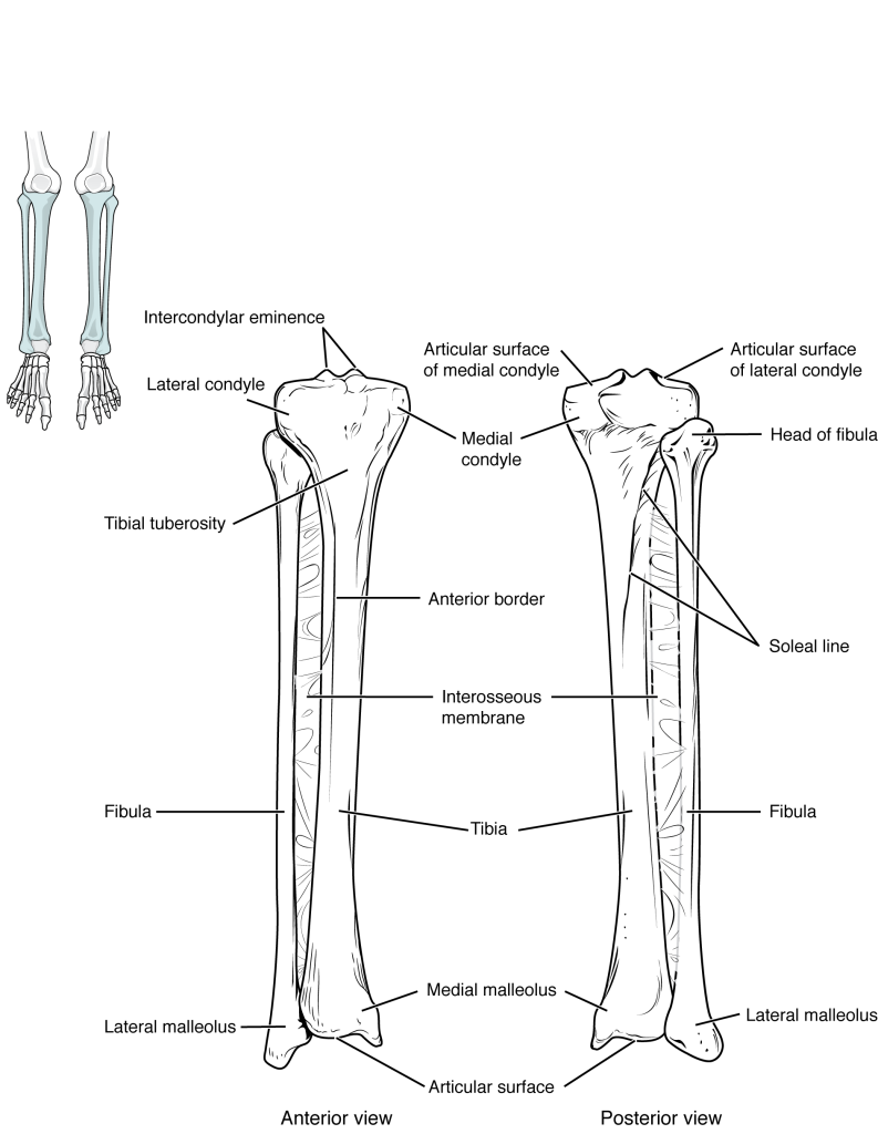
The proximal end of the tibia is greatly expanded. The two sides of this expansion form the medial and lateral condyles of the tibia and articulate with the femur to form the knee joint.
Fibula
The fibula is the slender bone located on the lateral side of the leg (see Figure 8.4.3). The fibula does not bear weight. It serves primarily for muscle attachments and thus is largely surrounded by muscles. Only the proximal and distal ends of the fibula can be easily palpated.
Tarsal Bones
The posterior half of the foot is formed by seven tarsal bones (Figure 8.4.4). The most superior bone is the talus. This has a relatively square-shaped, upper surface that articulates with the tibia and fibula to form the ankle joint. Three areas of articulation form the ankle joint: The talus bone articulates with the the tibia, the fibula and with the calcaneus (heel bone), the largest bone of the foot, which forms the heel. Body weight is transferred from the tibia to the talus to the calcaneus, which rests on the ground.

Metatarsal Bones
The foot is formed by the five metatarsal bones, which are located between the tarsal bones of the posterior foot and the phalanges of the toes (see Figure 8.4.4). These elongated bones are numbered 1–5, starting with the medial side of the foot. The first metatarsal bone is shorter and thicker than the others. The second metatarsal is the longest.
Phalanges
The toes contain a total of 14 phalanx bones (phalanges), arranged in a similar manner as the phalanges of the fingers (see Figure 8.4.4). The toes are numbered 1–5, starting with the big toe (hallux). The big toe has two phalanx bones, the proximal and distal phalanges. The remaining toes all have proximal, middle, and distal phalanges. A joint between adjacent phalanx bones is called an interphalangeal joint.
Chapter Review
The lower limb is divided into three regions. These are the thigh, located between the hip and knee joints; the leg, located between the knee and ankle joints; and distal to the ankle, the foot. There are 30 bones in each lower limb. These are the femur, patella, tibia, fibula, seven tarsal bones, five metatarsal bones, and 14 phalanges.
The femur is the single bone of the thigh. Its rounded head articulates with the acetabulum of the hip bone to form the hip joint.
The patella is a sesamoid bone located within a muscle tendon. It articulates with the patellar surface on the anterior side of the distal femur, thereby protecting the muscle tendon from rubbing against the femur.
The leg contains the large tibia on the medial side and the slender fibula on the lateral side. The tibia bears the weight of the body, whereas the fibula does not bear weight. The interosseous border of each bone is the attachment site for the interosseous membrane of the leg, the connective tissue sheet that unites the tibia and fibula.
The posterior foot is formed by the seven tarsal bones. The talus articulates superiorly with the tibia, the fibula to form the ankle joint. The talus articulates inferiorly with the calcaneus bone.
The five metatarsal bones form the anterior foot. The metatarsal heads, at their distal ends, articulate with the proximal phalanges of the toes. The big toe (toe number 1) has proximal and distal phalanx bones. The remaining toes have proximal, middle, and distal phalanges.
Glossary
- ankle joint
- joint that separates the leg and foot portions of the lower limb; formed by the articulations between the talus bone of the foot inferiorly, and the distal end of the tibia, medial malleolus of the tibia, and lateral malleolus of the fibula superiorly
- calcaneus
- heel bone; posterior, inferior tarsal bone that forms the heel of the foot
- femur
- thigh bone; the single bone of the thigh
- fibula
- thin, non-weight-bearing bone found on the lateral side of the leg
- foot
- portion of the lower limb located distal to the ankle joint
- greater trochanter
- large, bony expansion of the femur that projects superiorly from the base of the femoral neck
- hallux
- big toe; digit 1 of the foot
- head of the femur
- rounded, proximal end of the femur that articulates with the acetabulum of the hip bone to form the hip joint
- head of the fibula
- small, knob-like, proximal end of the fibula; articulates with the inferior aspect of the lateral condyle of the tibia
- head of the metatarsal bone
- expanded, distal end of each metatarsal bone
- hip joint
- joint located at the proximal end of the lower limb; formed by the articulation between the acetabulum of the hip bone and the head of the femur
- knee joint
- joint that separates the thigh and leg portions of the lower limb; formed by the articulations between the medial and lateral condyles of the femur, and the medial and lateral condyles of the tibia
- leg
- portion of the lower limb located between the knee and ankle joints
- lesser trochanter
- small, bony projection on the medial side of the proximal femur, at the base of the femoral neck
- ligament of the head of the femur
- ligament that spans the acetabulum of the hip bone and the fovea capitis of the femoral head
- metatarsal bone
- one of the five elongated bones that forms the anterior half of the foot; numbered 1–5, starting on the medial side of the foot
- metatarsophalangeal joint
- articulation between a metatarsal bone of the foot and the proximal phalanx bone of a toe
- neck of the femur
- narrowed region located inferior to the head of the femur
- patella
- kneecap; the largest sesamoid bone of the body; articulates with the distal femur
- phalanx bone of the foot
- (plural = phalanges) one of the 14 bones that form the toes; these include the proximal and distal phalanges of the big toe, and the proximal, middle, and distal phalanx bones of toes two through five
- proximal tibiofibular joint
- articulation between the head of the fibula and the inferior aspect of the lateral condyle of the tibia
- shaft of the femur
- cylindrically shaped region that forms the central portion of the femur
- shaft of the fibula
- elongated, slender portion located between the expanded ends of the fibula
- shaft of the tibia
- triangular-shaped, central portion of the tibia
- talus
- tarsal bone that articulates superiorly with the tibia and fibula at the ankle joint.
- thigh
- portion of the lower limb located between the hip and knee joints
- tibia
- shin bone; the large, weight-bearing bone located on the medial side of the leg
Solutions
Answers for Critical Thinking Questions
- The lower limb is divided into three regions. The thigh is the region located between the hip and knee joints. It contains the femur and the patella. The hip joint is formed by the articulation between the acetabulum of the hip bone and the head of the femur. The leg is the region between the knee and ankle joints, and contains the tibia (medially) and the fibula (laterally). The knee joint is formed by the articulations between the medial and lateral condyles of the femur, and the medial and lateral condyles of the tibia. Also associated with the knee is the patella, which articulates with the patellar surface of the distal femur. The foot is found distal to the ankle and contains 26 bones. The ankle joint is formed by the articulations between the talus bone of the foot and the distal end of the tibia, the medial malleolus of the tibia, and the lateral malleolus of the fibula. The posterior foot contains the seven tarsal bones, which are the talus, calcaneus, navicular, cuboid, and the medial, intermediate, and lateral cuneiform bones. The anterior foot consists of the five metatarsal bones, which are numbered 1–5 starting on the medial side of the foot. The toes contain 14 phalanx bones, with the big toe (toe number 1) having a proximal and a distal phalanx, and the other toes having proximal, middle, and distal phalanges.
- The talus bone articulates superiorly with the tibia and fibula at the ankle joint, with body weight passed from the tibia to the talus. Body weight from the talus is transmitted to the ground by both ends of the medial and lateral longitudinal foot arches. Weight is passed posteriorly through both arches to the calcaneus bone, which forms the heel of the foot and is in contact with the ground. On the medial side of the foot, body weight is passed anteriorly from the talus bone to the navicular bone, and then to the medial, intermediate, and lateral cuneiform bones. The cuneiform bones pass the weight anteriorly to the first, second, and third metatarsal bones, whose heads (distal ends) are in contact with the ground. On the lateral side, body weight is passed anteriorly from the talus through the calcaneus, cuboid, and fourth and fifth metatarsal bones. The talus bone thus transmits body weight posteriorly to the calcaneus and anteriorly through the navicular, cuneiform, and cuboid bones, and metatarsals one through five.
This work, Anatomy & Physiology, is adapted from Anatomy & Physiology by OpenStax, licensed under CC BY. This edition, with revised content and artwork, is licensed under CC BY-SA except where otherwise noted.
Images, from Anatomy & Physiology by OpenStax, are licensed under CC BY except where otherwise noted.
Access the original for free at https://openstax.org/books/anatomy-and-physiology/pages/1-introduction.
