Learning Objectives
By the end of this section, you will be able to:
- Describe how the features of the pelvis differ between the adult male and female pelvis
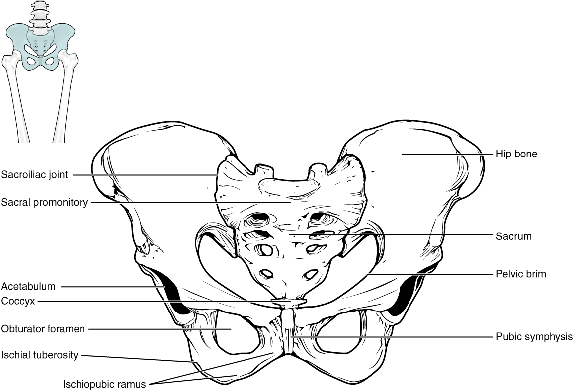
The two hip bones are together called the pelvic girdle (hip girdle) and serve as the attachment point for each lower limb. When the two hip bones are combined with the sacrum and coccyx of the axial skeleton, they are referred to as the pelvis. The right and left hip bones also converge anteriorly to attach to each other at the pubic symphysis (Figure 8.3.1).
Unlike the bones of the pectoral girdle, which are highly mobile to enhance the range of upper limb movements, the bones of the pelvis are strongly united to each other to form a largely immobile, weight-bearing structure. This is important for stability because it enables the weight of the body to be easily transferred laterally from the vertebral column, through the pelvic girdle and hip joints, and into the weight bearing lower limb(s). Thus, the immobility of the pelvis provides a strong foundation for the upper body as it rests on top of the mobile lower limbs.

Hip Bone
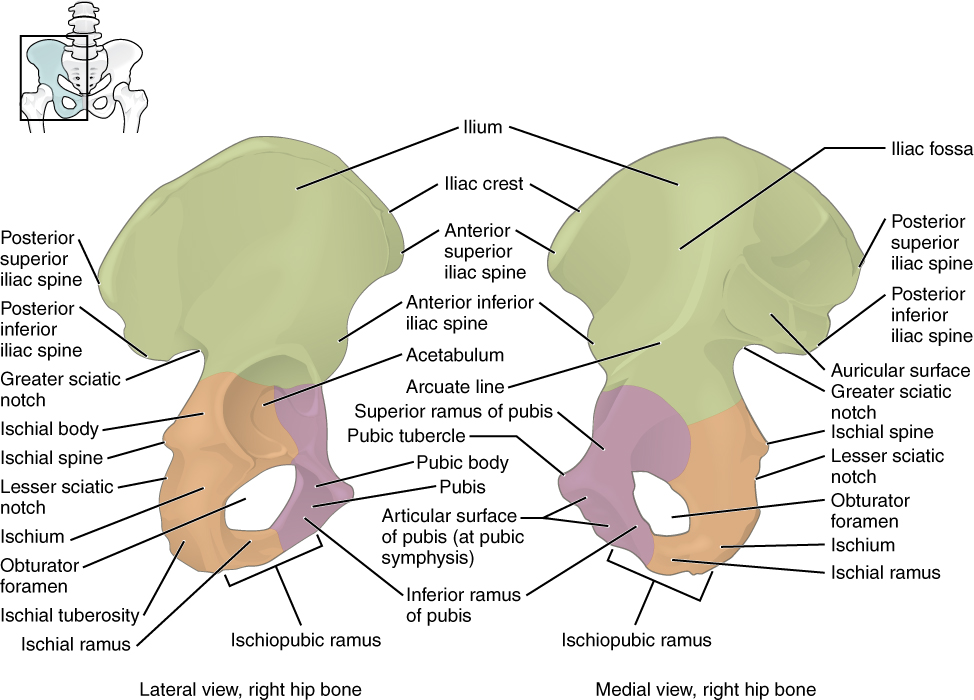
The hip (or coxal) bones form the pelvic girdle portion of the pelvis. The hip bones are large, curved bones that form the lateral and anterior aspects of the pelvis. Each adult hip bone is formed by three separate bones that fuse together during the late teenage years. These bony components are the ilium, ischium, and pubis (Figure 8.3.2). These names are retained and used to define the three regions of the adult hip bone.

The ilium is the fan-like, superior region that forms the largest part of the hip bone. It is firmly united to the sacrum at the largely immobile sacroiliac joint (see Figure 8.3.1). The ischium forms the posteroinferior region of each hip bone. It supports the body when sitting. The pubis forms the anterior portion of the hip bone. The pubis curves medially, where it joins to the pubis of the opposite hip bone at a specialized joint called the pubic symphysis.
Ilium
When you place your hands on your waist, you can feel the arching, superior margin of the ilium along your waistline (see Figure 8.3.2). This curved, superior margin of the ilium is the iliac crest.
Ischium
The ischium forms the posterolateral portion of the hip bone (see Figure 8.3.2). The large, roughened area is the ischial tuberosity. This serves as the attachment for the posterior thigh muscles and also carries the weight of the body when sitting. You can feel the ischial tuberosity if you wiggle your pelvis against the seat of a chair.
Pubis
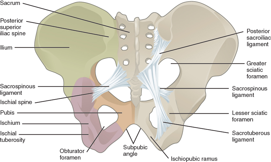
The pubis forms the anterior portion of the hip bone (see Figure 8.3.2). The enlarged medial portion of the pubis is the pubic body. The pubic body is joined to the pubic body of the opposite hip bone by the pubic symphysis. (Figure 8.3.3).
Pelvis
The pelvis consists of four bones: the right and left hip bones, the sacrum, and the coccyx (see Figure 8.3.1). The pelvis has several important functions. Its primary role is to support the weight of the upper body when sitting and to transfer this weight to the lower limbs when standing. It serves as an attachment point for trunk and lower limb muscles, and also protects the internal pelvic organs. When standing in the anatomical position, the pelvis is tilted anteriorly. In this position, the anterior superior iliac spines and the pubic tubercles lie in the same vertical plane, and the anterior (internal) surface of the sacrum faces forward and downward.
The three areas of each hip bone, the ilium, pubis, and ischium, converge centrally to form a deep, cup-shaped cavity called the acetabulum. This is located on the lateral side of the hip bone and is part of the hip joint. The large opening in the anteroinferior hip bone between the ischium and pubis is the obturator foramen. This space is largely filled in by a layer of connective tissue and serves for the attachment of muscles on both its internal and external surfaces.
Several ligaments unite the bones of the pelvis (Figure 8.3.3). The largely immobile sacroiliac joint is supported by a pair of strong ligaments that are attached between the sacrum and ilium portions of the hip bone. These ligaments help to support and immobilize the sacrum as it carries the weight of the body.

External Website

Watch this video for a 3-D view of the pelvis and its associated ligaments. What is the large opening in the bony pelvis, located between the ischium and pubic regions, and what two parts of the pubis contribute to the formation of this opening?

Comparison of the Female and Male Pelvis
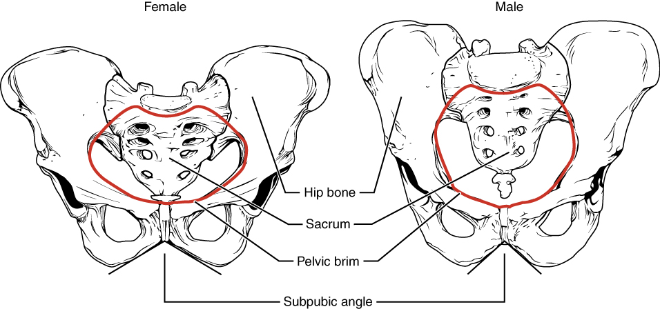
The differences between the adult female and male pelvis relate to function and body size. In general, the bones of the male pelvis are thicker and heavier, adapted for support of the male’s heavier physical build and stronger muscles. The greater sciatic notch of the male hip bone is narrower and deeper than the broader notch of females. Because the female pelvis is adapted for childbirth, it is wider than the male pelvis, as evidenced by the distance between the anterior superior iliac spines (see Figure 8.3.4). The female sacrum is wider, shorter, and less curved, giving the female pelvic inlet (pelvic brim) a more rounded or oval shape compared to males. Because of the obvious differences between female and male hip bones, this is the one bone of the body that allows for the most accurate sex determination.
Career Connection – Forensic Pathology and Forensic Anthropology
A forensic pathologist (also known as a medical examiner) is a medically trained physician who has been specifically trained in pathology to examine the bodies of the deceased to determine the cause of death. A forensic pathologist applies his or her understanding of disease as well as toxins, blood and DNA analysis, firearms and ballistics, and other factors to assess the cause and manner of death. At times, a forensic pathologist will be called to testify under oath in situations that involve a possible crime. Forensic pathology is a field that has received much media attention on television shows or following a high-profile death.
While forensic pathologists are responsible for determining whether the cause of someone’s death was natural, a suicide, accidental, or a homicide, there are times when uncovering the cause of death is more complex, and other skills are needed. Forensic anthropology brings the tools and knowledge of physical anthropology and human osteology (the study of the skeleton) to the task of investigating a death. A forensic anthropologist assists medical and legal professionals in identifying human remains. The science behind forensic anthropology involves the study of archaeological excavation; the examination of hair; an understanding of plants, insects, and footprints; the ability to determine how much time has elapsed since the person died; the analysis of past medical history and toxicology; the ability to determine whether there are any postmortem injuries or alterations of the skeleton; and the identification of the decedent (deceased person) using skeletal and dental evidence.
Due to the extensive knowledge and understanding of excavation techniques, a forensic anthropologist is an integral and invaluable team member to have on-site when investigating a crime scene, especially when the recovery of human skeletal remains is involved. When remains are bought to a forensic anthropologist for examination, he or she must first determine whether the remains are in fact human. Once the remains have been identified as belonging to a person and not to an animal, the next step is to approximate the individual’s age, sex, race, and height. The differences in the male and female pelvis aid in this identification process. The forensic anthropologist does not determine the cause of death, but rather provides information to the forensic pathologist, who will use all of the data collected to make a final determination regarding the cause of death.
Chapter Review
The pelvic girdle, consisting of two hip bones, serves to attach the lower limbs to the sacrum of the axial skeleton. The right and left hip bones converge anteriorly and articulate with each other at the pubic symphysis. The combination of the two hip bones, the sacrum, and the coccyx forms the pelvis. The pelvis has a pronounced anterior tilt. The primary function of the pelvis is to support the upper body and transfer body weight to the lower limbs. It also serves as the site of attachment for multiple muscles.
A hip bone consists of three regions: the ilium, ischium, and pubis. The ilium forms the large, fan-like region of the hip bone. The superior margin of this area is the iliac crest. The auricular surface of the ilium articulates with the sacrum to form the sacroiliac joint.
The posterolateral portion of the hip bone is the ischium. It has the expanded ischial tuberosity, which supports body weight when sitting.
The pubis forms the anterior portion of the hip bone. The body of the pubis articulates with the pubis of the opposite hip bone at the pubic symphysis.
The lateral side of the hip bone has the cup-like acetabulum, which is part of the hip joint. The large anterior opening is the obturator foramen. The sacroiliac joint is supported by the anterior and posterior sacroiliac ligaments.
The broad space of the upper pelvis is the greater pelvis, and the narrow, inferior space is the lesser pelvis. These areas are separated by the pelvic brim (pelvic inlet). The inferior opening of the pelvis is the pelvic outlet. Compared to the male, the female pelvis is wider to accommodate childbirth, has a larger subpubic angle, and a broader greater sciatic notch.
Interactive Link Questions
Watch this video for a 3-D view of the pelvis and its associated ligaments. What is the large opening in the bony pelvis, located between the ischium and pubic regions, and what two parts of the pubis contribute to the formation of this opening?
Answer: The obturator foramen is located between the ischium and the pubis. The superior and inferior pubic rami contribute to the boundaries of the obturator foramen.
Review Questions
Critical Thinking Questions
1. Describe the structure and function of the pelvic girdle.
2. Discuss the ways in which the female pelvis is adapted for childbirth.
Glossary
- acetabulum
- large, cup-shaped cavity located on the lateral side of the hip bone; formed by the junction of the ilium, pubis, and ischium portions of the hip bone
- anterior inferior iliac spine
- small, bony projection located on the anterior margin of the ilium, below the anterior superior iliac spine
- anterior sacroiliac ligament
- strong ligament between the sacrum and the ilium portions of the hip bone that supports the anterior side of the sacroiliac joint
- anterior superior iliac spine
- rounded, anterior end of the iliac crest
- arcuate line of the ilium
- smooth ridge located at the inferior margin of the iliac fossa; forms the lateral portion of the pelvic brim
- auricular surface of the ilium
- roughened area located on the posterior, medial side of the ilium of the hip bone; articulates with the auricular surface of the sacrum to form the sacroiliac joint
- coxal bone
- hip bone
- greater pelvis
- (also, greater pelvic cavity or false pelvis) broad space above the pelvic brim defined laterally by the fan-like portion of the upper ilium
- greater sciatic foramen
- pelvic opening formed by the greater sciatic notch of the hip bone, the sacrum, and the sacrospinous ligament
- greater sciatic notch
- large, U-shaped indentation located on the posterior margin of the ilium, superior to the ischial spine
- hip bone
- coxal bone; single bone that forms the pelvic girdle; consists of three areas, the ilium, ischium, and pubis
- iliac crest
- curved, superior margin of the ilium
- iliac fossa
- shallow depression found on the anterior and medial surfaces of the upper ilium
- ilium
- superior portion of the hip bone
- inferior pubic ramus
- narrow segment of bone that passes inferiorly and laterally from the pubic body; joins with the ischial ramus to form the ischiopubic ramus
- ischial ramus
- bony extension projecting anteriorly and superiorly from the ischial tuberosity; joins with the inferior pubic ramus to form the ischiopubic ramus
- ischial spine
- pointed, bony projection from the posterior margin of the ischium that separates the greater sciatic notch and lesser sciatic notch
- ischial tuberosity
- large, roughened protuberance that forms the posteroinferior portion of the hip bone; weight-bearing region of the pelvis when sitting
- ischiopubic ramus
- narrow extension of bone that connects the ischial tuberosity to the pubic body; formed by the junction of the ischial ramus and inferior pubic ramus
- ischium
- posteroinferior portion of the hip bone
- lesser pelvis
- (also, lesser pelvic cavity or true pelvis) narrow space located within the pelvis, defined superiorly by the pelvic brim (pelvic inlet) and inferiorly by the pelvic outlet
- lesser sciatic foramen
- pelvic opening formed by the lesser sciatic notch of the hip bone, the sacrospinous ligament, and the sacrotuberous ligament
- lesser sciatic notch
- shallow indentation along the posterior margin of the ischium, inferior to the ischial spine
- obturator foramen
- large opening located in the anterior hip bone, between the pubis and ischium regions
- pelvic brim
- pelvic inlet; the dividing line between the greater and lesser pelvic regions; formed by the superior margin of the pubic symphysis, the pectineal lines of each pubis, the arcuate lines of each ilium, and the sacral promontory
- pelvic girdle
- hip girdle; consists of a single hip bone, which attaches a lower limb to the sacrum of the axial skeleton
- pelvic inlet
- pelvic brim
- pelvic outlet
- inferior opening of the lesser pelvis; formed by the inferior margin of the pubic symphysis, right and left ischiopubic rami and sacrotuberous ligaments, and the tip of the coccyx
- pelvis
- ring of bone consisting of the right and left hip bones, the sacrum, and the coccyx
- posterior inferior iliac spine
- small, bony projection located at the inferior margin of the auricular surface on the posterior ilium
- posterior sacroiliac ligament
- strong ligament spanning the sacrum and ilium of the hip bone that supports the posterior side of the sacroiliac joint
- posterior superior iliac spine
- rounded, posterior end of the iliac crest
- pubic arch
- bony structure formed by the pubic symphysis, and the bodies and inferior pubic rami of the right and left pubic bones
- pubic body
- enlarged, medial portion of the pubis region of the hip bone
- pubic symphysis
- joint formed by the articulation between the pubic bodies of the right and left hip bones
- pubic tubercle
- small bump located on the superior aspect of the pubic body
- pubis
- anterior portion of the hip bone
- sacroiliac joint
- joint formed by the articulation between the auricular surfaces of the sacrum and ilium
- sacrospinous ligament
- ligament that spans the sacrum to the ischial spine of the hip bone
- sacrotuberous ligament
- ligament that spans the sacrum to the ischial tuberosity of the hip bone
- subpubic angle
- inverted V-shape formed by the convergence of the right and left ischiopubic rami; this angle is greater than 80 degrees in females and less than 70 degrees in males
- superior pubic ramus
- narrow segment of bone that passes laterally from the pubic body to join the ilium
Solutions
Answers for Critical Thinking Questions
- The pelvic girdle is very stable with large strong ligaments maintaining the integrity of the joints.
- Compared to the male, the female pelvis is wider to accommodate childbirth. Thus, the female pelvis has greater distances between the anterior superior iliac spines and between the ischial tuberosities. The greater width of the female pelvis results in a larger subpubic angle. The pelvic brim (pelvic inlet) of the female has a rounded or oval shape. The lesser pelvic cavity is wider and more shallow in females, and the pelvic outlet is larger than in males. Thus, the greater width of the female pelvis, with its larger pelvic inlet, lesser pelvis, and pelvic outlet, are important for childbirth because the baby must pass through the pelvis during delivery.
This work, Anatomy & Physiology, is adapted from Anatomy & Physiology by OpenStax, licensed under CC BY. This edition, with revised content and artwork, is licensed under CC BY-SA except where otherwise noted.
Images, from Anatomy & Physiology by OpenStax, are licensed under CC BY except where otherwise noted.
Access the original for free at https://openstax.org/books/anatomy-and-physiology/pages/1-introduction.

