Learning Objectives
By the end of this section, you will be able to:
- Describe the bones of the upper limb, including the bones of the arm, forearm, wrist, and hand
The upper limb is divided into three regions. These consist of the arm, located between the shoulder and elbow joints; the forearm, which is between the elbow and wrist joints; and the hand, which is located distal to the wrist. There are 30 bones in each upper limb. The humerus is the single bone of the arm, and the ulna (medially) and the radius (laterally) are the paired bones of the forearm. The base of the hand contains eight carpal bones, and the palm of the hand is formed by five metacarpal bones. The fingers and thumb contain a total of 14 phalanges.
Humerus
The humerus is the single bone of the arm region (Figure 8.2.1). At its proximal end is the head of the humerus. This is the large, round, smooth region that faces medially. The head articulates with the glenoid cavity of the scapula to form the glenohumeral (shoulder) joint (see Chapter 9).
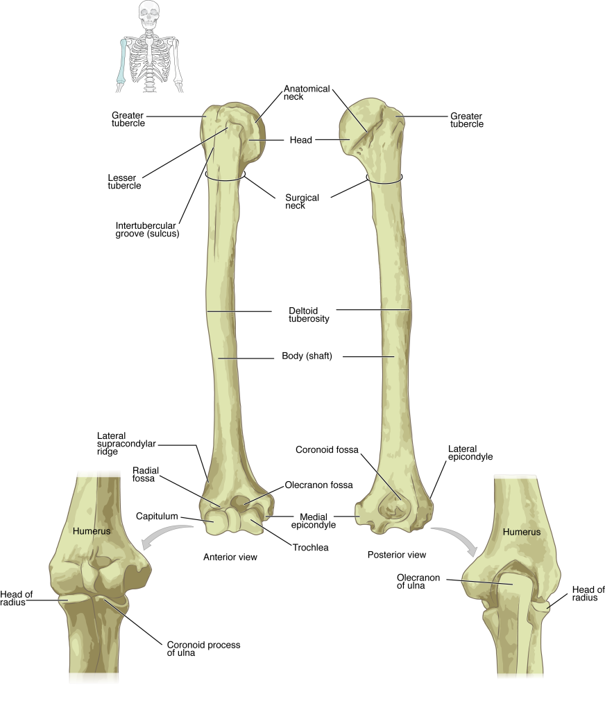
The distal end of the humerus has two articulation areas, which join the ulna and radius bones of the forearm to form the elbow joint.
Ulna
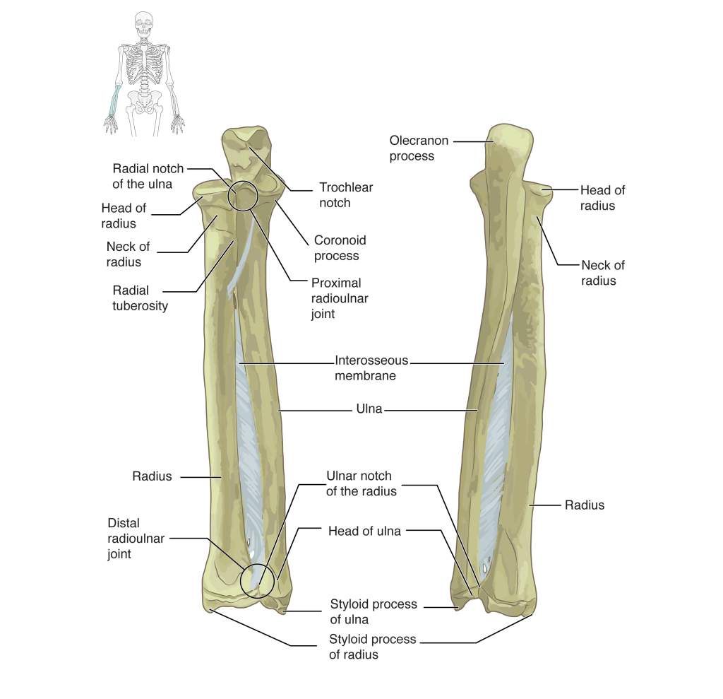
The ulna is the medial bone of the forearm. It runs parallel to the radius, which is the lateral bone of the forearm (Figure 8.2.2).
In the anatomical position, with the elbow fully extended and the palms facing forward, the arm and forearm do not form a straight line. Instead, the forearm deviates laterally by 5–15 degrees from the line of the arm. This deviation allows the forearm and hand to swing freely or to carry an object without hitting the hip.
Radius
The radius runs parallel to the ulna, on the lateral (thumb) side of the forearm (see Figure 8.2.2). The head of the radius is a disc-shaped structure that forms the proximal end.
Carpal Bones
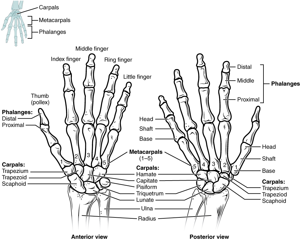
The wrist and base of the hand are formed by a series of eight small carpal bones (see Figure 8.2.3). The carpal bones are arranged in two rows, forming a proximal row of four carpal bones and a distal row of four carpal bones.
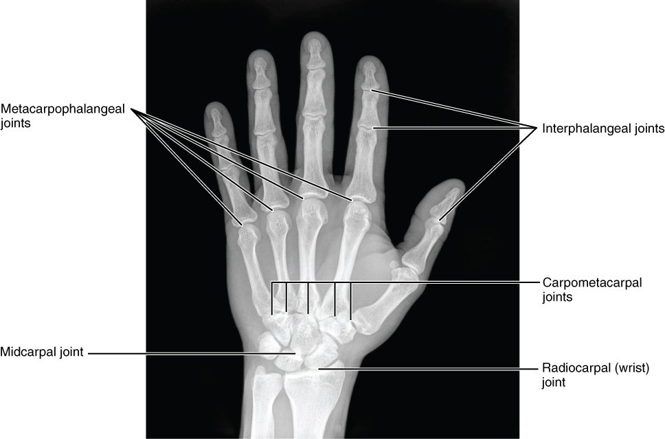
The carpal bones form the base of the hand. This can be seen in the radiograph (X-ray image) of the hand that shows the relationships of the hand bones to the skin creases of the hand (see Figure 8.2.4). Within the carpal bones, the four proximal bones are united to each other by ligaments to form a unit.
The four distal carpal bones are also held together as a group by ligaments. The proximal and distal rows of carpal bones articulate with each other to form the midcarpal joint (see Figure 8.2.4). Together, the radiocarpal and midcarpal joints are responsible for all movements of the hand at the wrist.
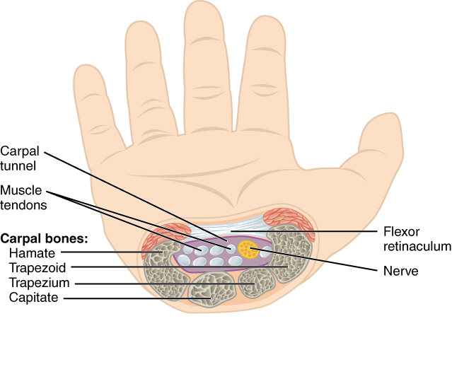
In the hand, the carpal bones form a U-shaped grouping. A strong ligament called the flexor retinaculum spans the top of this U-shaped area to maintain this grouping of the carpal bones (Figure 8.2.5). Together, the carpal bones and the flexor retinaculum form a passageway called the carpal tunnel. The tendons of nine muscles of the anterior forearm and an important nerve (the median nerve) pass through this narrow tunnel to enter the hand. Overuse of the muscle tendons or wrist injury can produce inflammation and swelling within this space. This produces compression of the nerve, resulting in carpal tunnel syndrome, which is characterized by pain or numbness, and muscle weakness in those areas of the hand supplied by this nerve.
Metacarpal Bones
The palm of the hand contains five elongated metacarpal bones. These bones lie between the carpal bones of the wrist and the bones of the fingers and thumb (see Figure 8.2.3). The metacarpal bones are numbered 1–5, beginning at the thumb.
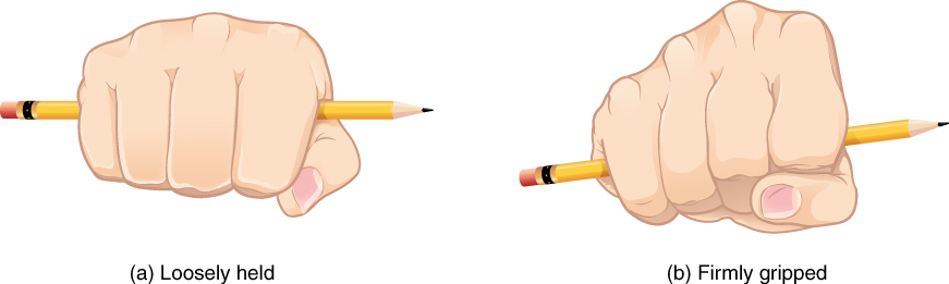
The first metacarpal bone, at the base of the thumb, is separated from the other metacarpal bones. This allows it a freedom of motion that is independent of the other metacarpal bones, which is very important for thumb mobility. The remaining metacarpal bones are united together to form the palm of the hand. The anterior movement of these bones, particularly the fifth metacarpal bone, increases the strength of contact for the medial hand during gripping actions.
Chapter Review
Each upper limb is divided into three regions and contains a total of 30 bones. The arm is the region located between the shoulder and elbow joints. This area contains the humerus. The proximal humerus consists of the head, which articulates with the scapula at the glenohumeral joint.
The forearm is the region of the upper limb located between the elbow and wrist joints. This region contains two bones, the ulna medially and the radius on the lateral (thumb) side. The elbow joint is formed by the articulation between the humerus and the ulna, plus the articulation between the humerus and the head of the radius.
The base of the hand is formed by eight carpal bones. The carpal bones are united into two rows of bones. The proximal and distal carpal rows articulate with each other at the midcarpal joint. The carpal bones, together with the flexor retinaculum, also form the carpal tunnel of the wrist.
The five metacarpal bones form the palm of the hand. The metacarpal bones are numbered 1–5, starting with the thumb side. The first metacarpal bone is freely mobile, but the other bones are united as a group. The digits are also numbered 1–5, with the thumb being number 1. The fingers and thumb contain a total of 14 phalanges (phalanx bones). The thumb contains a proximal and a distal phalanx, whereas the remaining digits each contain proximal, middle, and distal phalanges.
Glossary
- anatomical neck
- line on the humerus located around the outside margin of the humeral head
- arm
- region of the upper limb located between the shoulder and elbow joints; contains the humerus bone
- carpal bone
- one of the eight small bones that form the wrist and base of the hand
- carpal tunnel
- passageway between the anterior forearm and hand formed by the carpal bones and flexor retinaculum
- carpometacarpal joint
- articulation between one of the carpal bones in the distal row and a metacarpal bone of the hand
- distal radioulnar joint
- articulation between the head of the ulna and the ulnar notch of the radius
- elbow joint
- joint located between the upper arm and forearm regions of the upper limb
- flexor retinaculum
- strong band of connective tissue at the anterior wrist that spans the top of the U-shaped grouping of the carpal bones to form the roof of the carpal tunnel
- forearm
- region of the upper limb located between the elbow and wrist joints; contains the radius and ulna bones
- hand
- region of the upper limb distal to the wrist joint
- head of the humerus
- smooth, rounded region on the medial side of the proximal humerus; articulates with the glenoid fossa of the scapula to form the glenohumeral (shoulder) joint
- head of the radius
- disc-shaped structure that forms the proximal end of the radius; articulates with the capitulum of the humerus as part of the elbow joint, and with the radial notch of the ulna as part of the proximal radioulnar joint
- head of the ulna
- small, rounded distal end of the ulna; articulates with the ulnar notch of the distal radius, forming the distal radioulnar joint
- humerus
- single bone of the upper arm
- interphalangeal joint
- articulation between adjacent phalanx bones of the hand or foot digits
- metacarpal bone
- one of the five long bones that form the palm of the hand; numbered 1–5, starting on the lateral (thumb) side of the hand
- metacarpophalangeal joint
- articulation between the distal end of a metacarpal bone of the hand and a proximal phalanx bone of the thumb or a finger
- neck of the radius
- narrowed region immediately distal to the head of the radius
- radiocarpal joint
- wrist joint, located between the forearm and hand regions of the upper limb
- radius
- bone located on the lateral side of the forearm
- shaft of the humerus
- narrow, elongated, central region of the humerus
- shaft of the radius
- narrow, elongated, central region of the radius
- shaft of the ulna
- narrow, elongated, central region of the ulna
- ulna
- bone located on the medial side of the forearm
This work, Anatomy & Physiology, is adapted from Anatomy & Physiology by OpenStax, licensed under CC BY. This edition, with revised content and artwork, is licensed under CC BY-SA except where otherwise noted.
Images, from Anatomy & Physiology by OpenStax, are licensed under CC BY except where otherwise noted.
Access the original for free at https://openstax.org/books/anatomy-and-physiology/pages/1-introduction.
