Learning Objectives
By the end of this section, you will be able to:
Describe the functions of the skeletal system and define its two major subdivisions
- Discuss the functions of the skeletal system
- Describe and distinguish between the axial skeleton and appendicular skeleton
The skeletal system includes all of the bones, cartilages, and ligaments of the body that support and give shape to the body and body structures, whereas the skeleton consists of the bones of the body. For adults, there are 206 named bones in the skeleton. Younger individuals have higher numbers of bones because some bones fuse together during childhood and adolescence. The primary functions of the skeleton are to provide a rigid, internal structure that protects internal organs and supports the weight of the body, and to provide a structure upon which muscles can act to produce movements of the body. The bones of the skeleton also serve as the primary storage site for important minerals such as calcium and phosphate. The bone marrow found within bones stores fat and houses the blood-cell producing tissue of the body.
The skeleton is subdivided into two major divisions—the axial and appendicular.
The Axial Skeleton
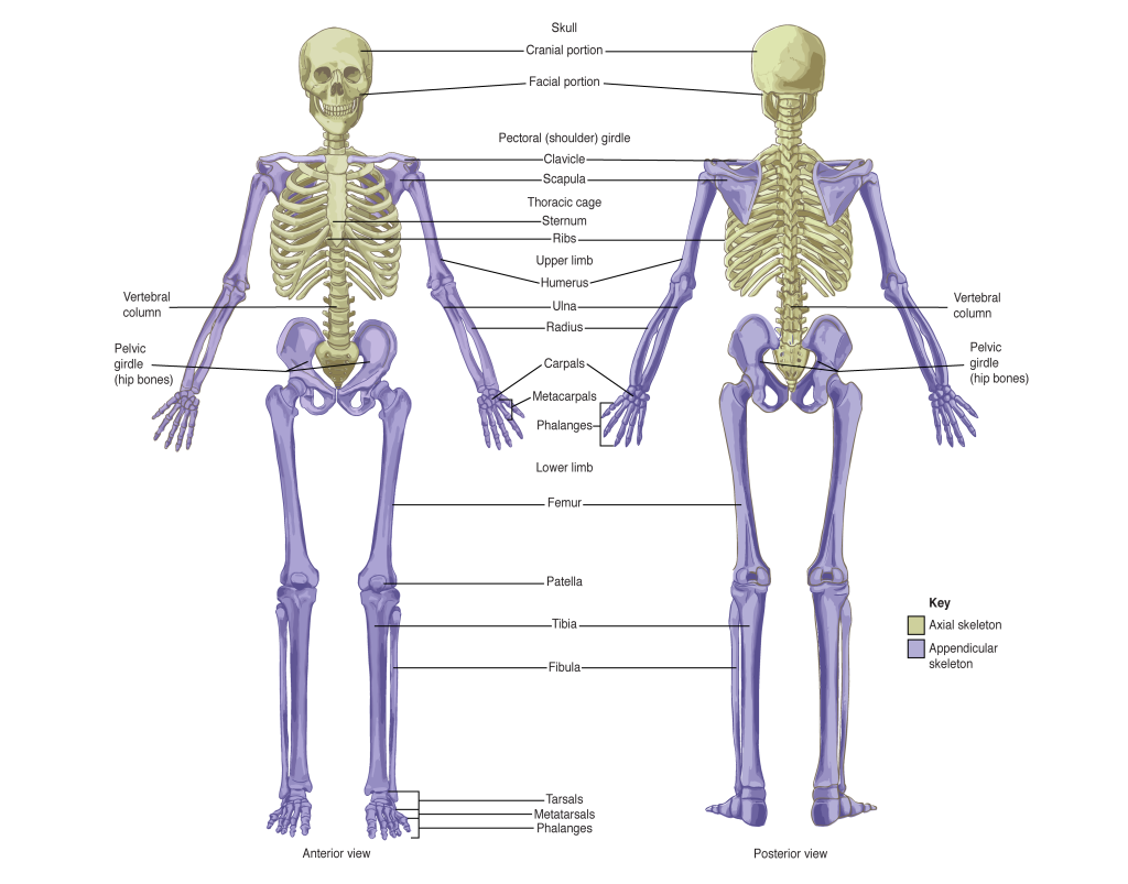
The axial skeleton forms the vertical, central axis of the body and includes all bones of the head, neck, chest, and back (Figure 7.1.1). It serves to protect the brain, spinal cord, heart, and lungs. It also serves as the attachment site for muscles that move the head, neck, and back, and for muscles that act across the shoulder and hip joints to move their corresponding limbs.
The axial skeleton of the adult consists of 80 bones, comprising the skull, the vertebral column, and the thoracic cage.

The Appendicular Skeleton
The appendicular skeleton includes all bones of the upper and lower limbs, plus the bones of the pectoral and pelvic girdles that attach each limb to the axial skeleton. There are 126 bones in the appendicular skeleton of an adult. The lower portion of the appendicular skeleton is specialized for stability during walking or running. In contrast, the upper portion of the appendicular skeleton has greater mobility and ranges of motion, features that allow you to lift and carry objects. The bones of the appendicular skeleton are covered in a separate chapter.
Chapter Review
The skeletal system includes all of the bones, cartilages, and ligaments of the body. It serves to support the body, protect the brain and other internal organs, and provides a rigid structure upon which muscles can pull to generate body movements. It also stores fat and the tissue responsible for the production of blood cells. The skeleton is subdivided into two parts. The axial skeleton forms a vertical axis that includes the head, neck, back, and chest. It has 80 bones and consists of the skull, vertebral column, and thoracic cage. The adult vertebral column consists of 24 vertebrae plus the sacrum and coccyx. The thoracic cage is formed by 12 pairs of ribs and the sternum. The appendicular skeleton consists of 126 bones in the adult and includes all of the bones of the upper and lower limbs plus the bones that anchor each limb to the axial skeleton.
Review Questions
Critical Thinking Question
1. Define the two divisions of the skeleton.
2. Discuss the functions of the axial skeleton.
Glossary
- appendicular skeleton
- all bones of the upper and lower limbs, plus the girdle bones that attach each limb to the axial skeleton
- axial skeleton
- central, vertical axis of the body, including the skull, vertebral column, and thoracic cage
- coccyx
- small bone located at inferior end of the adult vertebral column that is formed by the fusion of four coccygeal vertebrae; also referred to as the “tailbone”
- ear ossicles
- three small bones located in the middle ear cavity that serve to transmit sound vibrations to the inner ear
- hyoid bone
- small, U-shaped bone located in upper neck that does not contact any other bone
- ribs
- thin, curved bones of the chest wall
- sacrum
- single bone located near the inferior end of the adult vertebral column that is formed by the fusion of five sacral vertebrae; forms the posterior portion of the pelvis
- skeleton
- bones of the body
- skull
- bony structure that forms the head, face, and jaws, and protects the brain; consists of 22 bones
- sternum
- flattened bone located at the center of the anterior chest
- thoracic cage
- consists of 12 pairs of ribs and sternum
- vertebra
- individual bone in the neck and back regions of the vertebral column
- vertebral column
- entire sequence of bones that extend from the skull to the tailbone
Solutions
Answers for Critical Thinking Questions
- The axial skeleton forms the vertical axis of the body and includes the bones of the head, neck, back, and chest of the body. It consists of 80 bones that include the skull, vertebral column, and thoracic cage. The appendicular skeleton consists of 126 bones and includes all bones of the upper and lower limbs.
- The axial skeleton supports the head, neck, back, and chest of the body and allows for movements of these body regions. It also gives bony protections for the brain, spinal cord, heart, and lungs; stores fat and minerals; and houses the blood-cell producing tissue.
This work, Anatomy & Physiology, is adapted from Anatomy & Physiology by OpenStax, licensed under CC BY. This edition, with revised content and artwork, is licensed under CC BY-SA except where otherwise noted.
Images, from Anatomy & Physiology by OpenStax, are licensed under CC BY except where otherwise noted.
Access the original for free at https://openstax.org/books/anatomy-and-physiology/pages/1-introduction.

