13 The Brain
Original chapter by Diane Beck and Evelina Tapia adapted by the Queen’s University Psychology Department
This Open Access chapter was originally written for the NOBA project. Information on the NOBA project can be found below.
The human brain is responsible for all behaviors, thoughts, and experiences described in this textbook. This module provides an introductory overview of the brain, including some basic neuroanatomy, and brief descriptions of the neuroscience methods used to study it.
Learning Objectives
- Name and describe the basic function of the brain stem, cerebellum, and cerebral hemispheres.
- Name and describe the basic function of the four cerebral lobes: occipital, temporal, parietal, and frontal cortex.
- Describe a split-brain patient and at least two important aspects of brain function that these patients reveal.
- Distinguish between gray and white matter of the cerebral hemispheres.
- Name and describe the most common approaches to studying the human brain.
- Distinguish among four neuroimaging methods: PET, fMRI, EEG, and DOI.
- Describe the difference between spatial and temporal resolution with regard to brain function.
Introduction
Any textbook on psychology would be incomplete without reference to the brain. Every behavior, thought, or experience described in the other modules must be implemented in the brain. A detailed understanding of the human brain can help us make sense of human experience and behavior. For example, one well-established fact about human cognition is that it is limited. We cannot do two complex tasks at once: We cannot read and carry on a conversation at the same time, text and drive, or surf the Internet while listening to a lecture, at least not successfully or safely. We cannot even pat our head and rub our stomach at the same time (with exceptions, see “A Brain Divided”). Why is this? Many people have suggested that such limitations reflect the fact that the behaviors draw on the same resource; if one behavior uses up most of the resource there is not enough resource left for the other. But what might this limited resource be in the brain?
The brain uses oxygen and glucose, delivered via the blood. The brain is a large consumer of these metabolites, using 20% of the oxygen and calories we consume despite being only 2% of our total weight. However, as long as we are not oxygen-deprived or malnourished, we have more than enough oxygen and glucose to fuel the brain. Thus, insufficient “brain fuel” cannot explain our limited capacity. Nor is it likely that our limitations reflect too few neurons. The average human brain contains 100 billion neurons. It is also not the case that we use only 10% of our brain, a myth that was likely started to imply we had untapped potential. Modern neuroimaging (see “Studying the Human Brain”) has shown that we use all parts of brain, just at different times, and certainly more than 10% at any one time.
If we have an abundance of brain fuel and neurons, how can we explain our limited cognitive abilities? Why can’t we do more at once? The most likely explanation is the way these neurons are wired up. We know, for instance, that many neurons in the visual cortex (the part of the brain responsible for processing visual information) are hooked up in such a way as to inhibit each other (Beck & Kastner, 2009). When one neuron fires, it suppresses the firing of other nearby neurons. If two neurons that are hooked up in an inhibitory way both fire, then neither neuron can fire as vigorously as it would otherwise. This competitive behavior among neurons limits how much visual information the brain can respond to at the same time. Similar kinds of competitive wiring among neurons may underlie many of our limitations. Thus, although talking about limited resources provides an intuitive description of our limited capacity behavior, a detailed understanding of the brain suggests that our limitations more likely reflect the complex way in which neurons talk to each other rather than the depletion of any specific resource.
The Anatomy of the Brain
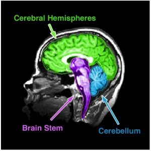
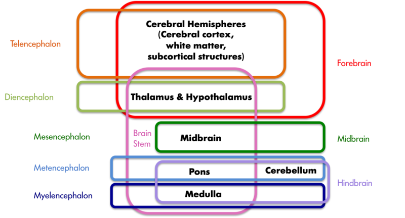
There are many ways to subdivide the mammalian brain, resulting in some inconsistent and ambiguous nomenclature over the history of neuroanatomy (Swanson, 2000). For simplicity, we will divide the brain into three basic parts: the brain stem, cerebellum, and cerebral hemispheres (see Figure 1). In Figure 2, however, we depict other prominent groupings (Swanson, 2000) of the six major subdivisions of the brain (Kandal, Schwartz, & Jessell, 2000).
Brain Stem
The brain stem is sometimes referred to as the “trunk” of the brain. It is responsible for many of the neural functions that keep us alive, including regulating our respiration (breathing), heart rate, and digestion. In keeping with its function, if a patient sustains severe damage to the brain stem he or she will require “life support” (i.e., machines are used to keep him or her alive). Because of its vital role in survival, in many countries a person who has lost brain stem function is said to be “brain dead,” although other countries require significant tissue loss in the cortex (of the cerebral hemispheres), which is responsible for our conscious experience, for the same diagnosis. The brain stem includes the medulla, pons, midbrain, and diencephalon (which consists of thalamus and hypothalamus). Collectively, these regions also are involved in our sleep–wake cycle, some sensory and motor function, as well as growth and other hormonal behaviors.
Cerebellum
The cerebellum is the distinctive structure at the back of the brain. The Greek philosopher and scientist Aristotle aptly referred to it as the “small brain” (“parencephalon” in Greek, “cerebellum” in Latin) in order to distinguish it from the “large brain” (“encephalon” in Greek, “cerebrum” in Latin). The cerebellum is critical for coordinated movement and posture. More recently, neuroimaging studies (see “Studying the Human Brain”) have implicated it in a range of cognitive abilities, including language. It is perhaps not surprising that the cerebellum’s influence extends beyond that of movement and posture, given that it contains the greatest number of neurons of any structure in the brain. However, the exact role it plays in these higher functions is still a matter of further study.
Cerebral Hemispheres
The cerebral hemispheres are responsible for our cognitive abilities and conscious experience. They consist of the cerebral cortex and accompanying white matter (“cerebrum” in Latin) as well as the subcortical structures of the basal ganglia, amygdala, and hippocampal formation. The cerebral cortex is the largest and most visible part of the brain, retaining the Latin name (cerebrum) for “large brain” that Aristotle coined. It consists of two hemispheres (literally two half spheres) and gives the brain its characteristic gray and convoluted appearance; the folds and grooves of the cortex are called gyri and sulci (gyrus and sulcus if referring to just one), respectively.
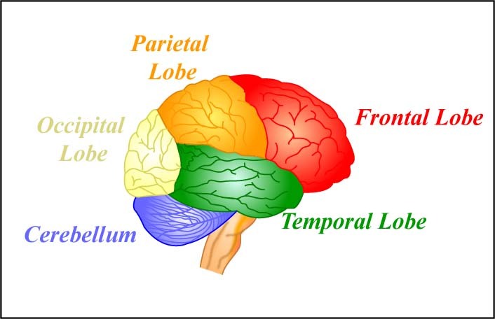
The two cerebral hemispheres can be further subdivided into four lobes: the occipital, temporal, parietal, and frontal lobes. The occipital lobe is responsible for vision, as is much of the temporal lobe. The temporal lobe is also involved in auditory processing, memory, and multisensory integration (e.g., the convergence of vision and audition). The parietal lobe houses the somatosensory (body sensations) cortex and structures involved in visual attention, as well as multisensory convergence zones. The frontal lobe houses the motor cortex and structures involved in motor planning, language, judgment, and decision-making. Not surprisingly then, the frontal lobe is proportionally larger in humans than in any other animal.
The subcortical structures are so named because they reside beneath the cortex. The basal ganglia are critical to voluntary movement and as such make contact with the cortex, the thalamus, and the brain stem. The amygdala and hippocampal formation are part of the limbic system, which also includes some cortical structures. The limbic system plays an important role in emotion and, in particular, in aversion and gratification.
A Brain Divided
The two cerebral hemispheres are connected by a dense bundle of white matter tracts called the corpus callosum. Some functions are replicated in the two hemispheres. For example, both hemispheres are responsible for sensory and motor function, although the sensory and motor cortices have a contralateral (or opposite-side) representation; that is, the left cerebral hemisphere is responsible for movements and sensations on the right side of the body and the right cerebral hemisphere is responsible for movements and sensations on the left side of the body. Other functions are lateralized; that is, they reside primarily in one hemisphere or the other. For example, for right-handed and the majority of left-handed individuals, the left hemisphere is most responsible for language.There are some people whose two hemispheres are not connected, either because the corpus callosum was surgically severed (callosotomy) or due to a genetic abnormality. These split-brain patients have helped us understand the functioning of the two hemispheres. First, because of the contralateral representation of sensory information, if an object is placed in only the left or only the right visual hemifield, then only the right or left hemisphere, respectively, of the split-brain patient will see it. In essence, it is as though the person has two brains in his or her head, each seeing half the world. Interestingly, because language is very often localized in the left hemisphere, if we show the right hemisphere a picture and ask the patient what she saw, she will say she didn’t see anything (because only the left hemisphere can speak and it didn’t see anything). However, we know that the right hemisphere sees the picture because if the patient is asked to press a button whenever she sees the image, the left hand (which is controlled by the right hemisphere) will respond despite the left hemisphere’s denial that anything was there. There are also some advantages to having disconnected hemispheres. Unlike those with a fully functional corpus callosum, a split-brain patient can simultaneously search for something in his right and left visual fields (Luck, Hillyard, Mangun, & Gazzaniga, 1989) and can do the equivalent of rubbing his stomach and patting his head at the same time (Franz, Eliason, Ivry, & Gazzaniga, 1996). In other words, they exhibit less competition between the hemispheres.
Gray Versus White Matter
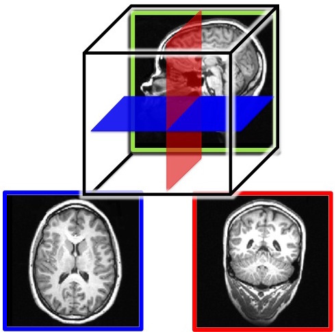
The cerebral hemispheres contain both grey and white matter, so called because they appear grayish and whitish in dissections or in an MRI (magnetic resonance imaging; see, “Studying the Human Brain”). The gray matter is composed of the neuronal cell bodies (see module, “Neurons”). The cell bodies (or soma) contain the genes of the cell and are responsible for metabolism (keeping the cell alive) and synthesizing proteins. In this way, the cell body is the workhorse of the cell. The white matter is composed of the axons of the neurons, and, in particular, axons that are covered with a sheath of myelin (fatty support cells that are whitish in color). Axons conduct the electrical signals from the cell and are, therefore, critical to cell communication. People use the expression “use your gray matter” when they want a person to think harder. The “gray matter” in this expression is probably a reference to the cerebral hemispheres more generally; the gray cortical sheet (the convoluted surface of the cortex) being the most visible. However, both the gray matter and white matter are critical to proper functioning of the mind. Losses of either result in deficits in language, memory, reasoning, and other mental functions. See Figure 3 for MRI slices showing both the inner white matter that connects the cell bodies in the gray cortical sheet.
Studying the Human Brain
How do we know what the brain does? We have gathered knowledge about the functions of the brain from many different methods. Each method is useful for answering distinct types of questions, but the strongest evidence for a specific role or function of a particular brain area is converging evidence; that is, similar findings reported from multiple studies using different methods. One of the first organized attempts to study the functions of the brain was phrenology, a popular field of study in the first half of the 19th century. Phrenologists assumed that various features of the brain, such as its uneven surface, are reflected on the skull; therefore, they attempted to correlate bumps and indentations of the skull with specific functions of the brain. For example, they would claim that a very artistic person has ridges on the head that vary in size and location from those of someone who is very good at spatial reasoning. Although the assumption that the skull reflects the underlying brain structure has been proven wrong, phrenology nonetheless significantly impacted current-day neuroscience and its thinking about the functions of the brain. That is, different parts of the brain are devoted to very specific functions that can be identified through scientific inquiry.
Neuroanatomy
Dissection of the brain, in either animals or cadavers, has been a critical tool of neuroscientists since 340 BC when Aristotle first published his dissections. Since then this method has advanced considerably with the discovery of various staining techniques that can highlight particular cells. Because the brain can be sliced very thinly, examined under the microscope, and particular cells highlighted, this method is especially useful for studying specific groups of neurons or small brain structures; that is, it has a very high spatial resolution. Dissections allow scientists to study changes in the brain that occur due to various diseases or experiences (e.g., exposure to drugs or brain injuries).Virtual dissection studies with living humans are also conducted. Here, the brain is imaged using computerized axial tomography (CAT) or MRI scanners; they reveal with very high precision the various structures in the brain and can help detect changes in gray or white matter. These changes in the brain can then be correlated with behavior, such as performance on memory tests, and, therefore, implicate specific brain areas in certain cognitive functions.
Changing the Brain
Some researchers induce lesions or ablate (i.e., remove) parts of the brain in animals. If the animal’s behavior changes after the lesion, we can infer that the removed structure is important for that behavior. Lesions of human brains are studied in patient populations only; that is, patients who have lost a brain region due to a stroke or other injury, or who have had surgical removal of a structure to treat a particular disease (e.g., a callosotomy to control epilepsy, as in split-brain patients). From such case studies, we can infer brain function by measuring changes in the behavior of the patients before and after the lesion.Because the brain works by generating electrical signals, it is also possible to change brain function with electrical stimulation. Transcranial magnetic stimulation (TMS) refers to a technique whereby a brief magnetic pulse is applied to the head that temporarily induces a weak electrical current in the brain. Although effects of TMS are sometimes referred to as temporary virtual lesions, it is more appropriate to describe the induced electricity as interference with neurons’ normal communication with each other. TMS allows very precise study of when events in the brain happen so it has a good temporal resolution, but its application is limited only to the surface of the cortex and cannot extend to deep areas of the brain. Transcranial direct current stimulation (tDCS) is similar to TMS except that it uses electrical current directly, rather than inducing it with magnetic pulses, by placing small electrodes on the skull. A brain area is stimulated by a low current (equivalent to an AA battery) for a more extended period of time than TMS. When used in combination with cognitive training, tDCS has been shown to improve performance of many cognitive functions such as mathematical ability, memory, attention, and coordination (e.g., Brasil-Neto, 2012; Feng, Bowden, & Kautz, 2013; Kuo & Nitsche, 2012).
Neuroimaging
Neuroimaging tools are used to study the brain in action; that is, when it is engaged in a specific task. Positron emission tomography (PET) records blood flow in the brain. The PET scanner detects the radioactive substance that is injected into the bloodstream of the participant just before or while he or she is performing some task (e.g., adding numbers). Because active neuron populations require metabolites, more blood and hence more radioactive substance flows into those regions. PET scanners detect the injected radioactive substance in specific brain regions, allowing researchers to infer that those areas were active during the task. Functional magnetic resonance imaging (fMRI) also relies on blood flow in the brain. This method, however, measures the changes in oxygen levels in the blood and does not require any substance to be injected into the participant. Both of these tools have good spatial resolution (although not as precise as dissection studies), but because it takes at least several seconds for the blood to arrive to the active areas of the brain, PET and fMRI have poor temporal resolution; that is, they do not tell us very precisely when the activity occurred.
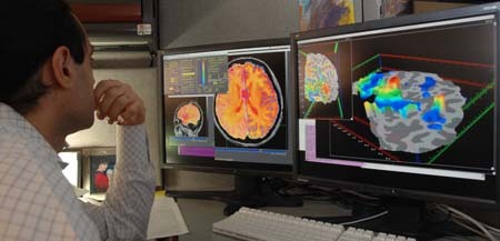
Electroencephalography (EEG), on the other hand, measures the electrical activity of the brain, and therefore, it has a much greater temporal resolution (millisecond precision rather than seconds) than PET or fMRI. Like tDCS, electrodes are placed on the participant’s head when he or she is performing a task. In this case, however, many more electrodes are used, and they measure rather than produce activity. Because the electrical activity picked up at any particular electrode can be coming from anywhere in the brain, EEG has poor spatial resolution; that is, we have only a rough idea of which part of the brain generates the measured activity.
Diffuse optical imaging (DOI) can give researchers the best of both worlds: high spatial and temporal resolution, depending on how it is used. Here, one shines infrared light into the brain, and measures the light that comes back out. DOI relies on the fact that the properties of the light change when it passes through oxygenated blood, or when it encounters active neurons. Researchers can then infer from the properties of the collected light what regions in the brain were engaged by the task. When DOI is set up to detect changes in blood oxygen levels, the temporal resolution is low and comparable to PET or fMRI. However, when DOI is set up to directly detect active neurons, it has both high spatial and temporal resolution.
Because the spatial and temporal resolution of each tool varies, strongest evidence for what role a certain brain area serves comes from converging evidence. For example, we are more likely to believe that the hippocampal formation is involved in memory if multiple studies using a variety of tasks and different neuroimaging tools provide evidence for this hypothesis. The brain is a complex system, and only advances in brain research will show whether the brain can ever really understand itself.
Unpacking Left Brain vs Right Brain
The concept of lateralization of function is more complicated than pop media presents. In this video, shared by Society for Neuroscience, Michael Colacci, a medical school student at Northwestern University Feinberg School of Medicine, unpacks some of the misunderstandings associated with lateralization of function.
Check Your Knowledge
To help you with your studying, we’ve included some practice questions for this module. These questions do not necessarily address all content in this module. They are intended as practice, and you are responsible for all of the content in this module even if there is no associated practice question. To promote deeper engagement with the material, we encourage you to create some questions of your own for your practice. You can then also return to these self-generated questions later in the course to test yourself.
Vocabulary
- Ablation
- Surgical removal of brain tissue.
- Axial plane
- See “horizontal plane.”
- Basal ganglia
- Subcortical structures of the cerebral hemispheres involved in voluntary movement.
- Brain stem
- The “trunk” of the brain comprised of the medulla, pons, midbrain, and diencephalon.
- Callosotomy
- Surgical procedure in which the corpus callosum is severed (used to control severe epilepsy).
- Case study
- A thorough study of a patient (or a few patients) with naturally occurring lesions.
- Cerebellum
- The distinctive structure at the back of the brain, Latin for “small brain.”
- Cerebral cortex
- The outermost gray matter of the cerebrum; the distinctive convolutions characteristic of the mammalian brain.
- Cerebral hemispheres
- The cerebral cortex, underlying white matter, and subcortical structures.
- Cerebrum
- Usually refers to the cerebral cortex and associated white matter, but in some texts includes the subcortical structures.
- Contralateral
- Literally “opposite side”; used to refer to the fact that the two hemispheres of the brain process sensory information and motor commands for the opposite side of the body (e.g., the left hemisphere controls the right side of the body).
- Converging evidence
- Similar findings reported from multiple studies using different methods.
- Coronal plane
- A slice that runs from head to foot; brain slices in this plane are similar to slices of a loaf of bread, with the eyes being the front of the loaf.
- Diffuse optical imaging (DOI)
- A neuroimaging technique that infers brain activity by measuring changes in light as it is passed through the skull and surface of the brain.
- Electroencephalography (EEG)
- A neuroimaging technique that measures electrical brain activity via multiple electrodes on the scalp.
- Frontal lobe
- The front most (anterior) part of the cerebrum; anterior to the central sulcus and responsible for motor output and planning, language, judgment, and decision-making.
- Functional magnetic resonance imaging (fMRI)
- Functional magnetic resonance imaging (fMRI): A neuroimaging technique that infers brain activity by measuring changes in oxygen levels in the blood.
- Gray matter
- The outer grayish regions of the brain comprised of the neurons’ cell bodies.
- Gyri
- (plural) Folds between sulci in the cortex.
- Gyrus
- A fold between sulci in the cortex.
- Horizontal plane
- A slice that runs horizontally through a standing person (i.e., parallel to the floor); slices of brain in this plane divide the top and bottom parts of the brain; this plane is similar to slicing a hamburger bun.
- Lateralized
- To the side; used to refer to the fact that specific functions may reside primarily in one hemisphere or the other (e.g., for the majority individuals, the left hemisphere is most responsible for language).
- Lesion
- A region in the brain that suffered damage through injury, disease, or medical intervention.
- Limbic system
- Includes the subcortical structures of the amygdala and hippocampal formation as well as some cortical structures; responsible for aversion and gratification.
- Metabolite
- A substance necessary for a living organism to maintain life.
- Motor cortex
- Region of the frontal lobe responsible for voluntary movement; the motor cortex has a contralateral representation of the human body.
- Myelin
- Fatty tissue, produced by glial cells (see module, “Neurons”) that insulates the axons of the neurons; myelin is necessary for normal conduction of electrical impulses among neurons.
- Nomenclature
- Naming conventions.
- Occipital lobe
- The back most (posterior) part of the cerebrum; involved in vision.
- Parietal lobe
- The part of the cerebrum between the frontal and occipital lobes; involved in bodily sensations, visual attention, and integrating the senses.
- Phrenology
- A now-discredited field of brain study, popular in the first half of the 19th century that correlated bumps and indentations of the skull with specific functions of the brain.
- Positron emission tomography (PET)
- A neuroimaging technique that measures brain activity by detecting the presence of a radioactive substance in the brain that is initially injected into the bloodstream and then pulled in by active brain tissue.
- Sagittal plane
- A slice that runs vertically from front to back; slices of brain in this plane divide the left and right side of the brain; this plane is similar to slicing a baked potato lengthwise.
- Somatosensory (body sensations) cortex
- The region of the parietal lobe responsible for bodily sensations; the somatosensory cortex has a contralateral representation of the human body.
- Spatial resolution
- A term that refers to how small the elements of an image are; high spatial resolution means the device or technique can resolve very small elements; in neuroscience it describes how small of a structure in the brain can be imaged.
- Split-brain patient
- A patient who has had most or all of his or her corpus callosum severed.
- Subcortical
- Structures that lie beneath the cerebral cortex, but above the brain stem.
- Sulci
- (plural) Grooves separating folds of the cortex.
- Sulcus
- A groove separating folds of the cortex.
- Temporal lobe
- The part of the cerebrum in front of (anterior to) the occipital lobe and below the lateral fissure; involved in vision, auditory processing, memory, and integrating vision and audition.
- Temporal resolution
- A term that refers to how small a unit of time can be measured; high temporal resolution means capable of resolving very small units of time; in neuroscience it describes how precisely in time a process can be measured in the brain.
- Transcranial direct current stimulation (tDCS)
- A neuroscience technique that passes mild electrical current directly through a brain area by placing small electrodes on the skull.
- Transcranial magnetic stimulation (TMS)
- A neuroscience technique whereby a brief magnetic pulse is applied to the head that temporarily induces a weak electrical current that interferes with ongoing activity.
- Transverse plane
- See “horizontal plane.”
- Visual hemifield
- The half of visual space (what we see) on one side of fixation (where we are looking); the left hemisphere is responsible for the right visual hemifield, and the right hemisphere is responsible for the left visual hemifield.
- White matter
- The inner whitish regions of the cerebrum comprised of the myelinated axons of neurons in the cerebral cortex.
References
- Beck, D. M., & Kastner, S. (2009). Top-down and bottom-up mechanisms in biasing competition in the human brain. Vision Research, 49, 1154–1165.
- Brasil-Neto, J. P. (2012). Learning, memory, and transcranial direct current stimulation. Frontiers in Psychiatry, 3(80). doi: 10.3389/fpsyt.2012.00080.
- Feng, W. W., Bowden, M. G., & Kautz, S. (2013). Review of transcranial direct current stimulation in poststroke recovery. Topics in Stroke Rehabilitation, 20, 68–77.
- Franz, E. A., Eliassen, J. C., Ivry, R. B., & Gazzaniga, M. S. (1996). Dissociation of spatial and temporal coupling in the bimanual movements of callosotomy patients. Psychological Science, 7, 306–310.
- Kandal, E. R., Schwartz, J. H., & Jessell, T. M. (Eds.) (2000). Principles of neural science (Vol. 4). New York, NY: McGraw-Hill.
- Kuo, M. F., & Nitsche, M. A. (2012). Effects of transcranial electrical stimulation on cognition. Clinical EEG and Neuroscience, 43, 192–199.
- Luck, S. J., Hillyard, S. A., Mangun, G. R., & Gazzaniga, M. S. (1989). Independent hemispheric attentional systems mediate visual search in split-brain patients. Nature, 342, 543–545.
- Swanson, L. (2000). What is the brain? Trends in Neurosciences, 23, 519–527.
How to cite this Chapter using APA Style:
Beck, D. & Tapia, E. (2019). The brain. Adapted for use by Queen’s University. Original chapter in R. Biswas-Diener & E. Diener (Eds), Noba textbook series: Psychology.Champaign, IL: DEF publishers. Retrieved from http://noba.to/jx7268sd
Copyright and Acknowledgment:
This material is licensed under the Creative Commons Attribution-NonCommercial-ShareAlike 4.0 International License. To view a copy of this license, visit: http://creativecommons.org/licenses/by-nc-sa/4.0/deed.en_US.
This material is attributed to the Diener Education Fund (copyright © 2018) and can be accessed via this link: http://noba.to/jx7268sd.
Additional information about the Diener Education Fund (DEF) can be accessed here.
