9 Respiratory Diseases of Sheep and Goats
| Course priority #1 | Course priority #2 | Course priority #3 | |
| Sheep and goats |
|
|
|
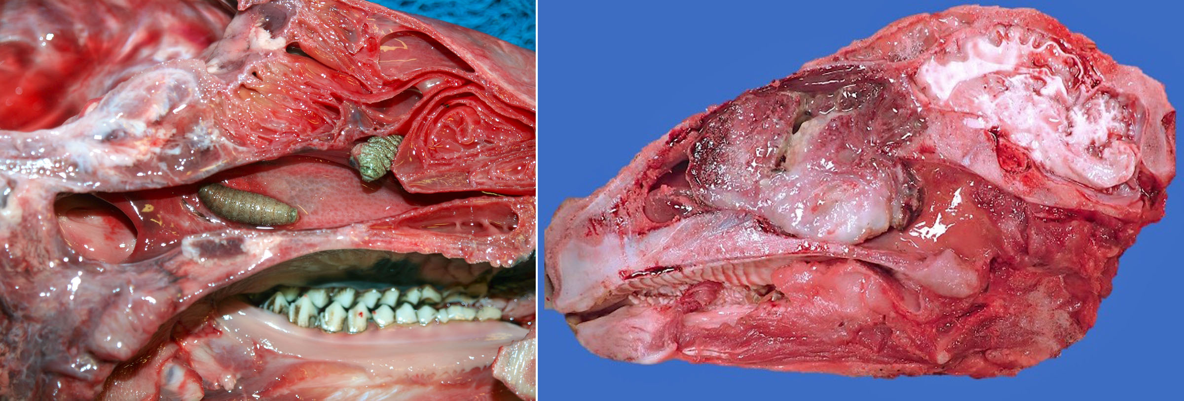
oestrus ovis nasal bots
Oestrus ovis adult flies deposit first-stage larvae that migrate through and develop in the nasal cavity. These can result in sneezing. They occasionally become imprisoned in nasal passages or sinuses as they outgrow their escape routes.
Enzootic nasal tumour
(aka Transmissible nasal tumour)
Nasal adenomas in sheep and goats are caused by a retrovirus named Enzootic nasal tumour virus, closely related to Jaagsiekte sheep retrovirus. Because the disease is contagious, multiple cases may occur within a flock.
Clinical signs are stertor, sneezing, or failure to thrive.
The tumour forms a large, expansile, usually non-invasive, white-tan mass obstructing the nasal passage. This occasionally deforms the face and is externally visible, but most cases would be missed if the nasal cavity were not examined.
Question:
List two ways that nasal tumours of dogs differ from those of sheep. (Answers are provided at the end of the chapter).
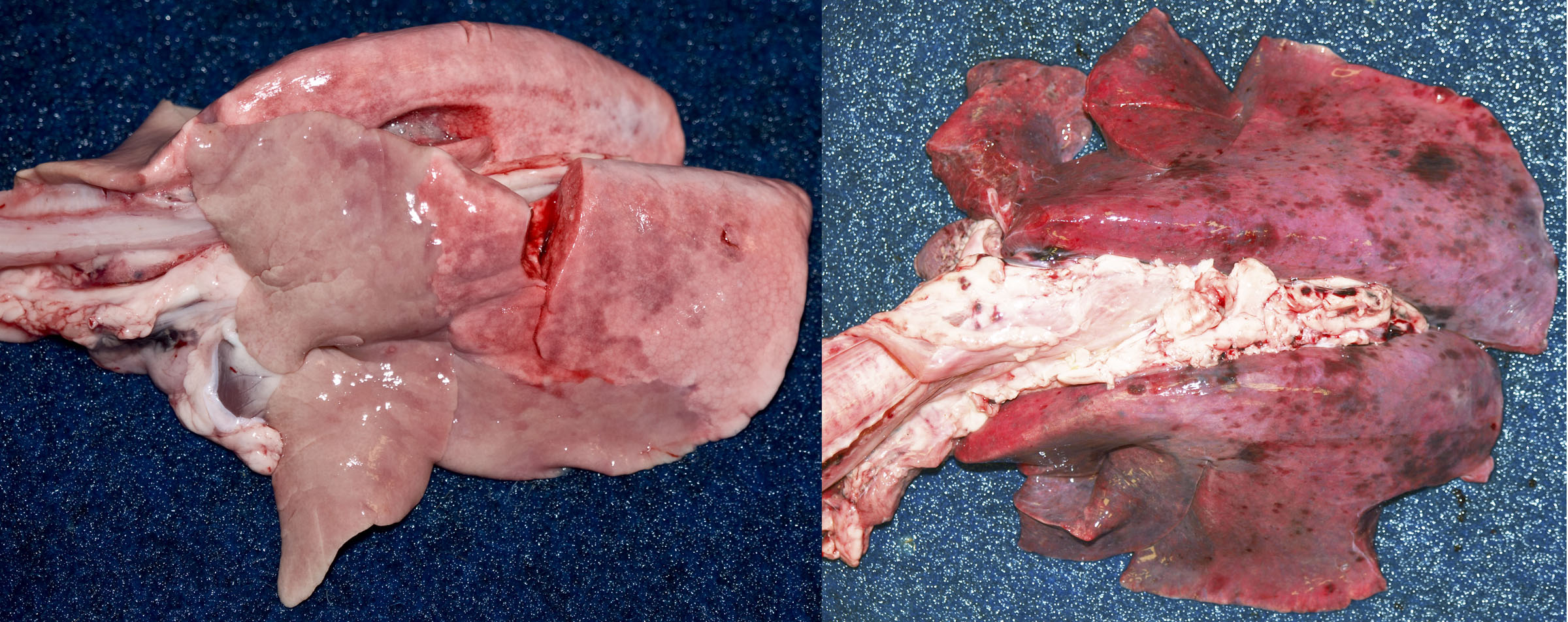
Pneumonic pasteurellosis
Mannheimia haemolytica, Bibersteinia trehalosi, Histophilus somni, and Pasteurella multocida cause similar diseases as in cattle. Predisposing causes include viral and mycoplasmal infections.
Septicemic pasteurellosis
Mannheimia haemolytica and Bibersteinia trehalosi (formerly Pasteurella trehalosi) are important causes of septicemia in lambs. Affected lambs are usually found dead, with widespread petechial hemorrhages in lung, muscle, lymph nodes, and serosal surfaces. In young lambs, Mannheimia haemolytica typically causes concurrent bronchopneumonia and septicemia with petechial hemorrhages and/or fibrinous exudates in body cavities. In weaned lambs (older lambs), Bibersteinia trehalosi typically causes widespread petechial hemorrhages from septicemia, but usually without pneumonia. In the latter, the digestive tract is thought to be a frequent portal of entry.
Question:
How do the lesions of pneumonic pasteurellosis differ from those of septicemic pasteurellosis in lambs? (Answers are provided at the end of the chapter).
Viral pneumonia
Respiratory syncytial viruses cause disease similar to BRSV.
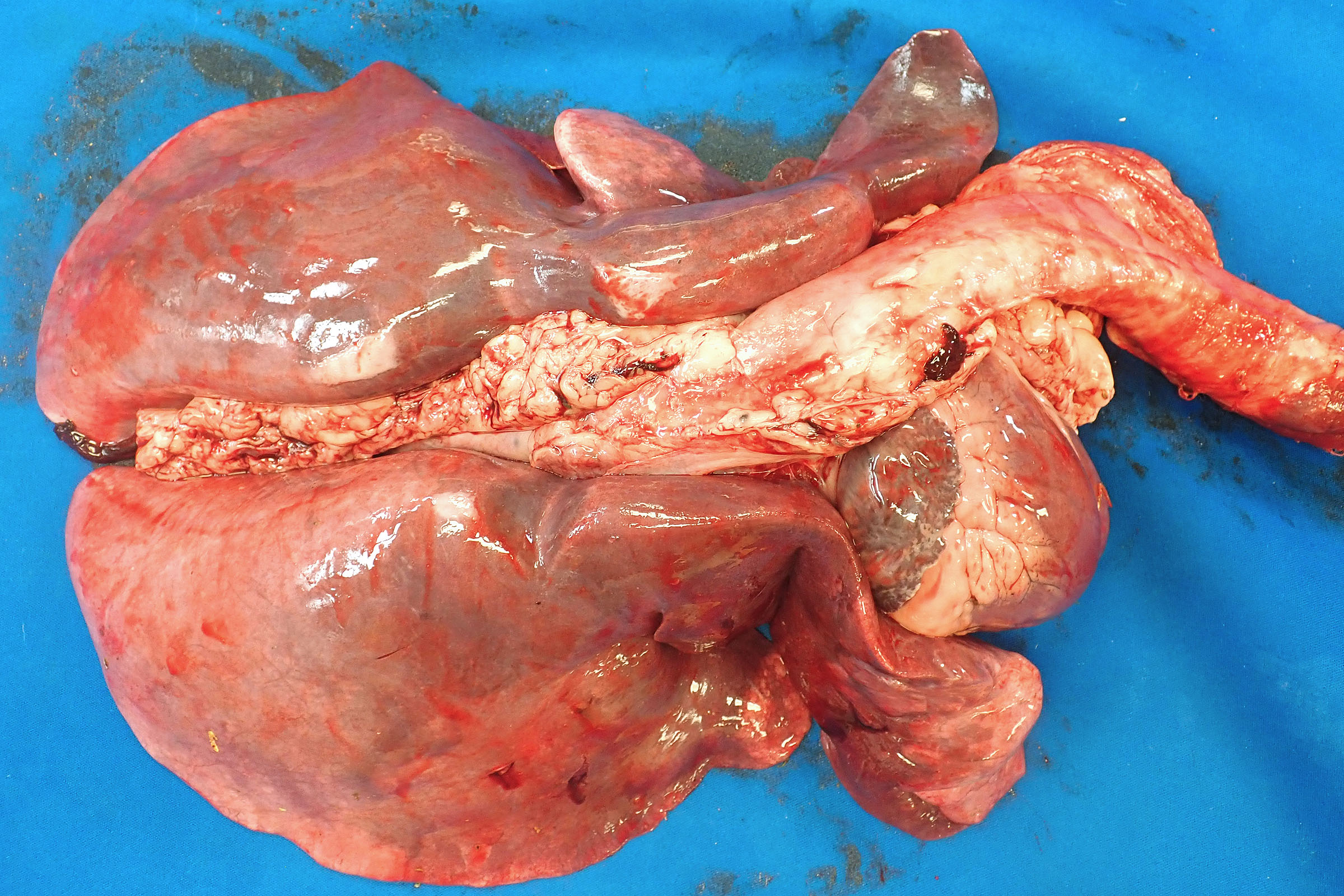
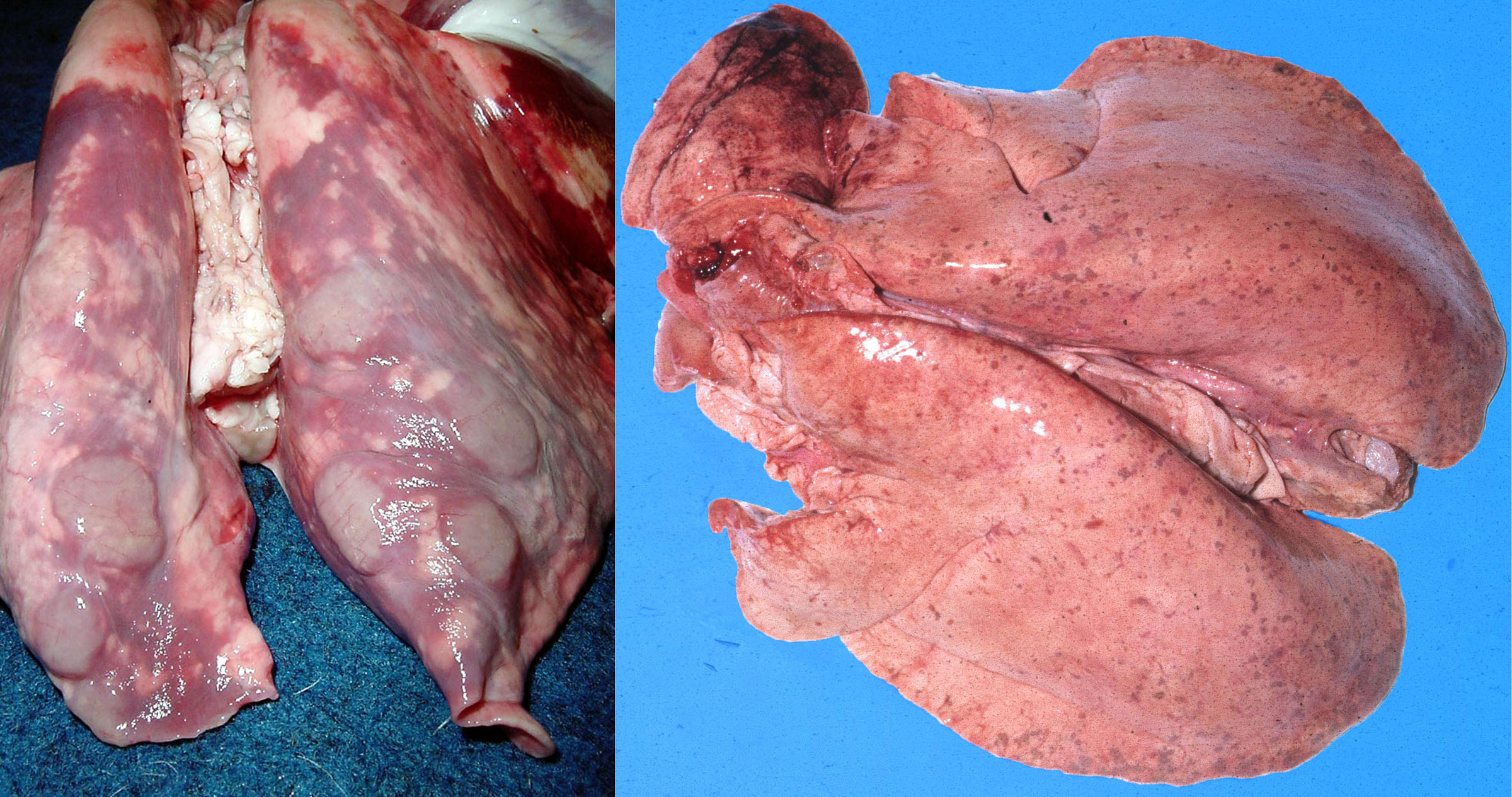
Lentiviral pneumonia
Lentiviruses are a subfamily of retroviruses. Lentiviruses cause maedi-visna in sheep (also known as ovine progressive pneumonia in the USA), and caprine arthritis-encephalitis in goats.
Maedi visna virus is transmitted to lambs by ingestion of infected colostrum, but disease manifests only in adulthood. This was the first-discovered of the “slow viral” infections, and the Icelandic names reflect this initial discovery. “Maedi” (dyspnea) refers to the pulmonary form. “Visna” (wasting) refers to the neurologic form, which is now rare.
The disease affects adult sheep usually more than 2 years old, causing chronic inexorably progressive weight loss and dyspnea. Gross lesions are of interstitial pneumonia: the lungs are diffusely pale, heavy, wet, and fail to collapse. Bronchial lymph nodes are enlarged. There may be polyarthritis and/or mastitis.
An ELISA test for maedi-visna is available, but is most useful for herd-level management of the disease rather than individual animal diagnosis. Diagnosis in individuals is usually based on gross and histologic lesions.
Caprine arthritis encephalitis (CAE) is the lentiviral disease of goats. Adult goats develop polyarthritis, mastitis, and/or respiratory disease. The respiratory disease is characterized by progressive cachexia and terminal dyspnea, and interstitial pneumonia is grossly similar to that described for maedi.
Neurologic or respiratory disease also affects kids. Note this difference: maedi-visna is a disease of adult sheep, whereas CAE may affect old goats or young kids.
Question:
Which could cause diffuse interstitial pneumonia in animals less than 6 months of age: Maedi visna virus or Caprine arthritis encephalitis virus, or both? (Answers are provided at the end of the chapter).
Ovine pulmonary adenocarcinoma
Ovine pulmonary adenocarcinoma is a form of lung cancer caused by “jaagsiekte sheep retrovirus”, which is closely related to enzootic nasal tumour virus. Jaagsiekte—an older name for the disease and still the official name for the causative virus—is an Afrikaans (South African Boer) word meaning “driving disease”, from the occurrence of disease when sheep are moved or “driven” from one area to another. The virus (JSRV) causes a pulmonary adenocarcinoma, and this primary neoplasm often metastasizes within the lung but not typically to other organs. The cancer-causing role of the virus’s envelope glycoprotein was discovered by none other than Dr. Sarah Wootton, a virologist in our department. This disease is important in many parts of the world, but rare or absent from Canada.
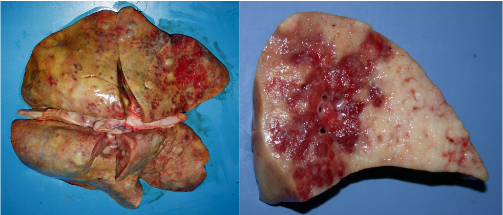
Muellerius capillaris
Infection with the nematode Muellerius capillaris is common in sheep, but usually of little clinical significance. It most commonly causes clinical disease in goats. The gross lesions are red to gray nodules in the dorsocaudal lung. The worms are not grossly visible but microscopic examination reveals numerous worms. In this way, Muellerius is comparable to Aleurostrongylus in cats. Other sheep lungworms include Dictyocaulus filaria and Protostrongylus rufescens; both are rare in Ontario.
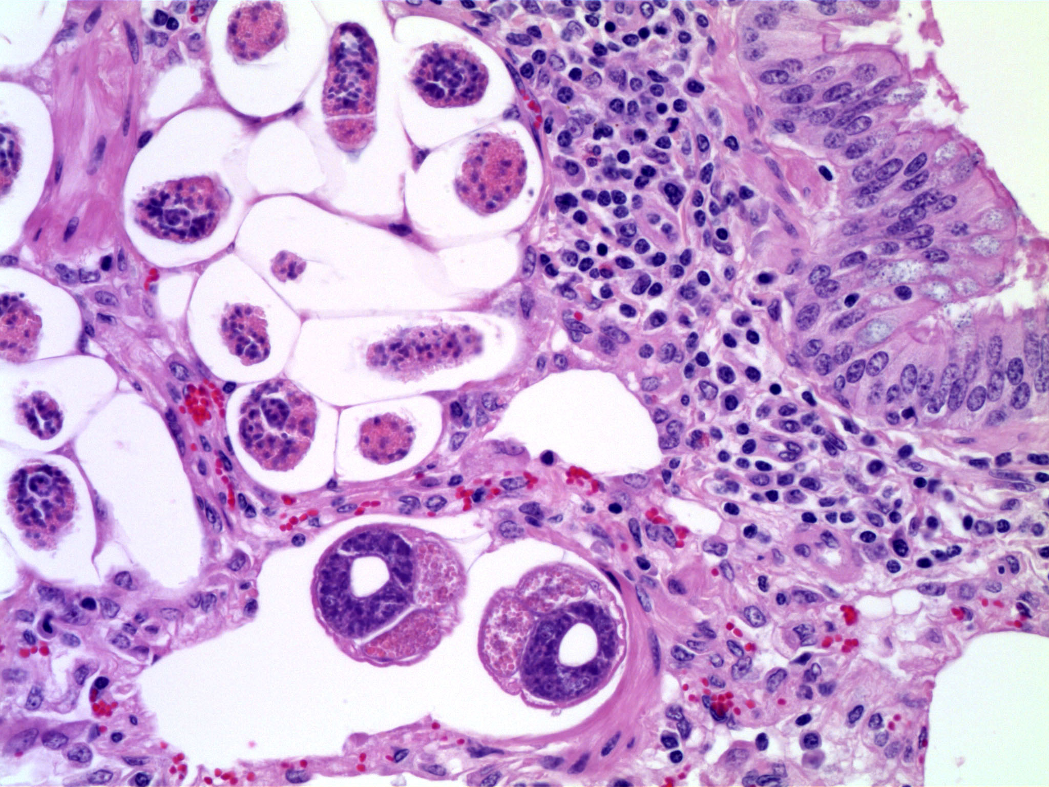
Question:
What is the common lungworm of sheep? Can you see the worms in the centre of the nodules, with the “naked eye”? (Answers are provided at the end of the chapter).
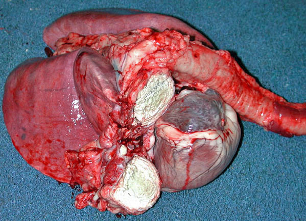
Caseous lymphadenitis
Many different bacteria can cause abscesses in lymph nodes of sheep, but “caseous lymphadenitis” is a specific contagious disease caused by Corynebacterium pseudotuberculosis. Abscesses develop in lymph nodes, lung, and other tissues, and often have a characteristic laminated appearance on cut section.
Peste des petits ruminants
PPR is an important disease of sheep in Africa. It is a reportable disease that is foreign to Canada, and unlikely to occur here. It is caused by a morbillivirus (Paramyxoviridae) named Peste des petits ruminants virus, which is closely related to rinderpest morbillivirus (as well as canine distemper and human measles). Rinderpest (now eradicated from the planet) causes lesions of the digestive tract, whereas PPRV causes digestive, upper respiratory, and lung lesions.
Answer:
List two ways that nasal tumours of dogs differ from those of sheep.
Sheep: infectious (retrovirus), expansile (benign).
Dogs: non-infectious, invasive (malignant).
Answer:
How do the lesions of pneumonic pasteurellosis differ from those of septicemic pasteurellosis in lambs?
Pneumonic pasteurellosis: cranioventral consolidation of the lung.
Septicemic pasteurellosis: widespread petechial hemorrhages in lung and other organs.
Answer:
Which could cause diffuse interstitial pneumonia in animals less than 6 months of age: Maedi visna virus or Caprine arthritis encephalitis virus, or both?
Only Caprine arthritis encephalitis virus affects youngsters. Maedi occurs only in adult sheep.
Answer:
What is the common lungworm of sheep? Can you see the worms in the centre of the nodules?
Muellerius capillaris; the worms are microscopic, not seen grossly.
