Tooth Morphology – Permanent Anterior Teeth
Understanding and Using Standard Diagrams of the Teeth
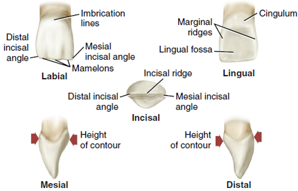
Each tooth must be “viewed” from all directions or surfaces when studying tooth morphology. When looking at diagrams of anterior teeth, you will typically refer to the tooth illustrated from five viewpoints.
As the tooth is described and landmarks are identified in this module, note which surface you are viewing. Often, labial or lingual surfaces will be compared and contrasted between teeth. For example, you will learn how the lingual anatomy differs between adjacent anterior teeth or the occlusal anatomy between adjacent posterior teeth. Any landmark or characteristic will be pointed out on the surface from which it is best viewed.
Permanent Anterior Dentition
The permanent anterior teeth include central incisors, lateral incisors and canines. The central incisors are closest to the midline, with the lateral incisors being adjacent to the central incisors, and the canines are adjacent to the lateral incisors. All anterior permanent teeth are succedaneous.
All anterior teeth have a cingulum on the lingual surface. The lingual surface has rounded, raised borders on the mesial and distal surfaces called marginal ridges. Some anterior teeth have a fossa.
An Interesting Fact About Maxillary Central Incisors
Did you know that in most people, the shape of the labial surface of the maxillary central incisors reflects the shape of their face? This means that when someone smiles, the maxillary central can appear to be oval, heart-shaped, round, square, or rectangular, depending on the general shape of their face.
Reflect: Look at the smiles of your friends and family and see if you can confirm the relationship between the shape of their face and the shape of their central incisors!
Maxillary Central Incisors – Teeth 11 and 21
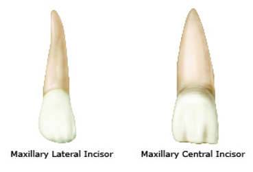
The maxillary central incisors have unique anatomic features. They are larger than the mandibular central incisors in all dimensions, especially in width (mesiodistally). The labial surfaces are more rounded from the incisal aspect. The root of the maxillary central incisors is shorter than the roots of the other permanent teeth.
Let’s take a closer look at the specific landmarks and characteristics found on a maxillary central incisor below.
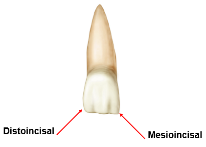
For maxillary central incisors teeth 11 and 21, the incisal ridge is an approximate right angle at the mesioincisal line angle. It has a slightly rounded upward slope at the distoincisal line angle which is shown in the image below.
See the hotspots in the image below to see the lingual surface of the maxillary central incisor.
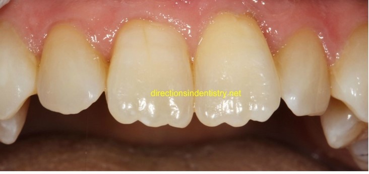
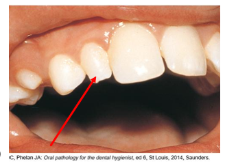
Newly erupted maxillary central incisors have mamelons, giving the appearance of a scalloped incisal edge shown in the image below.
What is a Mamelon?
When incisors first erupt into the oral cavity, they appear to have a scalloped incisal ridge, as shown previously and in the image below. With time and wear and tear, those scallops wear away into a more straight edge. That is why you often see mamelons in a child’s mouth but not in an adult’s. Mamelons represent areas on the crown corresponding to developmental centers as a tooth grows from a bud into its typical shape.
Maxillary Lateral Incisors – Teeth 12 and 22
Specific landmarks and characteristics found on maxillary lateral incisors. The lateral incisors vary in form more than any other tooth in the mouth, except the third molars, and are often congenitally missing. Because of the variations in form, the permanent maxillary lateral incisors present challenges during preventive, restorative, and orthodontic procedures.
Unattractive open contacts (spaces between teeth), called diastema, often occur in this area because of the variations in tooth size and position in the arch. The maxillary lateral incisor has a relatively smooth and straight single root that may curve slightly distally. Recognizing this feature is helpful in the mounting of radiographs.
When you compare a maxillary lateral incisor to a central incisor, you find that:
- Maxillary lateral incisors erupt after the maxillary central incisors.
- Maxillary lateral incisors are smaller in all crown dimensions.
- The roots are a bit longer and curve toward the distal.
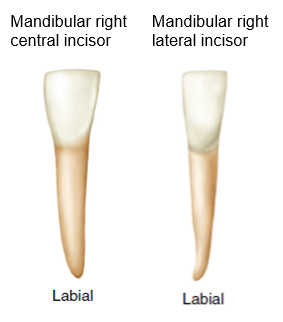
Mandibular Central Incisors – Teeth 31 and 41
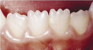
Permanent mandibular incisors are distinguished as the smallest and simplest teeth in the oral cavity. They are also most likely to erupt before the maxillary centrals. Let’s take a closer look at the specific landmarks and characteristics that differentiate a mandibular central incisor from a maxillary central incisor.
Mandibular Lateral Incisors – Teeth 32 and 42
The mandibular lateral incisors are slightly larger than the mandibular central incisors but otherwise similar to them. The lateral teeth usually erupt after the mandibular central incisors. The lateral incisors have a small, distally placed cingulum.
 |
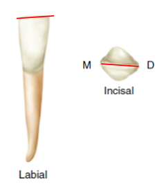 |
| The mandibular lateral incisors have slightly larger roots than the central incisors. There is less symmetry in these teeth than in the mandibular central incisors. | The mandibular lateral incisor has an incisal edge that slopes toward the distal surface of the crown. The slope is also visible from the incisal view. |
Activity 1: Maxillary Central and Lateral Incisors
Activity 2: Mandibular Central and Lateral Incisors
Permanent Canines
The canines are the cornerstones of the dental arches and are the longest, most stable teeth in the adult permanent dentition. When a person loses his or her teeth to periodontal disease, you will often observe that the canines are the last teeth remaining in the mouth.
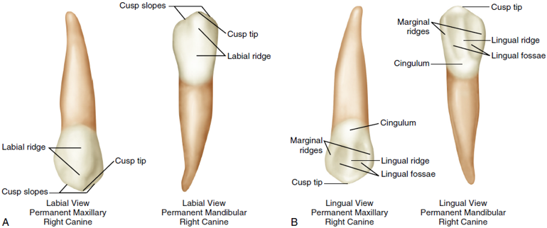
Patients may refer to the canines as “eye teeth,” they often exhibit a darker color than the incisors. Also, the canines are the only anterior teeth that have a cusp.
When you view canine teeth to identify their unique landmarks and characteristics, you will notice some terms that are different from the anatomical terms used to describe incisors. Because a canine tooth has a cusp instead of an incisal edge, the landmarks used to describe the labial and lingual surfaces are different. Look at the image to familiarize yourself with these differences.
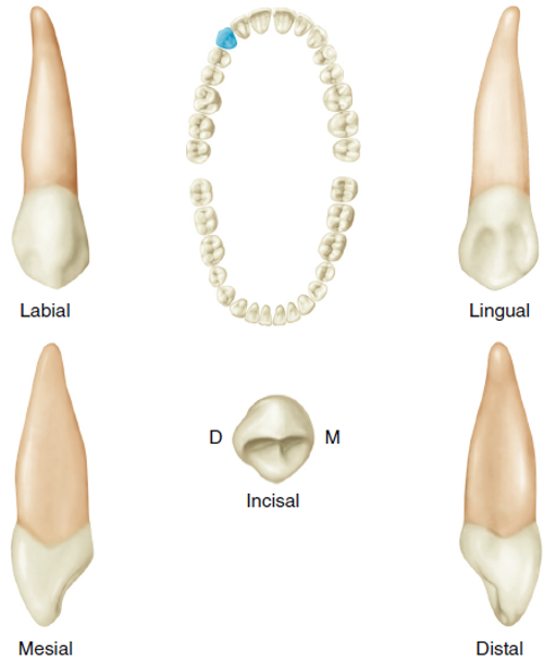
Maxillary Canines – Teeth 13 and 23
One of the first things you notice about the maxillary canines is that they have a pointed cusp instead of a straight incisal edge. The presence of this cusp relates to two facts concerning form and function:
- The function of a canine tooth is to grasp or hold large chunks of food.
- The canine tooth is positioned between the teeth that cut or bite food and the teeth that grind food.
Depending on our occlusion, the cusp point and cusp slopes may wear away somewhat as we age. However, the remaining anatomy of the maxillary canine usually stays intact and can be easily observed in the mouth.
Notice in the image below that:
- Maxillary canines have a large, well-developed cusp.
- The cusp tip is positioned directly over the root apex.
- The lingual features of the maxillary canines are prominent.
Because of the cusp, there are two cusp ridges, one mesial and one distal. The distal cusp ridge is longer, leading to an elevation of enamel, frequently referred to as a “dog ear.” This is an important anatomical landmark when positioning the x-ray machine for a canine radiograph.
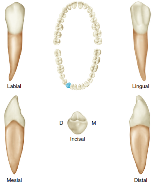
Mandibular Canines – Teeth 33 and 43
There is no doubt that mandibular canines look similar to maxillary canines. All of the landmarks and characteristics present on a maxillary canine are also present, to a lesser degree, on the mandibular canine. There are two ways to tell the difference between the maxillary and mandibular canines:
- The mesial line angle is very straight as viewed from the labial or lingual view.
- The cusp tip is positioned lingual to the root apex, almost like the tooth is “bending backward” a bit.
- The root tip of the mandibular canine will usually curve mesially, which is the opposite of the other teeth.
Look at the image below and observe these details.
Activity 3: Tooth Morphology for Canines
You have completed Module 8. Please return to Blackboard for the next steps.
Media Attributions
- Images from: Modern Dental Assisting, 13th and 14th Edition
(mor-FOL-uh-jee); Study of form and shape, as of the teeth.
(suk-se-DAY-nee-us); Permanent teeth that replace primary teeth.
(SING-gyoo-lum); Raised, rounded area on the cervical third of the lingual surface.
Rounded, raised border on the mesial and distal portions of the lingual surfaces of anterior teeth and the occlusal table of posterior teeth.
(MAM-uh-lon); Rounded enamel extension on the incisal ridges of incisors.
Ridge on permanent incisors that appears flattened on labial, lingual, or incisal view after tooth eruption.
(dye-uh-STEE-muh); Space between two teeth
Major elevation on the masticatory surfaces of canines and posterior teeth.
