Landmarks of the Face and Oral Cavity (Part A)
The Oral Cavity Proper
The oral cavity proper is the area inside the dental arches. Behind the last molar on each side is a space that links the vestibule and the oral cavity proper. If you close your teeth together, you can feel the areas of the oral cavity proper with your tongue.
Hard Palate
The hard palate separates the nasal cavity above from the oral cavity below. The nasal surfaces are covered with respiratory mucosa, and the oral surfaces are covered with oral mucosa. The mucosa of the hard palate is tightly bound to the underlying bone, which is why submucosal injections in this area can be extremely painful.
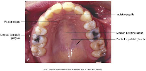
Behind the maxillary central incisors is the incisive papilla, a pear-shaped pad of tissue covering the incisive foramen. Located behind the incisive papilla are irregular, horizontal ridges of masticatory mucosa called palatal rugae. There is also the midline palatine raphe, which runs down the middle of the hard palate. With your tongue, feel your palate to feel some of the tissues and landmarks of the palate.
Soft Palate
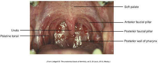
The soft palate is the movable posterior third of the palate located behind the hard palate. It has no bony support and hangs into the pharynx behind it. The soft palate ends with a pear-shaped hanging projection of tissue called the uvula.
The soft palate is supported posteriorly by two arches called fauces. The anterior arch goes from the soft palate down to the sides of the tongue and is called the anterior faucial pillar. The posterior arch is the free posterior border of the soft palate and is called the posterior faucial pillar. The opening between the two arches is called the isthmus of fauces, and the palatine tonsils are located here.
Below is an image displaying the surfaces of a soft palate.
Reflect: Does the tissue feel soft when gently touching your tongue? Does it feel muscular?
Test Your Knowledge
Tongue
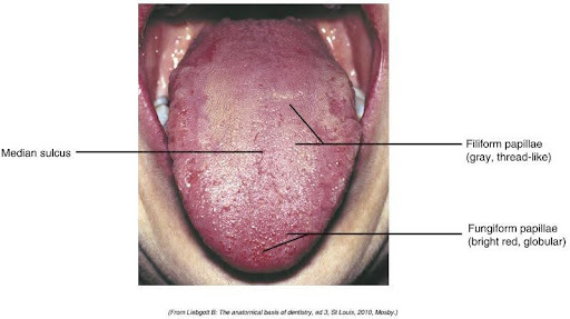
The tongue is made up mainly of muscles. It is covered on the top with a thick mucous membrane layer and thousands of tiny projections called papillae. Inside the papillae are sensory organs and nerves for taste and touch. The papillae will usually appear pinkish-white and be velvety smooth on the dorsal (top) surface of a healthy tongue.
The tongue is one of the body’s most versatile organs and is responsible for:
- speech
- manipulation and positioning of food
- sense of taste and touch
- swallowing
- cleansing of the oral cavity
Parts and Surfaces of the Tongue
The anterior two-thirds of the tongue is called the body. The root of the tongue is the posterior portion that turns vertically downward toward the pharynx. The dorsum comprises the superior (upper) and posterior roughened surfaces. Small papillae of various shapes and colours cover the dorsum surface.
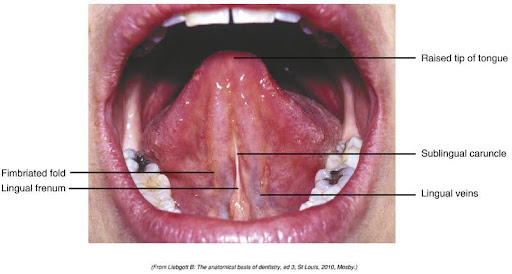
The sublingual surface of the tongue is covered with smooth, thin, transparent mucosa, and many underlying vessels can be seen. Two small papillae are on either side of the lingual frenum, just behind the mandibular central incisors. These papillae contain openings of the submandibular salivary ducts. Saliva enters the oral cavity through these ducts.
The lingual frenum is a thin fold of mucous membrane extending from the mouth’s floor to the tongue’s underside. On either side of the lingual surface are two smaller fimbriated folds.
Clinical Considerations: Lingual Frenum
When the lingual frenum is unusually short, it can severely limit the tongue’s ability to move. When this occurs, it may impact:
- speech
- an infant’s ability to latch on to a nipple from a breast or bottle
- The ability of an oral health care professional to take radiographs or impressions
This is commonly called “tongue-tied” and can be corrected with a surgical procedure called a lingual frenectomy, meaning the lingual frenum is cut.
Clinical Considerations: Gag Reflex
When working in a patient’s mouth, one must be careful not to trigger the gag reflex accidentally. The gag reflex may be the membranes of the soft palate, the fauces, and/or the posterior area of the tongue. Touching any of these areas may trigger the gag reflex, leading to the patient gagging and/or vomiting. Not all patients may gag. This is something that we should ask before any treatment.
Taste Buds
Taste buds are sensory organs that allow us to taste the flavours of food and warn us when the temperature of food is too hot. Taste buds are located on the dorsum of the tongue. To stimulate the taste buds to detect flavours, saliva is required. If saliva is reduced, our sense of taste is diminished.
Taste buds are located on the fungiform papillae and in the trough of the large vallate papillae, which form a V on the posterior portion of the tongue. The sense of touch is provided by numerous filiform papillae that cover the entire surface of the tongue; these papillae have no taste receptors.
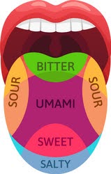
Although thousands of flavours are known, it is thought that only five primary tastes combine to create all flavours. These are salty, sweet, sour, bitter and umami.
Some substances may taste sweet when put into the mouth but taste bitter when they reach the back. An example of this is saccharin. Of the five primary tastes, the one that is the most easily distinguished is bitter. It is thought that this may be a protective mechanism as many deadly toxins taste bitter, so a person may spit them out before the toxin can do harm.
Teeth
People get two sets of teeth during their life. Teeth sit in bony sockets, or alveoli, within the alveolar process of the maxilla and mandible. The portion of the tooth visible in the oral cavity is called the crown, and a cuff of gingival tissue surrounds each tooth. Dental anatomy will be discussed in upcoming modules.
Test Your Knowledge
Activity 1: Landmarks of the Face and Oral Cavity Definitions
You have completed Module 5A. Please return to Blackboard for the next steps.
Media Attributions
- Images from: Modern Dental Assisting, 13th and 14th Edition
Space on the tongue side within the upper and lower dental arches.
Pear-shaped pad of tissue that covers the incisive foramen.
Pear-shaped projection at the end of the soft palate.
Anterior arch of the soft palate.
Posterior arch of the soft palate.
Opening between the two arches of the soft palate.
Knoblike projections on the tongue.
Largest papillae on the tongue, arranged in the form of a V.
Threadlike elevations that cover most of the tongue.
